
Definition
• PE: Thrombosis from a vein that embolizes to the pulmonary arterial system
• DVT: Thrombosis of lower extremity (popliteal, femoral [incl SFV], iliac) or upper extremity (basilic, cephalic) veins
Approach
• (1) Assess PreTP, (2) perform basic test (labs, CXR, ECG), (3) D-dimer, (4) imaging studies, to obtain (5) post-TP
• If HD stable perform Dx tests; if unstable, consider empiric tx ± thrombolytics if potential benefit outweighs risk of bleeding (Chest 2004;126(suppl):401S)
History (Chest 1991;100:598; Am J Card 1991;68:1723)
• RFs: Prior DVT/PE, hypercoagulable state, pregnancy/OCPs, malignancy, prolonged immobilization, recent surgery, FHx DVT/PE
• PE: Dyspnea (73%), pleuritic CP (66%), cough (37%), syncope, ↓ BP, PEA
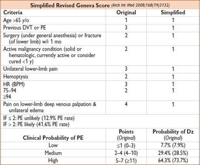
• Assess PreTP: May use PERC (to decide whether any testing is necessary) or Wells criteria (to decide whether D-dimer is sufficient w/u)
• PERC: If (1) low likely by gestalt + (2) “Yes” to all PERC criteria → 1.8% PE miss rate

Findings
• PE: Unexplained ↑ HR, ↑ RR, ↓ SpO2, fever, JVD
• DVT: Leg edema/pain. Clinical signs DVT ∼50% pts.
Evaluation (PE)
• Obtain ECG (sinus tachycardia, S1Q3T3 not sens/spec, simultaneous TWI in inferior & anterolateral leads), Hct, PT/PTT, Cr
• CXR: R/o other causes; “classic” PE CXR findings (Hampton’s hump & Westermark’s sign) not sens/spec
• D-dimer (sens 95–98%, spec 40–55%, ELISA or Rapid Quant. ELISA; Ann Intern Med 2004;140:589, NPV >99% for low pre-TP; JAMA 2006;295:172). Sens/spec vary by assay/cutoff.
• False+: Pregnancy, trauma, infection, malignancy, inflammatory conditions, surgery, ↑ age, SCD, AF, ACS, CVA, acute UGIB, DIC
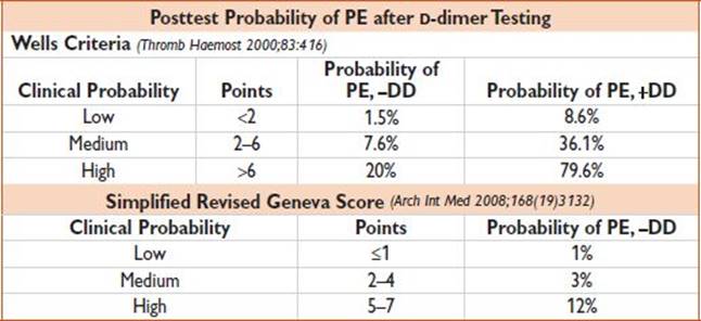
• In pts w/ a low PreTP for PE, a negative quantitative D-dimer assay result can be used to exclude PE
• In pts w/ an intermediate PreTP for PE, a negative quantitative D-dimer assay result may be used to exclude PE
• If D-dimer + → more testing (see below)
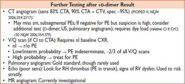
• For pts w/ low or PE unlikely PreTP of PE who require additional diagnostic testing (ie, +D-dimer or D-dimer not available), a negative, multidetector CT pulmonary angiogram alone can be used to excluse PE
• For pts w/ an intermediate or high probability for PE & a negative CTPA result in whom clinical concern for PE still exists & CT venogram has not already been performed, consider additional testing (ie, D-dimer, lower extremity imaging, VQ scanning, traditional pulmonary arteriography) to exclude VTE dz
• Risk stratify: ↑ HR, ↓ BP, ↓ SpO2, CTA RV/LV dimension >0.9, ↑ Tn or BNP, echo e/o RV dysfxn, D-dimer >4000 all predict bad outcomes
Evaluation (DVT)
• Assign clinical probability using Wells score
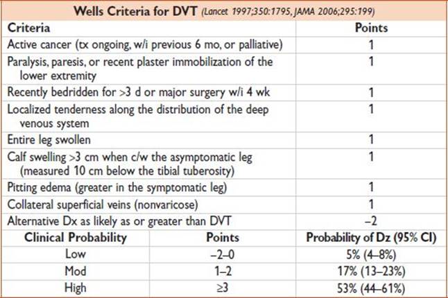
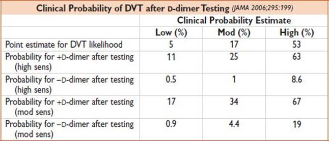
• D-dimer: Assays include ELISA & immunoturbidimetric methods (high sens) as well as whole-blood & quantitative latex agglutination assays (mod sens). There is a wide variation in the sens, nl reference ranges, & cut-off points among different assays.
• Compression US of proximal veins
• Whole-leg duplex ultrasound (“LENI”)
• CT venography
• See below for diagnostic algorithms in eval of DVT (Chest 2012;141(2)(suppl):e351S)
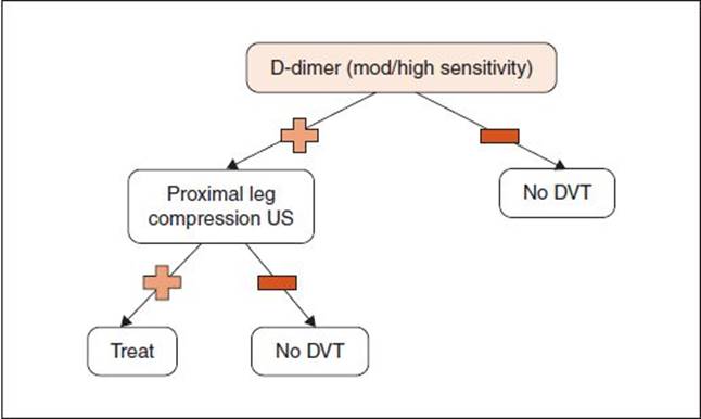
Figure 1.2 Low pretest probability of DVT.

Figure 1.3 Moderate pretest probability of DVT. A: Starting with D-dimer assessment. B: Starting with ultrasonography.
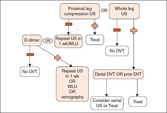
Figure 1.4 High pretest probability of DVT.
Treatment
• O2, monitor BP/rhythm, IV fluids for ↓ BP (preload dep)
• Anticoagulation (PE or DVT): Heparin (80 U/kg, 18 U/kg/h IV), enoxaparin (1 mg/kg SC). ?LMWH superior to heparin for DVT; LMWH may be superior to heparin for ↓ D/major bleeding in PE (Chest2004;126(suppl):401S; Ann Intern Med 2007;146:369), relative CI LMWH → renal insufficient, obesity (? unpredictable absorption)
• Thrombolytic: tPA (100 mg over 2 h), for extensive DVT/PE or high clinical suspicion & HD instability. No mortality benefit w/ RV dysfxn (on echo) alone (NEJM 2002;347:1143), so use is controversial.
• Administer thrombolytic therapy in HD unstable pts w/ confirmed PE for whom the benefits of tx outweigh the risks of life-threatening bleeding cx
• In centers w/ capability of surgical or mechanical thrombectomy, procedural intervention may be used as an alternative therapy
• Catheter or surgical thrombectomy (PE): For pts w/ HD instability & massive PE if (1) CI to lysis, (2) failed lysis w/ tPA, or (3) experienced center & +RV dysfxn. Consult cardiac surgery.
• IVC filter: When medical tx fails or is contraindicated. No long-term mortality benefit (NEJM 1998;338:409).
Disposition
• If PE negative → look for other causes
• If +DVT & no suspicion for PE → d/c home after period of observation w/ Lovenox & Lovenox teaching vs. admission
• Most pts w/ PE can be admitted to tele bed. If e/o HD instability or if thrombolytics given, then admit to ICU.
Pearls
• Atypical presentation is very common
• Consider massive PE (& potentially thrombolytics) in unexplained PEA arrest
• If you suspect ↓ CrCl in pt who may get CTA, begin renal pretreatment
• Check your hospital’s D-dimer assay → there are many & each has different sens/spec
Guideline: Fesmire FM, Brown MD, Espinosa JA. Critical issues in the evaluation and management of adult patients presenting to the emergency department with suspected pulmonary embolism. Ann Emerg Med. 2011;57(6):628–652.