
RHEUMATOLOGY
CRYOGLOBULINEMIA
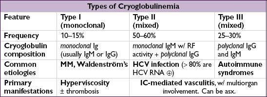
Definition & types (Lancet 2012;379:348)
• Proteins that precipitate on exposure to the cold and redissolve on rewarming, characterized by their composition
• Cryoglobulins=proteins that precipitate from serum and plasma when cooled
• Distinguish from cryofibrinogenemia=proteins (eg, fibrin, fibrinogen) that precipitate only from plasma; found in autoimmune dis, malignancies, infxns; unclear clinical significance
Etiologies
• Infections (types II & III): viral (HCV, HBV, HIV, HAV, EBV, CMV), bacterial (endocarditis, strep, etc.), fungal (coccidiomycosis, etc.) and parasitic (malaria, amoebiasis)
• Hematologic diseases
type I: MM, CLL, Waldenström’s
type II: B-cell lymphomas, solid organ malignancies
• Autoimmune syndromes (type III > II): Sjögren’s syndrome, SLE, RA, PAN
• Essential (idiopathic) in 10% of cases
• Renal transplant recipients (Clin Nephrol 2008;69:239)
Pathophysiology
• Chronic immune stimulation and/or lymphoproliferation → cryoglobulin generation
• Type I: cryo precipitation in microcirculation → hyperviscosity & vascular occlusion
• Types II/III: defective/insufficient immune complex (IC) clearance → IC-mediated inflammation of blood vessels w/ complement activation → vasculitis
Clinical manifestations (systemic sx usually due to type II > III)
• Most patients with cryoglobulinemia are asx
• Type I: hyperviscosity (cold worsens sx) → H/A, visual disturbance, livedo, digital ischemia
• Type II: vasculitis (sx not affected by cold exposure)
General: weakness, low-grade fever
Dermatologic (54–80%): lower extremity purpura, livedo reticularis, leg ulcers
Joint (44–70%): symmetric, migratory arthralgias of small or medium joints
Renal (50%): glomerulonephritis (proteinuria, hematuria, ARF, HTN, edema)
Neurologic (17–60%): peripheral neuropathy (polyneuropathy > mononeuritis multiplex)
Hematologic: anemia, thrombocytopenia, ↑ risk of B-cell lymphoma
GI (5%): abdominal pain, hepatosplenomegaly, abnormal LFTs
Diagnostic studies
• ✓ Cryoglobulins; must keep blood warmed to 37°C at all times en route to lab; early cooling causes false  cryoglobulin, loss of RF and ↓↓ complement
• Cryocrit is quantification of cryoprotein, does not always correlate w/ disease activity
• False ↑ in WBC or plt on automated CBC, due to cryoprecipitation
• Type I: ✓ serum viscosity, symptomatic if ≥4.0 centipoise; complement levels normal
• Type II: ↓ C4 levels, variable C3 levels, ↑ ESR,  rheumatoid factor (RF)
✓ HCV, HBV, & HIV serologies in all Pts w/ mixed cryoglobulinemia
Bx of affected tissue: hyaline thrombi; vasculitis w/ mixed inflammatory infiltrates of small vessels; leukocytoclastic vasculitis in purpuric lesions
Treatment (Autoimmun Rev 2011;10:444; Arth Rheum 2012;64:604; Blood 2012;119:5996)
• Treat underlying disorder:
Lymphoproliferative disease: chemotherapy and/or radiation
HCV: antivirals ± immunosuppression for severe disease
Connective tissue-related disease: DMARD/steroids ± rituximab
• Type I: Plasma exchange if hyperviscosity
• Type II: NSAIDs for control of mild symptoms for Pts w/ normal renal function
Rituximab or cyclophosphamide for major organ involvement
Plasmapheresis or plasma exchange in severe, life-threatening disease