PHOTO INSERTS
Radiology

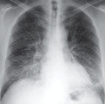
1 Normal PA CXR. The convex right cardiac border is formed by the right atrium (straight arrows), and the curved arrows indicate the location of the superior vena cava. The left cardiac and great vessels border what might be considered as four skiing moguls. From cephalad to caudad, the moguls are the aortic arch, the main and left pulmonary arter-ies, the left atrial appendage, and the left ventricle. (Radiology 101, 3rd ed, 2009.)

2 Normal lateral CXR. (Radiology 101, 3rd ed, 2009.)

3 COPD: with hyperlucent, overinflated lungs and flat diaphragms. (Radiology 101, 3rd ed, 2009.)
4 Interstitial pulmonary edema: with Kerley A, B, and C lines and cephalization of the vascular markings. (Fund. Diag. Radiology 3rd ed, 2006.)

5 Alveolar pulmonary edema. (Fund. Diag. Radiology 3rd ed, 2006.)

6 Right upper lobe pneumonia. (Radiology 101, 3rd ed, 2009.)
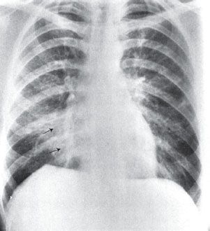
7 Right middle lobe pneumonia. (Radiology 101, 3rd ed, 2009.)

8 Right lower lobe pneumonia (PA). (Radiology 101, 3rd ed, 2009.)
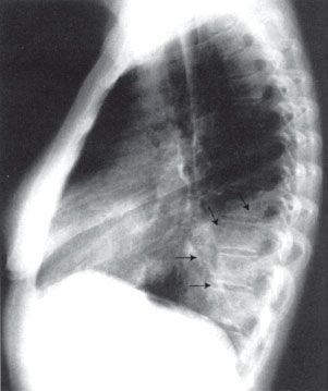
9 Right lower lobe pneumonia (lateral). (Radiology 101, 3rd ed, 2009.)

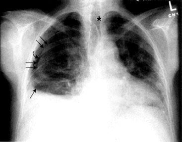
10 Bilateral pleural effusions (curved arrows) and enlarged azygous vein (straight arrow) (PA). (Radiology 101, 3rd ed, 2009.)

11 Bilateral pleural effusions (curved arrows) (lateral). (Radiology 101, 3rd ed, 2009.)
12 Pneumothorax. (Radiology 101, 3rd ed, 2009.)

13 Normal chest CT at level of pulmonary arteries (parenchymal windows).
(Radiology 101, 3rd ed, 2009.)
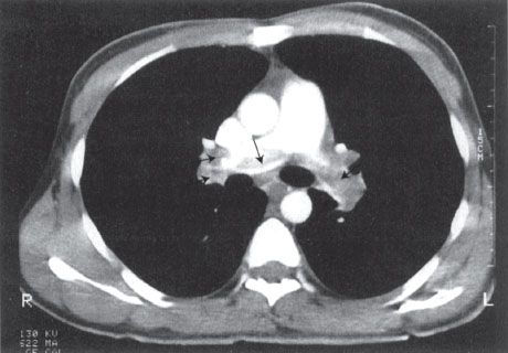
14 Bilateral PE (mediastinal windows). (Radiology 101, 3rd ed, 2009.)

15 Sarcoidosis with perilymphatic nodules. (Fund. Diag. Radiology 3rd ed, 2006.)

16 Idiopathic pulmonary fibrosis. (Fund. Diag. Radiology 3rd ed, 2006.)

17 Normal abdomen CT at level of liver & spleen. (Radiology 101, 3rd ed, 2009.)

18 Normal abdomen CT at level of pancreas. (Radiology 101, 3rd ed, 2009.)