PHOTO INSERTS
Echocardiography

1 Parasternal long-axis view allows visualization of the right ventricle (RV), ven- tricular septum (VS), posterior wall (PW) aortic valve cusps, left ventricle (LV), mitral valve, left atrium (LA), and ascending thoracic aorta (Ao). *Pulmonary artery. (Top: From Mayo Clinic Proceedings. [Tajik AJ, Seward JB, Hagler DJ, et al. Two-dimensional real-time ultrasonic imaging of the heart and great vessels: Technique, image orientation, structure identification, and validation. Mayo Clinic Proceedings, 1978;53:271–303], with permission. Bottom: From Oh JK, Seward JB, Tajik AJ. The Echo Manual, 3rd ed. Philadelphia: Lippincott Williams & Wilkins, 2006. By permission of Mayo Foundation for Medical Education and Research. All rights reserved.)
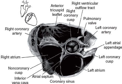
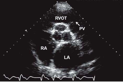
2 Parasternal short-axis view at the level of the aorta: LA, left atrium; PV, pul- monary valve; RA, right atrium; RVOT, right ventricular outflow tract. (Top: From Mayo Clinic Proceedings. [Tajik AJ, Seward JB, Hagler DJ, et al. Two-dimensional real-time ultrasonic imaging of the heart and great vessels: Technique, image orientation, structure identification, and validation. Mayo Clinic Proceedings, 1978;53:271–303], with permission. Bottom: From Oh JK, Seward JB, Tajik AJ. The Echo Manual, 3rd ed. Philadelphia: Lippincott Williams & Wilkins, 2006. By permission of Mayo Foundation for Medical Education and Research. All rights reserved.)
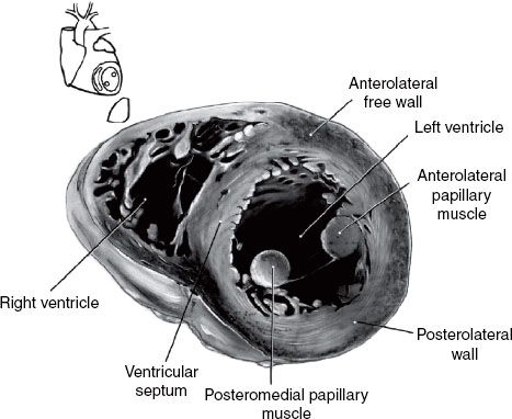
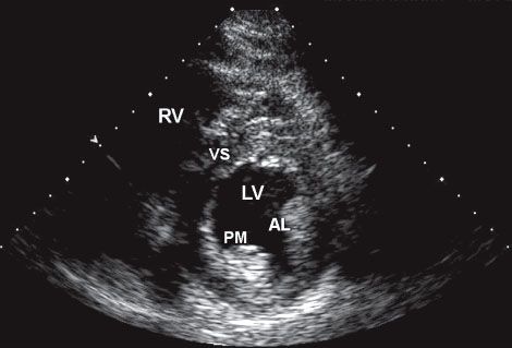
3 Parasternal short-axis view at the level of the papillary muscles: AL, anterolateral papillary muscle; PM, posteromedial papillary muscle; RV, right ventricle; VS, ventricular septum; LV, left ventricle. (Top: From Mayo Clinic Proceedings. [Tajik AJ, Seward JB, Hagler DJ, et al. Two-dimensional real-time ultrasonic imaging of the heart and great vessels: Technique, image orientation, structure identification, and validation. Mayo Clinic Proceedings, 1978;53:271–303], with permission. Bottom: From Oh JK, Seward JB, Tajik AJ. The Echo Manual, 3rd ed. Philadelphia: Lippincott Williams & Wilkins, 2006. By permission of Mayo Foundation for Medical Education and Research. All rights reserved.)
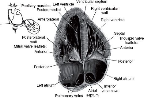
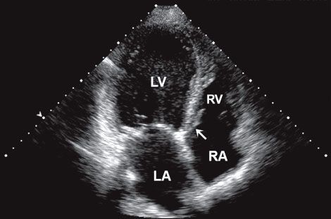
4 Apical four-chamber view: Note that at some institutions the image is re- versed so that the left side of the heart appears on the right side of the screen. LA, left atrium; LV, left ventricle; RA, right atrium; RV, right ventricle. (Top: From Mayo Clinic Proceedings. [Tajik AJ, Seward JB, Hagler DJ, et al. Two-dimensional real-time ultrasonic imaging of the heart and great vessels: Technique, image orientation, structure identification, and validation. Mayo Clinic Proceedings, 1978;53:271–303], with permission. Bottom: From Oh JK, Seward JB, Tajik AJ. The Echo Manual, 3rd ed. Philadelphia: Lippincott Williams & Wilkins, 2006. By permission of Mayo Foundation for Medical Education and Research. All rights reserved.)
Coronary Angiography