
PULMONARYQ
PLEURAL EFFUSION
Pathophysiology
• Systemic factors (eg, ↑ PCWP, ↓ oncotic pressure) → transudative effusion
• Local factors (ie, Δ pleural surface permeability) → exudative effusion
Transudates
• Congestive heart failure (40%): 80% bilateral, ± cardiomegaly on CXR occasionally exudative (esp. after aggressive diuresis or if chronic), but ~75% of exudative effusions in CHF Pts found to have non-CHF cause (Chest 2002;122:1518)
• Constrictive pericarditis (knock on exam, calcification or thickening on imaging)
• Cirrhosis (“hepatic hydrothorax”): diaphragmatic defect w/ passage of ascitic fluid often right-sided (2/3) & massive (even w/o marked ascites)
• Nephrotic syndrome: usually small, bilateral, asymptomatic (r/o PE b/c hypercoag)
• Other: PE (usually exudate), malignancy (lymphatic obstruction), myxedema, CAPD
Exudates
• Lung parenchymal infection (25%)
bacterial (parapneumonic): can evolve along spectrum of exudative (but sterile) → fibropurulent (infected fluid) → organization (fibrosis & formation of rigid pleural peel). Common causes: Strep pneumo, Staph aureus, Strep milleri, Klebsiella, Pseudomonas, Haemophilus, Bacteroides, Peptostreptococcus, mixed flora in aspiration pneumonia.
mycobacterial: >50% lymphs 80% of the time, ADA >40, pleural bx ~70% Se
fungal, viral (usually small), parasitic (eg, amebiasis, echinococcosis, paragonimiasis)
• Malignancy (15%): primary lung cancer most common, metastases (esp. breast, lymphoma, etc.), mesothelioma (✓ serum osteopontin levels; NEJM 2005;353:15)
• Pulmonary embolism (10%): effusions in ~40% of PEs; exudate (75%) > transudate (25%); hemorrhagic—must have high suspicion b/c presentation highly variable
• Collagen vascular disease: RA (large), SLE (small), Wegener’s, Churg-Strauss
• Gastrointestinal diseases: pancreatitis, esophageal rupture, abdominal abscess
• Hemothorax (Hcteff/Hctblood >50%): trauma, PE, malignancy, coagulopathy, leaking aortic aneurysm, aortic dissection, pulmonary vascular malformation
• Chylothorax (triglycerides >110): thoracic duct damage due to trauma, malignancy, LAM
• Other:
post-CABG: left-sided; initially bloody, clears after several wks
Dressler’s syndrome (pericarditis & pleuritis post-MI), uremia, postradiation therapy
Asbestos exposure: benign;  eosinophils
Drug-induced (eg, nitrofurantoin, methysergide, bromocriptine, amiodarone): eos
Uremia; post-XRT; sarcoidosis
Meigs’ syndrome = benign ovarian tumor → ascites & pleural effusion
Yellow-nail syndrome: yellow nails, lymphedema, pleural effusion, bronchiectasis
Diagnostic studies
• Thoracentesis (NEJM 2006;355:e16)
Indications: all effusions >1 cm in decubitus view if suspect due to CHF, can diurese and see if effusions resolve (75% do so in 48 h) asymmetry, fever, chest pain or failure to resolve → thoracentesisparapneumonics should be tapped ASAP (cannot exclude infxn clinically)
Diagnostic studies: ✓ total protein, LDH, glucose, cell count w/ differential, Gram stain & culture, pH; remaining fluid for additional studies as dictated by clinical scenario
Complications: PTX (5–10%), hemothorax (~1%), re-expansion pulm edema (if >1.5 L removed), spleen/liver lac.; post-tap CXR not routinely needed (Annals 1996;124:816)
↓ PTX w/ U/S and experienced supervisor (Chest 2009;135:1315; Archives 2010;170:332)
• Transudate vs. exudate (Annals 1972;77:507)
Light’s criteria: exudate = TPeff/TPserum >0.5 or LDHeff/LDHserum >0.6 or LDHeff >2/3 ULN of LDHserum; 98% Se, 83% Sp; best Se of all methods (Chest 1995;107:1604); however, will misidentify 25% of transudates as exudates; ∴ if clinically suspect transudate but meets criterion for exudate, confirm w/ test w/ higher Sp
exudative criteria w/ better Sp: serum-effusion alb gradient ≤1.2, Se 87%, Sp 92%; serum-effusion TP gradient ≤3.1, Se 84%, Sp 91%; choleff >45 mg/dL and LDHeff >200, 90% Se, 98% Sp (no serum required)
CHF effusions: TP may ↑ with diuresis or chronicity → “pseudoexudate”; alb gradient ≤1.2, choleff >60 mg/dL (Se 54%, Sp 92%) or clin judgment to distinguish (Chest 2002;122:1524)
• Complicated vs. uncomplicated parapneumonic (Chest 1995;108:299)
complicated = Gram stain or culture or pH <7.2 or glucose <60
complicated parapneumonic effusions usually require drainage to achieve resolution
empyema = frank pus, also needs drainage to achieve resolution
• Additional pleural fluid studies (NEJM 2002;346:1971)
NT-proBNP ≥1,500 pg/mL has 91% Se & 93% Sp for CHF (Am J Med 2004;116:417)
WBC & diff.: exudates tend to have ↑ WBC vs. transudates but nonspecific neutrophils → parapneumonic, PE, pancreatitis lymphocytes (>50%) → cancer, TB, rheumatologic eos (>10%) → blood, air, drug rxn, asbestos, paragonimiasis, Churg-Strauss, PE
RBC: Hcteff 1–20% → cancer, PE, trauma; Hcteff/Hctblood >50% → hemothorax
AFB: yield in TB 0–10% w/ stain, 11–50% w/ culture, ~70% w/ pleural bx
adenosine deaminase (ADA): seen w/ granulomas, >70 suggests TB, <40 excludes TB
cytology: ideally ≥150 mL and at least 60 mL should be obtained (Chest 2010;137:68)
glucose: <60 mg/dL → malignancy, infection, RA
amylase: seen in pancreatic disease and esophageal rupture (salivary amylase)
rheumatoid factor, CH50, ANA: limited utility in dx collagen vascular disease
triglycerides: >110 → chylothorax, 50–110 → ✓ lipoprotein analysis for chylomicrons
cholesterol: >60; seen in chronic effusions (eg, CHF, RA, old TB)
creatinine: effusion/serum ratio >1 → urinothorax
fibulin-3: ↑ plasma and/or effusion levels → mesothelioma (NEJM 2012;367:1417)
• Chest CT; pleural biopsy; VATS
• Undiagnosed persistent pleural effusions (Clin Chest Med 2006;27:309)
Transudative: most commonly CHF or hepatic hydrothorax. ✓ s/s CHF or cirrhosis, NT-proBNPeff; consider intraperitoneal injection of technetium-99m sulfur colloid
Exudative (ensure using Sp test listed above): most commonly malig, empyema, TB, PE. ✓ s/s malig, chest CT (I+), ADA or IFN-g release assay; consider thoracoscopy.
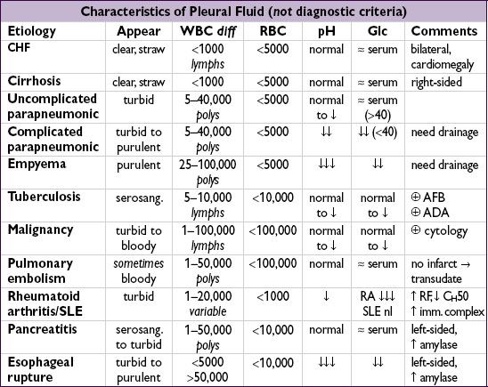
Treatment
• Symptomatic effusion: therapeutic thoracentesis, treat underlying disease process
• Parapneumonic effusion (Chest 2000;118:1158)
uncomplicated → antibiotics for pneumonia
>1/2 hemithorax or complicated or empyema → tube thoracostomy (otherwise risk of organization and subsequent need for surgical decortication)
loculated→ tube thoracostomy or VATS; intrapleural t-PA + DNase ↓ need for surgical referral (NEJM 2011;365:518)
• Malignant effusion: serial thoracenteses vs. tube thoracostomy + pleurodesis (success rate ~80–90%) vs. indwelling pleural catheter ( JAMA 2012;307:2383); choice of pleurodesis agent (talc, bleo, doxy) controversial; systemic steroids & pH <7.2 a/w ↑ pleurodesis failure rate
• TB effusions: effusion will often resolve spontaneously; however, treat Pt for active TB
• Hepatic hydrothorax
Rx: Δ pressure gradient (ie, ↓ ascitic fluid volume, NIPPV)
avoid chest tubes; prn thoracenteses, pleurodesis, TIPS or VATS closure of diaphragmatic defects if medical Rx fails; NIPPV for acute short-term management
spontaneous bacterial empyema (SBEM) can occur (even w/o SBP being present), ∴ thoracentesis if suspect infection
transplant is definitive treatment and workup should begin immediately