
HEMATOLOGY-ONCOLOGY
LEUKEMIA
ACUTE LEUKEMIA
Definition
• Clonal proliferation of hematopoietic progenitor with ↓ ability to differentiate into mature elements → ↑ blasts in bone marrow and periphery → ↓ RBCs, platelets and neutrophils
Epidemiology and risk factors
• Acute myelogenous leukemia (AML): ~14,000 cases/y; median age 66 y; >80% of adult acute leukemia cases
• Acute lymphocytic leukemia (ALL): ~6000 cases/y; median age 14 y; bimodal with 2nd peak in adults
• Risk factors: radiation, chemo (alkylating agents, topo II inhib), benzene, smoking
• Acquired hematopoietic diseases: MDS, MPN (esp. CML), aplastic anemia, PNH
• Inherited: Down’s & Klinefelter’s, Fanconi’s anemia, Bloom syndrome, ataxia telangiectasia
Clinical manifestations
• Cytopenias → fatigue (anemia), infection (neutropenia), bleeding (thrombocytopenia)
• More common in AML (esp. monocytic leukemias):
leukostasis (when blast count >50,000/µL): occluded microcirculation → local hypoxemia and hemorrhage → dyspnea, hypoxia, headache, blurred vision, TIA/CVA; look for hyperviscosity retinopathy (vascular engorgement, exudates, hemorrhage)
DIC (esp. with APL)
leukemic infiltration of skin, gingiva (esp. with monocytic subtypes)
chloroma: extramedullary tumor of leukemic cells, virtually any location
• More common in ALL:
bone pain, lymphadenopathy, hepatosplenomegaly (also seen in monocytic AML)
CNS involvement (up to10%): cranial neuropathies, N/V, headache anterior mediastinal mass (esp. in T-cell); tumor lysis syndrome (qv)
Diagnostic evaluation (Blood 2009;114:937)
• Peripheral smear: anemia, thrombocytopenia, variable WBC (50% p/w ↑ WBC) + circulating blasts (seen in >95%;  Auer Rods in AML)
• Bone marrow: hypercellular with >20% blasts; cytogenetics, flow cytometry
• Presence of certain cytogenetic anomalies, eg, t(15;17), t(8;21), inv(16) or t(16;16), are sufficient for dx of AML regardless of the blast count
• ✓ for tumor lysis syndrome (rapid cell turnover): ↑ UA, ↑ LDH, ↑ K, ↑ PO4, ↓ Ca
• Coagulation studies to r/o DIC: PT, PTT, fibrinogen, D-dimer, haptoglobin, bilirubin
• LP (w/ co-admin of intrathecal chemotherapy to avoid seeding CSF w/ circulating blasts) for Pts w/ ALL (CNS is sanctuary site) and for Pts w/ AML w/ CNS sx
• TTE if prior cardiac history or before use of anthracyclines
• HLA typing of Pt, siblings and parents for potential allogeneic HSCT candidates
ACUTE MYELOGENOUS LEUKEMIA (AML)
Classification (FAB no longer used clinically; Blood 2009;114:937)
• Features used to confirm myeloid lineage and subclassify AML to guide treatment: morphology: blasts, granules, ± Auer rods (eosinophilic needle-like inclusions) cytochemistry: myeloperoxidase and/or nonspecific esterase
• Immunophenotype: myeloid antigens → CD13, CD33, CD117; monocytic antigens → CD11b, CD64, CD14, CD15
• Cytogenetics: important for prognosis. Intermed. risk = no favorable/unfavorable features.


Treatment (Blood 2010;115:453; JNCCN 2012;10:984; Lancet 2013;381:484)
• Induction chemo followed by consolidation Rx
• Induction chemo: “7 + 3” = cytarabine × 7 d + ida/daunorubicin × 3 d. Cytarabine dose: continuous intermed.  high dose (NEJM 2011;364:1027). Daunorubicin dose: age <60 → high (90 mg/m2); age>60 → standard (60 or 45 mg/m2) (NEJM 2009;361:1249). Gemtuzumab ozogamicin (ɑ-CD33) ? benefit in fav/int risk AML (Lancet 2012;379:1508)
• ✓ for complete remission (CR) = normal peripheral counts, <5% BM blasts CR ≠ cure; ∴ must always f/u induction with consolidation Rx
• If CR: consolidation Rx based on risk stratification (age, genetics, PS): chemo (eg, high dose cytarabine) if favorable risk; otherwise → allo-HSCT ( JAMA 2009;301:2349)
• If  CR: reinduction with similar chemotherapy (“5 + 2”) or alternative regimen
• If relapse after CR: salvage chemo → allogeneic HSCT (↓ intensity conditioning if >60 y)
• Supportive care: hydration + allopurinol or rasburicase for tumor lysis prophylaxis; transfusions; antibiotics for fever and neutropenia; antifungals for prolonged fever & neutropenia; hydroxyurea ± leukophoresis for leukostasis
Prognosis
• CR achieved in 70–80% of Pts <60 y and in 40–50% for Pts >60 y
• Overall survival depends on prognostic factors: ranges from ~50% for Pts <60 y w/ favorable prognostic factors to <10% for Pts >60 y w/ poor prognostic factors
• Poor prognostic factors: age >60, unfavorable cytogenetics (see above), FLT3-ITD , poor performance score, antecedent MDS/MPN, therapy-related AML; genetic profiling (NEJM 2012;366:1079)
Acute promyelocytic leukemia (APL) (Blood 2009;113:1875)
• Rare disease w/ only ~1000 cases/y in the U.S. but biologically and clinically distinct
• Atypical promyelocytes (large, granular cells; bilobed nuclei) in blood and bone marrow
• Defined by translocation of retinoic acid receptor: t(15;17); PML-RARɑ (>95% of cases)
• Medical emergency with DIC and bleeding common; supportive care measures crucial
• Remarkable responses to all-trans-retinoic acid (ATRA), which induces differentiation, and arsenic trioxide (ATO); early initiation of ATRA is critical as soon as APL suspected; ATO highly active as first-line therapy or in treatment of refractory disease.
• Induction regimen: anthracycline + ATRA ± cytarabine → CR in ~90%; or ATRA + ATO alone (ASH 2012; JCO 2009;27:504)
• Differentiation (ATRA) syndrome: ~25% of Pts; fever, pulm infiltrates, SOB, edema, HoTN, AKI; tx w/ dexamethasone 10 mg bid, supportive care (eg, diuresis) (Blood 2008;113:775)
• Consolidation Rx: eg, ATO → anthracycline + ATRA (Blood 2010;116:3751)
• Role of maintenance Rx (eg, ATRA + 6MP + MTX) currently controversial
• Best prognosis of all AMLs: >90% cure; WBC >10,000/µL = ↓ prognosis (Blood 2000;96:1247)
ACUTE LYMPHOBLASTIC LEUKEMIA (ALL)
Classification
• Lymphoblastic neoplasms may present as acute leukemia (ALL) with >20% BM blasts or as lymphoblastic lymphoma (LBL) w/ mass lesion & <20% BM blasts. ALL and LBL are considered the same disease with different clinical presentations.
• Morphology: no granules (granules seen in myeloid lineage)
• Cytochemistry: terminal deoxynucleotidyl transferase (TdT) in 95% of ALL
• Cytogenetics (Blood 2010;115:206): t(9;22) = Philadelphia chrom (Ph) ~25% of adults w/ ALL
• Immunohistochemistry: 3 major phenotypes (Burkitt’s usually treated differently)

Treatment (NEJM 2006;354:166; JCO 2011;29:532)
• Induction chemo: multiple acceptable regimens including combination of anthracycline, vincristine, steroids, cyclophosphamide, ± asparaginase
• CNS prophylaxis: intrathecal MTX/cytarabine ± cranial irradiation or systemic MTX
• Postremission therapy options:
consolidation/intensification chemo (~7 mo) followed by maintenance chemo (~2–3 y) high-dose chemo w/ allo HSCT considered for all Pts in CR1 w/ available donor pediatric regimens in young adults (<30 y); consider allo SCT for all Pts <50 (controversial)
• If relapse → salvage chemo followed by allogeneic HSCT if able
• Ph t(9;22) → add imatinib or dasatinib, followed by allogeneic HSCT
• MLL-AF4 t(4;11) or hypodiploidy (<44 chromosomes) → consider for allogeneic HSCT
• Infusion of chimeric antigen receptor–modified T cells promising (NEJM 2013;368:1509)
Prognosis
• CR achieved in >80% of adults
• Cure achieved in 50–60% if good prog. factors vs. 10–30% w/ poor prog. factors
• Good prognostic factors: younger age, WBC <30,000/µL, T-cell immunophenotype, absence of Ph chromosome or t(4;11), early attainment of CR
• Gene expression patterns may be useful in predicting chemo resistance (NEJM 2004;351:533)
CHRONIC MYELOGENOUS LEUKEMIA (CML)
Definition (Blood 2009;114:937)
• Myeloproliferative neoplasm with clonal overproduction of hematopoietic myeloid stem cells that can differentiate
• Philadelphia chromosome (Ph) = t(9;22) → BCR-ABL fusion → ↑ Abl kinase activity
BCR-ABL required for Dx of CML
• “Atypical CML” (BCR-ABL ) now considered a separate disease and reclassified as MDS/MPN (see “Myelodysplastic Syndromes”)
Epidemiology and risk factors
• ~5400 new cases/y in U.S.; median age ~64 at presentation; ~15% of adult leukemias
• ↑ risk with irradiation; no clear relation to cytotoxic drugs
Clinical manifestations
• Triphasic clinical course; 85% present in the chronic phase
• Chronic phase: often asymptomatic but common features are fatigue, malaise, weight loss, night sweats, abdominal fullness (splenomegaly 50%)
• Accelerated phase: refractory leukocytosis, thrombocytopenia and worsening sx → fever, wt loss, ↑ splenomegaly, bone pain, bleeding, infections, pruritus (basophilia)
• Blastic phase acute leukemia → severe constitutional symptoms, infection, bleeding and possible leukostasis (see “Acute Leukemia”)
Diagnostic evaluation
• Peripheral smear: leukocytosis (often >100,000/µL), left-shifted with all stages of myeloid maturation; anemia, thrombocytosis, basophilia
• Bone marrow: hypercellular, ↑ myeloid to erythroid ratio, ↓ leuk alkaline phosphatase
• Chronic: <10% blasts (peripheral or BM)
• Accelerated: 10–20% blasts, >20% basos, plts <100k, ↑ spleen size, karyotypic prog.
• Blastic: >20% blasts (2⁄3 myeloid, 1⁄3 lymphoid), may see extramedullary leukemia
Treatment (NEJM 2010;362:2260; Blood 2011;118:1208 & 2012;120:1390)
• Tyrosine kinase inhibitor (TKI): imatinib, dasatinib, nilotinib, bosutinib, ponatinib are selective inhibitors of BCR-ABL (JCO 2010;28:428; Blood 2012;120:1390).
Imatinib, nilotinib, & dasatinib approved as initial Rx.
Resistance = recurrent dis. on TKI, often result of BCR-ABL mutation or amplification.
Nilotinib, dasatinib, bosutinib, & ponatinib approved for resistant disease, w/ only ponatinib effective on T315I resistance mutation (NEJM 2012;367:2075).
Side effects include nausea, diarrhea, muscle cramps, cytopenias, ↓ PO4, ↑ QT, rarely CHF; dasatinib also a/w pericardial & pleural effusions, nilotinib w/ ↑ bili & lipase.
• Chronic phase: TKI; continued indefinitely in responders (Blood 2012;120:1390)
• Accelerated phase: TKI upfront, consider allogeneic HSCT
• Blastic phase: TKI + HSCT vs. ALL or AML induction (based on cell type) + HSCT
• Allogeneic HSCT: consider for Pts w/ available donor who present in accelerated or blastic phase; reasonable option for Pts with relapsed/refractory disease to TKIs

Prognosis
• Chronic phase CML Rx’d w/ imatinib: 89% overall survival, 95% survival free of CML-related deaths, 7% progression to blast phase at 5 y (NEJM 2006;355:2408)
• Accelerated phase CML Rx’d w/ imatinib: ~50% overall survival at 4 y (Cancer 2005;103:2099)
• Poor prognostic factors: ↑ age, ↑ platelet count, ↑ spleen size, ↑ percentage of blasts
CHRONIC LYMPHOCYTIC LEUKEMIA (CLL)
Definition (NEJM 2005;352:804; Blood 2008;111:5446)
• Monoclonal accumulation of functionally incompetent mature B lymphocytes
• CLL (>5000/µL malignant cells) & small lymphocytic lymphoma (SLL; <5000/µL malignant cells, but + LAN ± splenomegaly) now classified as same disease
• Monoclonal B lymphocytosis (<5000/µL, nodes <1.5 cm, nl RBC and Plt counts): observe
Epidemiology and risk factors
• ~16,000 new cases/y; median age at dx is 72 y; most common adult leukemia
• ↑ incidence in 1st-degree relatives; no known association with radiation, chemicals, drugs
Clinical manifestations
• Symptoms: often asx & identified when CBC reveals lymphocytosis; 10–20% p/w fatigue, malaise, night sweats, weight loss (ie, lymphoma “B” sx)
• Signs: lymphadenopathy (80%) and hepatosplenomegaly (50%)
• Autoimmune hemolytic anemia (AIHA) (~7%) or thrombocytopenia (ITP) (~1–2%)
• Hypogammaglobulinemia ± neutropenia → ↑ susceptibility to infections
• Bone marrow failure in ~13%; monoclonal gammopathy in ~5%
• Aggressive transformation: ~5% develop Richter’s syndrome = transformation into high-grade lymphoma (usually DLBCL) and sudden clinical deterioration
Diagnostic evaluation (see “Lymphoma” for general approach)
• Peripheral smear: lymphocytosis (>5000/µL, mature-appearing small cells) “smudge” cells from damage to abnl lymphs from shear stress of making blood smear
• Flow cytometry: clonality with dim surface Ig (sIg); CD5+, CD19+, CD20(dim), CD23+. CD38+ or ZAP70+ a/w unmutated Ig variable heavy chain region & worse prognosis.
• Bone marrow: normo- or hypercellular; infiltrated w/ small B-cell lymphocytes (≥30%)
• Lymph nodes: infiltrated w/ small lymphocytic or diffuse small cleaved cells = SLL
• Genetics: del 11q22-23 & 17p13 unfavorable; trisomy 12 neutral; del 13q14 and mut IgVH favorable. Nine significantly mutated genes, including TP53, NOTCH1, MYD88 and SF3B1. Key role for spliceosome mutations (NEJM 2011;365:2497; JCI 2012;122:3432).
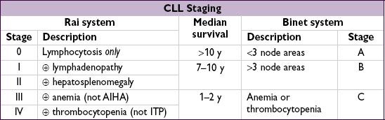
Treatment
• Treatment is primarily palliative → early stage disease can be followed w/o Rx
• Indications for treatment: Rai stages III/IV, Binet stage C, disease-related sx, progressive disease, AIHA or ITP refractory to steroids, recurrent infections
• Options:
purine analogues: fludarabine (“F”), pentostatin (“P”)
alkylating agents: cyclophosphamide (“C”), bendamustine (“B”), CVP, CHOP; ? chlorambucil for elderly (lower response vs. F, but survival; NEJM 2000;343:1750)
± monoclonal Ab against CD20 (rituximab, “R”) or CD52 (alemtuzumab, esp. w/ 17p-) combination regimens (eg, FR, FCR, BR) superior to monoRx (Lancet 2007;370:230)
• Novel Rx refractory dis.: ofatumumab (ɑ-CD20), ibrutinib (BTK inhib), CAL101 (PI3K inhib)
• Consider allo-HSCT in p53 mut or refractory CLL (BBMT 2009;15:53; BJH 2012;158:174)
• Supportive care: PCP, HSV, VZV prophylaxis; CMV monitoring for Pts receiving anti-CD52; AIHA/ITP → steroids; recurrent infections → IVIg
Prognosis (NEJM 2004;351:893; JCO 2006;24:4634 & 2010;28:4473; Blood 2008;111:865)
• Survival varies substantially. Median overall survival ~10 y (Am J Hematol 2011;12:985)
• Favorable prognosis: 13q14 deletion (~50% of CLL cases)
• Factors a/w worse prognosis include:
unfavorable cytogenetics (eg, 17p-/TP53 mutation)
unmutated (<2% c/w germline) IgVH gene (<8–10 y vs. >20–25 y if mutated)
high (>20–30%) Zap-70 expression (part of T cell receptor; correlated w/ unmutated IgVH)
CD38 >30% or CD49d <30% (correlated with unmutated IgVH)
higher β2-microglobulin levels (correlate with disease stage and tumor burden)