
Vickie McDonald1 & Samuel J. Machin2
1University College London Hospitals NHS Foundation Trust, London, UK
2Haemostasis Research Unit, Department of Haematology, University College London, London, UK
Normal haemostasis
Haemostasis is a complex process involving the interaction of many components – blood vessels, platelets, coagulation factors, coagulation factor inhibitors and fibrinolytic enzymes – that ultimately leads to clot formation followed by resolution. In a normal individual there is a constant balance between procoagulant and anticoagulant activities.
The generation of thrombin is key to successful haemostasis (Figure 25.1). Historically it was thought that two initiating coagulation cascades, the intrinsic and extrinsic pathways, ultimately led to generation of thrombin, which in turn converted fibrinogen to fibrin. We now know that the coagulation process is a complex network of positive and negative feedback loops that are explained better by the ‘cell based’ model of coagulation [1]. This describes three overlapping phases: initiation, amplification and propagation. Initiation of coagulation occurs when exposure of tissue factor (TF) on damaged endothelial cells/activated monocytes leads to the generation of activated factor VII (FVIIa) and formation of TF-VIIa complex (under normal circumstances, approximately 1–2% of circulating plasma factor VII circulates in the activated form). The TF-VIIa complex activates factors IX (FIXa) and X (FXa) and trace amounts of thrombin are subsequently generated by FXa. Concurrent to this, platelets adhere to the subendothelial matrix and are activated, providing a phosphilipid surface for coagulation factor activity. VWF is bound to and released from endothelial cells leading to further platelet recruitment and activation.
Fig 25.1 The procoagulant pathway. The diamond represents the tenase complex and the circle represents the prothrombinase complex.

During amplification and propagation, the small amounts of thrombin generated activate factors V, VIII and XI. This allows formation of the ‘tenase’ complex (FIXa/FVIIIa) to generate further FXa and the ‘prothrombinase’ complex (FXa/FVa), which leads to the explosive generation of thrombin from prothrombin and, ultimately, by activation of fibrin and factor XIII, production of a crosslinked stable clot.
The initial TF-VIIa complex is quickly inhibited by tissue factor pathway inhibitor (TFPI); however, by this time, positive feedback loops between thrombin and factors XI, V and VIII are sufficient to amplify the signal and propagate the clot formation. Factor XI is also activated by factor XIIa formed from the HMWK-prekallikrein complex on endothelial cells, but this contribution to physiological haemostasis is not significant.
There are inbuilt mechanisms to control the procoagulant response. These include TFPI, which inactivates the TF-FVIIa complex, antithrombin, which complexes with and inactivates FIXa, FXa, FXIa and thrombin, the protein C and S pathways, which inactivate FVa and FVIIIa, and thrombomodulin, which binds to thrombin and alters its substrate specificity for factors V, VIII and fibrinogen. Fibrinolysis is also part of the normal haemostatic response. Circulating plasminogen is activated to form the serine protease plasmin, which digests crosslinked fibrin to form D-dimers and other fibrinogen fragments.
Investigation of abnormal haemostasis
Abnormalities in the haemostatic system may be congenital or acquired and clinical presentation can vary from asymptomatic to life-threatening haemorrhage. A careful history should be taken focusing on personal or family bleeding history, bleeding following dental work, surgery or childbirth and objective evidence of excess bleeding such as development of anaemia, requirement for transfusion or surgical intervention.
The initial laboratory investigation of patients with abnormal haemostasis should include a platelet count, prothrombin time (PT), activated partial thromboplastin time (APTT) and Clauss fibrinogen. Additional screening tests that can also be performed are listed in Table 25.1. If one/more of these tests are abnormal, further specialized investigations should be performed in order to define precisely the defect and its severity.
Table 25.1 Simple laboratory haemostasis screening tests.
|
System |
Test |
Factor |
|
Coagulation |
PT |
II, V, VII, X |
|
APTT |
VIII, IX, XI, XII |
|
|
INR – only in patients receiving oral anticoagulation |
||
|
TT |
Fibrinogen, heparin |
|
|
Clauss fibrinogen |
Fibrinogen |
|
|
Platelets |
Platelet count |
|
|
Blood film inspection |
||
|
Platelet function (using PFA-100™ which measures in vitro ‘high shear’ bleeding time) |
||
|
Fibrinolysis |
D-dimers |
|
|
Euglobulin clot lysis time |
||
|
Global |
Thromboelastogram |
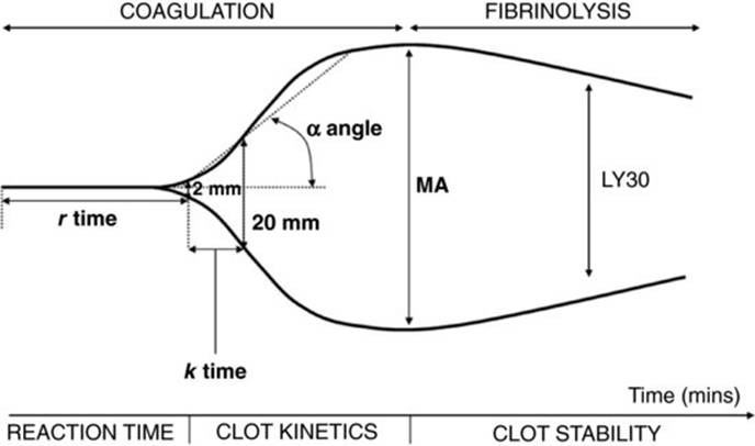
In high dependency units, the availability of near patient testing devices to rapidly assess coagulation (PT and APTT) and overall global haemostasis (thromboelastogram, TEG®) potentially allows rapid treatment decisions to be made without sending a citrated sample to the laboratory. TEG® measures the changes in elastic shear stresses seen during clot formation and subsequent fibrinolysis. A stationary pin attached to a torsion wire is immersed in a rotating cup that contains whole blood and as clot forms the pin and cup rotate together. The magnitude of pin movement (in mm) is proportional to the strength of the clot. Five major parameters are measured (Figure 25.2):
1. Reaction time (r), which is the time from adding the sample to first measurable clot (predetermined as 2 mm). It is shortened by hypercoagulable states and increased by coagulation factor deficiencies/heparin effect.
2. Time k, which is the time to achieve a predesignated clot strength (designated as 20 mm amplitude). This parameter is most reflective of fibrinogen function.
3. Alpha (α) angle, which is the slope of the trace between R and K and reflects the speed of fibrin accumulation and polymerization.
4. Maximum amplitude (MA) is the highest vertical amplitude of the TEG® tracing and is an indication of platelet function. Affected by thrombocytopenia, abnormal platelet function and problems with the interaction between fibrinogen and platelets.
5. LY30 is the rate of amplitude reduction 30 minutes after the MA is reached. This is reflective of clot stability, in particular the degree of fibrinolysis and breakdown of the clot.
Fig 25.2 Schematic representation of thromboelastography (TEG®).
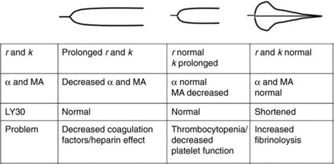
Schematic representations of abnormal traces and the causes are shown in Figure 25.3. In a modified version of TEG® called ROTEM®, it is the pin rather than the cup that oscillates but similar measurements are given.
Fig 25.3 Schematic representation of common abnormalities seen in TEG®.
It is important to note that in some disorders (e.g. mild haemophilia or von Willebrand disease, or VWD), tests such as the APTT may not be overly prolonged, so if a bleeding disorder is strongly suspected from the patient's history and the clinical picture, specific factor assays and/or immunological tests should be performed regardless of the ‘screening’ test result.
Inherited haemostatic defects
Haemophilia A
This disorder results in reduced or absent activity of factor VIII. The factor VIII gene is on the long arm of the X-chromosome and there is often a family history of haemophilia. However, up to one-third of cases are as a result of new mutations. Some female carriers may be symptomatic.
Haemophilia A is classified into mild, moderate or severe according to factor VIII activity (FVIII:C) (Table 25.2). The minimal effective level for haemostasis is generally about 25–30%.
Table 25.2 Clinical manifestations and treatment of haemophilia A and B.
|
Factor level (% normal) |
Clinical manifestation |
Treatment |
|
<1% (severe disease) |
Usual age of onset <1 year |
Regular FVIII or IX prophylaxis in children and some adults |
|
Post-traumatic bleeding |
||
|
Crippling joint deformity if inadequate treatment |
||
|
1–5% (moderate disease) |
Usual age of onset <2 year |
Some patients may need regular factor VIII or IX prophylaxis |
|
6–40% (mild disease) |
Usual age of onset >2 year |
Regular prophylaxis usually not required |
Investigations
Laboratory abnormalities seen in haemophilia A include:
· prolonged APTT;
· reduction of FVIII:C;
· normal VWF activity (it is important to measure VWF activity in order to exclude VWD, which will also give low factor VIII levels).
Management
The mainstay of treatment is to raise the FVIII:C sufficiently to prevent or arrest spontaneous and traumatic bleeds or to cover surgery. There are a number of products currently available, including:
· recombinant factor VIII preparations;
· plasma-derived factor VIII concentrates (which vary in degree of purity);
· DDAVP (for mild disease only – baseline factor VIII above 15%);
· tranexamic acid.
Recombinant products are the initial product of choice to prevent spontaneous joint bleeds in children with severe haemophilia (prophylaxis), as well as treatment of bleeds in previously untreated patients because of the lack of risk of transmission of infection [2]. Plasma-derived factor concentrates undergo donor screening and specific double viral inactivation procedures, but transmission of some viruses, such as human parvovirus B19, and new emerging infections, such as prion disease, remain a theore- tical risk.
There has been concern that patients treated with recombinant factor VIII products have a higher incidence of inhibitor development than those treated with plasma-derived factor concentrates. However, the evidence is conflicting and at present recombinant products are the recommended first line for previously untreated patients. The choice of product for previously treated patients will depend on factors such as previous response to treatment, a history of inhibitor development and whether they have been previously exposed to plasma products.
Patients with moderate/severe haemophilia will require factor VIII concentrates for bleeding, prior to invasive procedures, surgery, etc. One unit factor VIII/kg body weight will result in an increase in plasma factor VIII level by 2%. The amount of factor VIII concentrate required is calculated according to the formula:

The plasma half-life of factor VIII is 8–12 hours, and thus repeated doses at 12-hourly intervals are usually needed. Alternatively, a continuous infusion of factor VIII can be given for surgery. For major soft tissue bleeds, levels above 50% are generally sufficient; however, for major surgery, a preoperative level of 100% is necessary and thereafter levels of 50–100% are sufficient for adequate wound healing. Factor VIII:C can be measured before and after doses of concentrate to ensure appropriate levels have been achieved.
Mild haemophilia A should be treated with DDAVP (with or without tranexamic acid) where possible [2]. DDAVP (0.3 μg/kg body weight) is given intravenously, subcutaneously or alternatively a 300 μg dose (for adults) can be administered via intranasal spray. This dose typically increases the levels of factor VIII and VWF 3–5 times above baseline. Hyponatraemia and water intoxication are side effects of this drug, and hence it is not recommended for patients with cardiac failure or children under 2 years of age. It is also thought to have thrombogenic potential and should be used with caution in the elderly or those with known vascular disease. The response to DDAVP should be assessed in all patients prior to its use to treat bleeding or cover invasive procedures to ensure that an adequate increase in factor VIII levels is achieved.
Tranexamic acid reduces fibrinolysis and is of particular use in patients with bleeding from mucosal surfaces, such as epistaxis, oral bleeding or menorrhagia. It is given as an adjunct to DDAVP to reduce bleeding. It should be avoided in patients with haematuria to avoid the complication of clot retention. It is usually given for 7–10 days to allow adequate healing.
Haemophilia B
This X-linked recessive disorder results in a deficiency of factor IX. The clinical features are identical to those of haemophilia A (Table 25.2).
Investigations
Laboratory abnormalities seen in haemophilia B include:
· prolonged APTT;
· reduction of factor IX coagulant activity.
Management
The main types of products that are currently used for treatment include:
· recombinant factor IX products;
· high purity plasma-derived factor IX concentrates.
The product of choice for prophylaxis, treatment of bleeding or cover for surgical procedures in previously untreated patients is recombinant factor IX [2]. If unavailable, then high purity plasma-derived factor IX concentrates should be used. Prothrombin complex concentrates (PCC), containing factors II, VII, IX and X, have been used in the past but are not recommended now due to their prothrombotic effects.
The dosage of factor IX required can be calculated according to the formula:

The plasma half-life of factor IX is 18–30 hours and therefore if repeated doses are needed, they should be given every 12–24 hours or by continuous infusion.
The choice of product for patients who have required previous treatment with factor concentrates depends on the history of inhibitor development and response to treatment. There is some evidence to suggest that plasma-derived products have different pharmacokinetic properties to recombinant products; therefore, the response to treatment should be monitored closely if switching from one product to another.
Treatment of patients with inhibitors
Patients with haemophilia can develop inhibitory antibodies to factors VIII or IX. Inhibitor development is often heralded by increased frequency of bleeding or loss of response to factor VIII. It is diagnosed by measuring factor VIII levels before and after a dose of factor VIII concentrate and by a Bethesda inhibitor assay.
For patients with haemophilia A, if the inhibitor is of low titre (i.e. <5 Bethesda units), then bleeding episodes can be treated with higher than normal doses of human factor VIII [3]. If the inhibitor is of high titre (i.e. >10 Bethesda units), human factor VIII is ineffective to control bleeding and the use of recombinant FVIIa or FEIBA® (Baxter) is recommended. For major haemorrhage, recombinant FVIIa (dose of 70–90 μg/kg initially every 2 hours) is generally recommended as first-line therapy (if available). Eradication of inhibitors with ‘immune tolerance induction’ using factor VIII concentrates alone or together with immunosuppression is considered the best long-term treatment option for these patients [3].
For patients with haemophilia B, recombinant factor VIIa is used for bleeding [3]. Immune tolerance using factor IX concentrates can be attempted, although this is more difficult than in haemophilia A. Up to 50% of haemophilia B patients who develop an inhibitor have anaphylaxis or severe allergic reactions to factor IX concentrates.
Von Willebrand disease (VWD)
This is the most common of the inherited bleeding disorders and is due to a quantitative and/or qualitative defect in the VWF protein. VWF has two main functions: it promotes the adhesion of platelets to the subendothelium by binding to the platelet receptor glycoprotein Ib and it protects factor VIII:C from proteolytic degradation by forming a noncovalent association. Patients who are blood group O have lower levels of VWF than other blood groups.
VWD is classified into three different types (Table 25.3) [4]. Clinical symptoms vary; some patients may be asymptomatic whereas others will have haemophilia-like bleeding. Laboratory abnormalities seen in VWD include (variably):
· prolonged PFA-100™ closure time;
· reduction of VWF antigen (VWF:Ag);
· reduction of VWF ristocetin cofactor activity (VWF:RiCoF);
· reduction of FVIII:C (which can cause prolonged APTT);
· abnormal VWF multimers in some subtypes.
Table 25.3 Variants of von Willebrand disease.
|
Type 1 |
Autosomal dominant inheritance |
|
Partial quantitative deficiency of VWF |
|
|
Normal VWF multimers |
|
|
Mild bleeding disorder which decreases during pregnancy, elderly |
|
|
Type 2 |
Autosomal dominant inheritance |
|
Qualitative deficiency of VWF |
|
|
Numerous subtypes |
|
|
Abnormal VWF multimers |
|
|
Generally mild bleeding disorder |
|
|
Type 3 |
Autosomal recessive inheritance |
|
Severe quantitative deficiency of VWF |
|
|
Severe haemophilia-like bleeding disorder |
The goal of therapy in patients with VWD is to correct the dual defect of haemostasis, i.e. the abnormal platelet adhesion and the abnormal coagulation due to low FVIII levels. Treatment differs for the various types of VWD [5]:
· Type 1. To reduce exposure to blood components, DDAVP is the treatment of choice and a dose of 0.3 μg/kg body weight is usually given intravenously or subcutaneously. Intranasal doses (300 μg for adults or 150 μg for children) can also be given. These doses give a two- to fivefold increase in endogenous VWF and FVIII:C levels. The choice of route of administration depends on the patient and the nature of the bleeding or surgery. It is important to test an individual's response to DDAVP prior to using it to ‘cover’ procedures. Tranexamic acid is often also given either alone for minor bleeding/procedures or in conjunction with DDAVP.
· Types 2 and 3. VWF ‘replacement therapy’ is generally required. At present there are no recombinant VWF concentrates available so either a factor VIII concentrate rich in VWF or a purified VWF concentrate is the treatment of choice, preferably those with double viral inactivation steps.
In the past, cryoprecipitate was used to treat patients with VWD; however, it is now unacceptable to use such untreated plasma derivatives when there are ‘safer’ alternatives available.
Other inherited disorders
Hereditary deficiencies of other coagulation factors are rare. Factor XI deficiency is particularly common amongst Ashkenazi Jews and is transmitted as an autosomal recessive trait. There is a poor correlation between factor XI levels and bleeding tendency, which usually presents following surgery or dental procedures. If available, factor XI concentrates should be given to treat bleeding; if not, then FFP should be administered. There have been concerns about the potential thrombogenicity of factor XI concentrates, so peak levels should ideally not exceed 70 IU/dL [6].
Cryoprecipitate can be used for fibrinogen deficiency/dysfibrinogenaemias, but fibrinogen concentrates should be used in preference if they are available because they undergo additional viral inactivation steps [2]. Deficiencies of factors II, V, VII, X and XIII can all be treated with FFP, but if more specific therapies are available they should be used in preference. Currently, there are specific factor concentrates for factors VII and XIII. Prothrombin complex concentrates (PCCs) contain factors II, IX and X with variable amounts of VII and are used in conditions associated with deficiencies of one/more of these factors (e.g. treatment of overdosage with warfarin). They can be given to patients with factors II or X deficiency (although thromboembolic risks should be considered). Factor V-deficient patients are treated with FFP and it is recommended that virally inactivated plasma is used.
Patients with deficiencies of ‘contact factors’ (factor XII, prekallikrein and high-molecular-weight kininogen) do not bleed excessively and do not require any treatment.
Acquired haemostatic defects
Disseminated intravascular coagulation (DIC)
This is a complex disorder resulting from inappropriate and excessive activation of the haemostatic system that can be manifested by both thrombotic and haemorrhagic pathology. DIC may be acute (uncompensated) with decreased levels of haemostatic components or chronic (compensated) with normal or sometimes elevated levels of coagulation factors.
The main triggering mechanism for DIC is the exposure of blood to a source of tissue factor that initiates coagulation, e.g. on the surface of endothelial cells or monocytes stimulated by endotoxins/cytokines as a result of sepsis, on the surface of damaged cells (placental abruption, cerebral trauma) or from malignant cells.
The final consequence of coagulation activation is thrombin generation and fibrin formation, which may result in microthrombus formation (e.g. gangrene of fingers, toes and renal failure). Secondary activation of the fibrinolytic pathway occurs with subsequent lysis of fibrin and the formation of crosslinked complexes such as D-dimers. Raised levels of these fibrin degradation products (FDP) further add to the bleeding diathesis as they inhibit the action of thrombin and also inhibit platelet function by binding to the platelet membrane.
Hepatic synthesis of coagulation factors is unable to compensate fully for the ongoing consumption of clotting factors, so there is a reduction in levels of particularly factors V, VIII, XIII and fibrinogen. In addition, a consumptive thrombocytopenia develops. This combination of coagulation factor deficiency, thrombocytopenia and the inhibitory actions of raised FDPs causes the generalized and continued bleeding tendency characteristic of DIC. The main causes of DIC are listed in Table 25.4.
Table 25.4 Main causes of DIC.
|
Condition |
Examples |
|
Infection |
Septicaemia, viraemia |
|
Malignancy |
Leukaemia (especially acute promyelocytic) |
|
Metastatic carcinomas |
|
|
Obstetric disorders |
Septic abortion |
|
Placenta praevia and abruptio placentae |
|
|
Eclampsia |
|
|
Amniotic fluid embolism |
|
|
Trauma |
Extensive surgical trauma |
|
Fat embolism |
|
|
Shock |
Burns |
|
Heat stroke |
|
|
Liver disease |
Acute hepatic necrosis |
|
Transplantation |
Tissue rejection |
|
Extracorporeal circulation |
Cardiac bypass surgery |
|
Extensive intravascular haemolysis |
ABO-incompatible transfusion |
|
Certain snake bites |
|
|
Vascular abnormalities |
Kasabach–Merrit syndrome |
In order of frequency the following laboratory abnormalities are seen in DIC [7]:
· fall in platelet count/thrombocytopenia: ∼50% of patients have a platlelet count ≤ 50 × 109/L;
· increased FDPs: raised D-dimers, increased fibrin monomers;
· prolonged PT and APTT: in 50–60% cases of DIC;
· reduced fibrinogen levels;
· anaemia, fragmented red cells, raised reticulocyte count.
In order to help with the diagnosis of DIC in the clinical setting, scoring systems such as that from the International Society for Thrombosis and Haemostasis (ISTH) have been devised [8] (Table 25.5). The most important aspect of management is removal/alleviation of the underlying trigger as well as treatment of any associated infection, hypovolaemia, etc. Obstetric emergencies should be attended to immediately. Abnormalities of laboratory tests in the absence of bleeding are not a reason to treat with plasma products. The current BCSH guidelines suggest the following therapies if the patient is bleeding, at high risk of bleeding or requires surgical intervention [7].
· Platelet concentrates if the platelet count is ≤50 × 109/L.
· FFP if the PT or APTT are prolonged. Almost all procoagulant factors and inhibitors are contained within FFP. Standard doses of 15 mL/kg should be given, but patients often need up to 30 mL/kg. PCCs can be considered if the patient is at risk of fluid overload but they do not contain all clotting factors, e.g. no factor V.
· Fibrinogen replacement with either cryprecipitate or fibrinogen concentrates if plasma levels are <1.0 g/L. Cryoprecipitate contains fibrinogen in a ‘concentrated’ form and two pooled packs (adult therapeutic doses) are the standard dose. Fibrinogen concentrates are virally inactivated plasma-derived preparations that also have the advantage of being highly concentrated; 3–4 g of concentrate will raise the fibrinogen level by 1 g/L.
Table 25.5 International Society of Thrombosis and Haemostasis Diagnostic Scoring system for overt DIC.
|
Risk assessment: · Platelet count: >100 × 109/L = 0, <100 × 109/L = 1, <50 × 109/L = 2 · Elevated fibrin marker (e.g. D-dimer, fibrin degradation products): no increase = 0, moderate increase = 2, strong increase = 3 · Prolonged PT: <3 s = 0, >3 but <6 s = 1, >6 s = 2 · Fibrinogen level: >1 g/L = 0, <1 g/L = 1 Calculate score: |
Following initial replacement therapy, any further treatment should be guided by the clinical and laboratory response with suggested threshold values: platelets >50 × 109/L, fibrinogen >1.0 g/L and the maintenance of the PT and APTT <1.5 times the mean control.
Heparin anticoagulation may also be useful in situations where initial replacement therapy has failed to control excessive bleeding or when DIC is complicated by microvascular thrombosis or large vessel thrombosis. Low dose continuous intravenous therapy (500–1000 IU/h) is one suggested regimen. Critically ill patients with DIC who are not bleeding should receive heparin thromboprophylaxis.
Specific clotting factor inhibitor concentrates (e.g. activated protein C or antithrombin) may have a role in the management of certain groups of patients (e.g. those who do not respond to simple replacement therapy, overwhelming sepsis and meningococcaemia).
Purpura fulminans is the condition of DIC associated with skin ecchymoses and necrosis. Primary disease is usually associated with varicella zoster infection whereas secondary disease is precipitated by overwhelming bacterial sepsis (e.g. meningococcal). It is more common in children and primary purpural fulminans often has associated low protein S levels. Therapy is controversial; however, in primary disease associated with low protein S levels, plasma exchange or plasma infusions to keep the protein S levels >25% have been used [9]. Other treatment modalities such as steroids or intravenous immunoglobulin have also been used but the efficacy is unclear. Heparin has been reported to reduce the skin necrosis.
Trauma
It is estimated that approximately 10 000 people per year die following trauma in England and Wales and 30–40% do so due to uncontrolled haemorrhage. By the time the patient reaches hospital, a coagulopathy has often already set in and needs to be corrected promptly to prevent further haemorrhage and allow treatment of injuries. The coagulopathy is multifactorial with the leading causes being:
· consumption of clotting factors and platelets;
· dilution of clotting factors due to fluid resuscitation/massive transfusion;
· acidosis leading to clotting factor dysfunction;
· hypothermia leading to clotting factor dysfunction;
· DIC, particularly in those with brain injuries.
The combination of acidosis, hypothermia and coagulopathy is referred to as the ‘lethal triad’. Early recognition of the condition is imperative using standard coagulation testing, but there are limitations in this setting and the value of newer tests of global haemostasis, such as TEG® and ROTEM®, is being explored. Blood component replacement remains the cornerstone of management. The target for red cell replacement is usually Hb >8 g/dL and for platelets is >50–75 × 109/L. FFP transfusion is likely to be needed once one blood volume has been transfused and is usually given at a dose of 15 mL/kg. In addition, if the fibrinogen level is <1 g/L then fibrinogen concentrates or cryoprecipitate can be given. Recombinant FVIIa has also been used in the trauma setting. There is only evidence for its benefit in blunt trauma in clinical studies [10]. In addition the results of the CRASH-2 trial have showed that early administration of tranexamic acid to trauma patients significantly reduces mortality from bleeding [11].
Massive transfusion
The management of massive blood loss and transfusion is discussed in Chapter 26.
Liver disease
All coagulation factors (except VWF) and protease inhibitors are synthesized by hepatocytes. The liver also removes activated intermediates of coagulation from the bloodstream. In liver disease a hypocoagulable state may result from a number of mechanisms – reduced synthesis of coagulation factors; cholestasis and subsequent malabsorption resulting in vitamin K deficiency; and an acquired ‘dysfibrinogenaemia’. The platelet count is often reduced due to hypersplenism.
Laboratory abnormalities seen in liver disease include prolonged PT, APTT and thrombin time (TT); the latter may result from low fibrinogen concentration or dysfibrinogenaemia. A prolonged reptilase time in spite of a normal fibrinogen concentration implies a dysfibrinogenaemia and elevated D-dimers.
Coagulation abnormalities occur quite frequently in patients with severe liver disease but they are not always associated with bleeding. Bleeding is often precipitated by an event such as surgery or liver biopsy and is rarely attributable to the haemostatic defect alone. If there is bleeding (or a very strong possibility that bleeding will occur), then FFP is indicated. Large volumes of FFP are often required to control the bleeding/correct the defect, and this can be problematic in patients who may already have an expanded plasma volume. Complete normalization of a prolonged PT is often not possible and the use of PCCs may be considered. However, one must be aware of the potential risks of inducing thrombosis or DIC in these patients, particularly since they already suffer from impaired clearance of activated clotting factors and reduced levels of antithrombin. Vitamin K in doses of 10–20 mg may produce some improvement in the coagulation abnormalities. Since thrombocytopenia and platelet function defects are also a feature of hepatic disease, platelet concentrates may also need to be given to maintain a platelet count above 50 × 109/L. For patients undergoing liver biopsies, the prothrombin time should be corrected to within 2–3 seconds of the upper limit of normal.
Uraemia
The haemostatic defect is mainly due to platelet dysfunction and a defect in platelet–vessel wall interactions. Many qualitative platelet defects can be demonstrated in vitro, including impaired aggregation in response to agonists as well as storage pool defects. However, these abnormalities do not appear to correlate well with clinical bleeding. It is also thought that plasma from uraemic patients contains an inhibitor that interferes with normal VWF–platelet interaction.
Dialysis is useful in reversing the haemostatic defects in uraemia – although this may not correct them entirely. Anaemia (particularly when the haematocrit is <20%) should be corrected by either blood transfusion or erythropoietin as this improves platelet function and shortens bleeding time. Infusions of DDAVP (0.3–0.4 μg/kg) have been used successfully to provide short-term correction of the bleeding time and decreased symptoms of bleeding.
Complications of anticoagulant and thrombolytic drugs
Vitamin K antagonists
Coumarin and phenindione derivatives act by blocking the γ-carboxylation of glutamic acid residues of vitamin K-dependent coagulation factors, resulting in decreased biological activity of factors II, VII, IX and X, as well as proteins C and S. The INR monitors their effect on the haemostatic system. Some clinical situations may be associated with an increased risk of bleeding during anticoagulation and these are listed in Table 25.6.
Table 25.6 Conditions associated with increased risk of bleeding during anticoagulation with vitamin K antagonists.
|
Age (possible) |
|
Uncontrolled hypertension |
|
Alcoholism |
|
Liver disease |
|
Vitamin K deficiency |
|
Poor drug or clinic visit compliance |
|
Active major bleeding |
|
Previous intracranial bleeding |
|
Potential bleeding lesion (e.g. aneurysm, internal ulcer) |
|
Thrombocytopenia |
|
Platelet dysfunction (e.g. use of aspirin) |
Management of excessive anticoagulation depends on the INR level and whether there is minor or major bleeding [12]. It should be noted that the risk of major bleeding from warfarin is around 2% per year, with a case fatality of 20%. Therefore, in the event of major bleeding, prompt appropriate action is required. In the absence of haemorrhage, warfarin should be stopped for a few days and recommenced when the INR falls into the desired range. Small doses of vitamin K (1–2.5 mg) may be given intravenously/orally if the INR >5.0, as there is a significantly greater risk of serious haemorrhage at this level.
If the patient is bleeding, then the anticoagulant effect should be reversed. Vitamin K 5–10 mg should be given intravenously and will have an initial onset of action after 4–6 hours. The action of vitamin K is, however, not maximal for at least 24 hours and therefore additional measures are required.
· Prothrombin complex concentrates (PCCs) – Beriplex®, CSL Behring, Octaplex®, Octapharma – which contain factors II, VII, IX and XI, are now recommended as the first line for warfarin reversal when available. Ideal dosing is unclear. Two regimens are currently in use: either dosing calculated on 50 IU FIX/kg body weight or alternatively fixed dosing regimens giving either 500 IU or 1000 IU. Whilst a dose of 50 IU FIX/kg will effectively reverse anticoagulation, it should be remembered that clinical assessment still remains paramount as INR correction is quickly achieved by the correction of FVII levels alone. The disadvantage of these concentrates is that they carry the potential risk of inducing thromboembolism as they often contain activated coagulation components. Therefore, when using these products, caution should be exercised, especially in high risk groups.
· In the absence of PCCs, FFP (12–15 mL/kg) will immediately supply the necessary coagulation factors. However, there are some potential problems with this type of therapy. Very large amounts of plasma (1–2 L) may need to be infused in order to correct the coagulopathy, and even though the INR may correct into the normal range, this is misleading since it is not sensitive to factor IX – the concentration of which is only minimally increased by treatment with FFP. The levels of individual clotting factors will typically remain <20% after FFP infusion.
Haemorrhage occurring in a warfarinized patient with an INR in the therapeutic range should be managed as above and repeat dosing may be required due to the short duration of action of both PCC and FFP. Red cell and platelet transfusion may become necessary if major bleeding occurs. Additional investigations to exclude any underlying local lesions should also be remembered.
New oral anticoagulants
The last few years has seen the development and introduction of new oral anticoagulants that have the advantage of not requiring routine monitoring. The drugs that are currently in use or advanced stages of development are the direct thrombin inhibitor dabigatran and the FXa inhibitors Rivaroxaban and Apixaban [13]. They have fewer drug interactions than warfarin, but the increased bleeding risk with nonsteroidal anti-inflammatory drugs still applies. There are no ideal tests for overanticoagulation; the dilute thrombin time, ecarin clotting time or anti-Xa assay using the relevant drug can be used. There are no specific reversal agents for these drugs and PCCs have been used to treat bleeding with Xa inhibitors while rVIIa has been used to treat bleeding with both IIa and Xa inhibitors. Dialysis will also remove the drug from the body in cases of intractable, severe haemorrhage.
Thrombolytic agents
These agents generally cause a state of systemic lysis. However, the degree to which this is affected varies according to the particular drug used. Streptokinase has a greater effect on the laboratory markers of systemic lysis than does tissue plasminogen activator, but this does not appear to correlate with the incidence of bleeding.
Laboratory tests such as the thrombin time and fibrinogen levels will detect the presence of a systemic lytic state, but they do not predict the likelihood of haemorrhage, and nowadays most protocols use fixed-dose schedules.
Haemorrhage complicating these agents is most commonly local (e.g. at the site of catheterization in the groin); however, intracranial or gastrointestinal bleeding may occur. Measures such as pressure packs will often control local bleeding; more serious bleeding usually necessitates discontinuing thrombolysis. Most agents have a short half-life (minutes) and so the fibrinolytic state will reverse within a few hours of drug cessation. The exception to this is APSAC (acylated plasminogen-streptokinase activator complex), which has a half-life of 90 minutes. In the case of life-threatening haemorrhage, infusions of cryoprecipitate or FFP can be given to reverse the hypocoagulable state. Antifibrinolytic drugs such as epsilon-aminocaproic acid may/may not provide some additional benefit.
Vitamin K deficiency
Conditions that impair vitamin K absorption (e.g. biliary tract obstruction) as well as haemorrhagic disease of the newborn can result in a coagulopathy similar to that seen with warfarin overdosage. Any serious/life-threatening bleeding should be treated in the same manner.
Cardiopulmonary bypass
Haemostatic disturbances that occur during cardiopulmonary bypass are usually due to platelet dysfunction. If there is persistent bleeding (despite adequate platelet transfusion) and a coagulopathy other than that caused by heparin has been demonstrated, then FFP should be used.
Acquired prothrombotic conditions treated with plasma products
Thrombotic thrombocytopenic purpura
Patients with acute thrombotic thrombocytopenic purpura (TTP) require plasma exchange with FFP to achieve remission. Large daily doses of FFP are needed, usually in the order of 3 L/day. Solvent–detergent plasma should be used if available to reduce the risk of virus transmission. FFP contains ADAMTS13, the metalloproteinase enzyme that is deficient or inhibited in TTP. ADAMTS13 degrades ultralarge multimers of VWF that cause the excessive platelet activation and consumption in this condition. The reduced activity of protein S in SD-treated FFP has been associated with the development of venous thromboembolism in patients with TTP; this risk is small and SD plasma should still be used in preference to standard FFP. Methylene blue-treated plasma is not recommended because it has been shown to be less effective than solvent–detergent plasma in these patients. Patients with acute idiopathic TTP often require immunosuppression to maintain remission. Rituximab is increasingly being used in TTP and reduces the total amount of plasma received by patients by reducing the number of relapses [14].
Inherited deficiencies of inhibitors of coagulation
Previously, FFP has been used as a source of antithrombin, protein C and protein S for patients with inherited deficiencies of these inhibitors who may be receiving heparin therapy for spontaneous thrombosis or who are undergoing surgery. Now that specific concentrates are being manufactured (antithrombin and protein C), FFP should be used only when these are not available.
Key points
1. Basic initial screening tests for haemostasis include the platelet count, prothrombin time, activated partial thromboplastin time and fibrinogen level.
2. In inherited bleeding disorders, recombinant products should be used where available.
3. The mainstay of treatment of DIC remains management of the underlying cause. In bleeding patients, prompt administration of FFP and cryoprecipitate with regular laboratory monitoring is required.
4. Appropriate guidelines (e.g. those provided by the BCSH) should be followed when managing major haemorrhage, aiming for the following parameters: Hb >8 g/dL; platelets >50 × 109/L; PT and APTT <1.5 × mean control; fibrinogen >1.0 g/L.
5. PCCs should be the first choice for urgent reversal of vitamin K antagonists along with vitamin K. FFP may be used if PCCs are contraindicated or unavailable.
6. Patients with TTP should receive plasma exchange with solvent–detergent plasma or standard FFP if not available. Methylene blue treated plasma should not be used.
References
1. Hoffman M. A cell-based model of coagulation and the role of factor VIIa. Blood Rev 2003; 17 (Suppl. 1): S1–S5.
2. Keeling D, Tait C & Makris M. Guideline on the selection and use of therapeutic products to treat haemophilia and other hereditary bleeding disorders. A United Kingdom Haemophilia Center Doctors' Organisation (UKHCDO) guideline approved by the British Committee for Standards in Haematology. Haemophilia 2008; 14: 671–684.
3. Hay CR, Brown S, Collins PW, Keeling DM & Liesner R. The diagnosis and management of factor VIII and IX inhibitors: a guideline from the United Kingdom Haemophilia Centre Doctors' Organisation. Br J Haematol 2006; 133: 591–605.
4. Laffan M, Brown SA, Collins PW et al. The diagnosis of von Willebrand disease: a guideline from the UK Haemophilia Centre Doctors' Organization. Haemophilia 2004; 10: 199–217.
5. Pasi KJ, Collins PW, Keeling DM et al. Management of von Willebrand disease: a guideline from the UK Haemophilia Centre Doctors' Organization. Haemophilia 2004; 10: 218–231.
6. Bolton-Maggs PH, Perry DJ, Chalmers EA et al. The rare coagulation disorders – review with guidelines for management from the United Kingdom Haemophilia Centre Doctors' Organisation. Haemophilia 2004; 10: 593–628.
7. Levi M, Toh CH, Thachil J & Watson HG. Guidelines for the diagnosis and management of disseminated intravascular coagulation. British Committee for Standards in Haematology. Br J Haematol 2009; 145: 24–33.
8. Toh CH & Hoots WK. The scoring system of the Scientific and Standardisation Committee on Disseminated Intravascular Coagulation of the International Society on Thrombosis and Haemostasis: a 5-year overview. J Thromb Haemost 2007; 5: 604–606.
9. Chalmers E, Cooper P, Forman K et al. Purpura fulminans: recognition, diagnosis and management. Arch Dis Child 2011; 96: 1066–1071.
10. Hauser CJ, Boffard K, Dutton R et al. Results of the CONTROL trial: efficacy and safety of recombinant activated Factor VII in the management of refractory traumatic hemorrhage. J Trauma 2010; 69: 489–500.
11. Shakur H, Roberts I, Bautista R et al. Effects of tranexamic acid on death, vascular occlusive events, and blood transfusion in trauma patients with significant haemorrhage (CRASH-2): a randomised, placebo-controlled trial. Lancet 2010; 376: 23–32.
12. Keeling D, Baglin T, Tait C et al. Guidelines on oral anticoagulation with warfarin – fourth edition. Br J Haematol 2011; 154: 311–324.
13. Garcia D, Libby E & Crowther MA. The new oral anticoagulants. Blood 2010; 115: 15–20.
14. Scully M, McDonald V, Cavenagh J et al. A phase 2 study of the safety and efficacy of rituximab with plasma exchange in acute acquired thrombotic thrombocytopenic purpura. Blood 2011; 118: 1746–1753.
Further reading
British Committee for Standards in Haematology. Guidelines for the use of fresh frozen plasma, cryoprecipitate and cryosupernatant. Br J Haematol 2004; 126: 11–28.
British Committee for Standards in Haematology. Guidelines on the assessment of bleeding risk prior to surgery or invasive procedures. Br J Haematol 2008; 140: 496–504.
Paediatric Working Party of the UK Haemophilia Doctors Organization. The management of haemophilia in the fetus and neonate. Br J Haematol 2011; 154: 208–215.
Richards M, Williams M, Chalmers E, Liesner R, Collins P, Vidler V & Hanley J on behalf of the Paediatric Working Party of the UK Haemophilia Doctors' Organization. Guideline on the use of prophylactic factor VIII concentrate in children and adults with severe haemophilia A. Br J Haematol 2010; 149: 498–507.
Tripodi A & Mannucci PM. Mechanisms of disease: the coagulopathy of chronic liver disease. N Engl J Med 2001; 365: 147–156.