
Beverley J. Hunt1 & John R. Hess2
1 Kings College, London, UK and Departments of Haematology, Pathology and Rheumatology, Guy’s and St Thomas’ NHS Foundation Trust, London, UK
2 Departments of Pathology and Medicine, University of Maryland School of Medicine, Baltimore, MD, USA
Definition and burden of massive blood loss
Massive blood loss is defined either as the cause of acute haemorrhagic mortality or, arbitrarily, as replacement of the patient's blood volume in less than 24 hours. Uncontrolled haemorrhage is the major cause of death in the developing world through injury and obstetric bleeding. Worldwide, injury is the leading cause of death among those aged 5–44 years, leading to 5 million deaths annually [1]. About 1.6 million people die as a result of intentional acts of interpersonal, collective or self-directed violence every year. Road traffic injuries are the ninth leading cause of death globally and are predicted to rise to the third leading cause of death and disability by 2020. More than 90% of trauma deaths occur in low income and middle income countries. Uncontrolled haemorrhage causes about one-third of in-hospital trauma deaths and can contribute to deaths from later multiorgan failure.
In the USA, where most injured patients are treated in trauma centres, injury is the leading cause of death in the 1–44 year age group and the third leading cause of death overall. Haemorrhage is the cause of 30 to 40% of injury deaths and most such hospital deaths occur within hours of admission [2].
Obstetric haemorrhage remains the leading cause of maternal mortality worldwide [3].
· One woman dies from obstetric haemorrhage every 4 minutes, 140 000–160 000 each year.
· The latest Confidential Enquires into Maternal Deaths in the UK, 2006–2008, ‘Saving Mothers’ Lives', indicates that obstetric haemorrhage still accounts for maternal deaths every year, with ‘early warning signs of impending maternal collapse … unrecognised’ [4].
· The European Project and Haemorrhage Reduction (EUPHRATES) observed considerable variation between the 14 participant European countries in the medical policies for immediate management of obstetric haemorrhage.
Massive blood loss in situations such as liver transplantation can be predicted, and thus sophisticated monitoring and management protocols can be employed [5]. Best practice in managing massive blood loss as shown by ‘Saving Mothers Lives’ is not always followed [4]. This seems, in part, to be due to poor understanding of the appropriate use of monitoring, blood components and pharmacologic agents.
Management is aimed at limiting blood loss and the correction of tissue hypoxia and coagulation abnormalities. This requires a multidisciplinary approach including the control of pain, ventilation and temperature, rapid control of bleeding, and blood component or pharmacologic treatment of coagulation disorders. This chapter aims to describe the appropriate use of blood components. The use of pharmacologic agents is covered in Chapter 37. It is important to recognize that the evidence base to support current recommendations is modest and evolving rapidly .
Physiological response of coagulation to blood loss
During traumatic, surgical and obstetric haemorrhage blood loss depletes blood coagulation factors and platelets [6]. It may also be associated with widespread tissue factor exposure leading to massive thrombin activation, with both early fibrin clot formation and increased tissue plasminogen activator production leading to simultaneous coagulation factor consumption and fibrinolytic activation [7]. This activation and loss of coagulation factors and fibrinolytic activation will consume and deplete haemostatic factors. Haemostasis is also strongly influenced by body temperature and acidosis. In hypothermia, a coagulation screen or thromboelastography performed at 37°C will underestimate the extent of any coagulopathy. Platelet function is profoundly disturbed at temperatures below 33°C. Metabolic acidosis from tissue hypoxia interferes with plasma coagulation reducing procoagulant activity by two-thirds at pH 7. Early abnormalities of coagulation in massive blood loss are independent predictors of mortality. Key factors in the development of coagulopathy include:
· injury severity;
· haemorrhagic shock;
· blood loss;
· haemodilution;
· acidosis;
· hypothermia;
· hypocalcaemia;
· coagulation factor and platelet consumption;
· fibrinolytic activation.
These factors can cause an acute coagulopathy of trauma and combine with further resuscitative haemodilution and heat loss to form a vicious cycle [8]. If the lethal triad of hypothermia, acidosis and coagulopathy is present, surgical control of bleeding alone is unlikely to be successful and is associated with high mortality.
Management of bleeding
The general principles are to achieve rapid control of bleeding and restoration of tissue perfusion (Table 26.1) [9]. Mild hypotension is well tolerated for short periods, whereas overly aggressive early resuscitation may move more blood through the vascular deficits and worsen coagulopathy. Adequate analgesia is necessary to control pain and prevent tachycardia, which can be misinterpreted as a sign of hypovolaemia.
Table 26.1 The changing principles of massive blood loss management.
|
Over the last 5 years there have been changing views about the haemostatic management of massive blood loss. · Retrospective data from battle fields has suggested survival is improved if haemostatic blood components are given upfront; this is known as ‘amage control resuscitation’. · Fibrinogen is important. In vitro and mainland Europe anecdotal experience suggests the previous triggers for fibrinogen replacement were too low. · The use of prothrombin complex concentrates (factors II, VII, IX and X) and factor XII concentrates has entered clinical practice without being properly trialled. · Aprotinin has been withdrawn but tranexamic acid has been shown to reduce mortality and is safe. · Analysis of rFVIIa off-licence use has shown a 5% risk of arterial thrombosis. Unfortunately: · The use of TEG/ROTEM still has not been adequately validated. · It is the authors' experience that haemostatic management of bleeding is poorly taught and remains poorly managed in many areas. |
Heart rate, blood pressure and urine output are useful but nonreliable signs for the initial assessment of the degree of blood loss, especially in young people where blood pressure is usually preserved until very late. Combinations of clinical signs and measurements such as the shock index (heart rate divided by systolic blood pressure) are more useful, especially when measured repeatedly [7]. Peripheral pulses are lost before the femoral pulse, which is lost before the carotid.
The clinical signs of shock are the ‘three windows on the microcirculation’:
· mental status/level of consciousness (cerebral perfusion) – agitation, confusion, somnolence or lethargy;
· peripheral perfusion – cold and clammy skin, delayed capillary refill, tachycardia;
· renal perfusion – urine output (<0.5 mL/kg/h).
These clinical findings help to differentiate whether a patient is ‘haemodynamically normal’ or ‘apparently haemodynamically stable’ but in compensated shock. Arterial blood gas analysis can measure lactate or base deficit, which are highly sensitive measures of persistent shock. Clinical scores based on heart rate, systolic blood pressure, injury mechanism and base deficit, lactate, pH or haemoglobin concentration have high predictive power for the requirements for massive transfusion.
In the most seriously injured, attempts to correct all injuries at initial surgery leads to prolonged operations with hypothermia and coagulopathy complicating the process. ‘Damage control’ surgery aims to shunt major injured vessels, tie off other sources of active bleeding, pack oozing, tie off gut and drain biliary and urinary sources of contamination [9]. Such patients are taken to intensive care units with their wounds packed open for warming, resuscitation and preparation for their next surgery. Ultimately, severely injured patients treated with staged surgeries have lower mortality.
Fluid management
Traditional injury treatment guidelines generally employed early and aggressive fluid administration to restore blood volume and achieve a prompt restoration of blood pressure [10]. However, some studies have shown increased mortality rates with rapid infusion of fluids compared to more modest volumes and immediate compared with delayed administration. The concept of low volume fluid resuscitation or ‘permissive hypotension’ avoids the detrimental effects of early aggressive resuscitation while maintaining a level of tissue perfusion that, although decreased from normal, is adequate for short periods. This approach is contraindicated in brain and spinal cord injuries and its effectiveness still needs to be confirmed in randomized clinical trials.
Over the last decade, the fluids themselves have performed poorly in clinical trials. Saline and Ringer's lactate have been shown to be inflammatory [11]. Hypertonic saline was not better than equivalent volumes of normal saline in a large resuscitation trial. Dextrans and hydroxyethyl starch have been shown to interfere with platelet function and a large trial of albumin showed no benefit compared to cheaper crystalloid solutions. All of these fluids dilute the coagulation system and the colloids interfere with protein–protein interactions as well. Dextrans and starch can potentiate fibrinolysis and interfere with platelet function.
Crystalloid fluids are inexpensive and relatively safe and are probably the treatment of choice for patients with mild and many moderate injuries. They are the preferred fluids for keeping IV lines open for drug and blood administration.
Prevention of hypothermia and strategies for re-warming
In general, the greater the degree of hypothermia, the greater is the risk of uncontrolled bleeding. When hypothermia is associated with severe injury, mortality rates up to 100% have been reported [12]. The effects of hypothermia include altered platelet function, impaired coagulation factor function (as a rule of thumb, a 1°C drop in temperature is associated with a 10% drop in function), enzyme inhibition and fibrinolysis. Preventive measures include covering the patient to avoid additional heat loss, increasing ambient temperature, forced air re-warming, giving warm fluid therapy and, in extreme cases, extracorporeal re-warming devices.
Blood component use
In the Maryland Shock-Trauma Center, only 9% of the patients used blood components and only 1.7% were massively transfused [13]. Most patients do well receiving blood following conventional transfusion triggers based on laboratory tests. For a small fraction of the most seriously bleeding treatment must be started quickly and in ways that ensure adequate amounts of all components [14].
For the majority of moderately or more severely injured patients, a coagulation screen and full blood count should be performed as soon as possible to guide the use of blood components with frequent repeat testing determined by the rate of blood loss. The results may be misleading if the patient is hypothermic.Near-patient haemostatic testing with thromboelastography is a possible alternative in high use centres, where the recognition of platelet dysfunction and severe fibrinolysis can be hastened.
For severely injured patients needing immediate blood transfusion, giving all components simultaneously in unit ratios approaching 1 unit of plasma and 1 unit of platelets for each unit of red cells (1:1:1) can maximally deliver a haemoglobin of 10 g/dL, a platelet concentration of 90 × 109/L and coagulation factor levels at least two thirds of normal (a PT ratio less than 1.4) [8]. Storage-related decreases in vivo red cell and platelet survival reduce these values further. In patients bleeding and being transfused so rapidly that it is impossible to obtain concurrent laboratory measures, giving blood components at a 1:1:1 ratio assures at least adequate amounts of all components [8]. Using this initial approach, several groups have reported marked reductions in massive transfusion and improved survival, although there is no high quality evidence base to support this practice and other groups advocate giving red cells and plasma in a lower ratio such as 1:2 [15].
Use of red cell transfusion in trauma
Red cell transfusion is recommended to maintain Hb above 8 g/dL. No prospective randomized trial comparing restrictive and liberal transfusion regimens in massive transfusion exists. As noted above, the composition of currently available blood components and products may preclude such a trial in the acutely injured [8]. Among 203 trauma patients who survived 24 hours and were haemodynamically stable, a reanalysis of the Transfusion Requirements in Critical Care (TRICC) trial showed no benefit of trying to obtain a higher haemoglobin concentration [16].
Stored red cells have reduced 2,3-diphosphoglycerate, reduced membrane flexibility, fail to secrete ATP to modulate capillary diameter, and contain lipid and protein breakdown products that can reduce their effectiveness [17]. For all of these reasons, achieving haemorrhage control quickly and limiting transfusion in the postresuscitation period seem excellent practices.
There are no evidence-based guidelines for using red cell transfusion in obstetric haemorrhage, but full blood count monitoring to limit both haemorrhage and transfusion appears justified.
Platelets
Platelets are recommended by all guidelines in the management of massive blood loss when the platelet counts fall below 50 × 109/L. A higher target concentration of ≥ 100 × 109/L is for those with neurologic injury or polytrauma. One platelet apheresis concentrate will increase the platelet count by 30–50 × 109/L in normal-sized adults, depending on the platelet dose provided by the blood supplier. The platelet count should be checked 10–15 minutes after platelet infusion to ensure the adequacy of therapy. A poor increment of less than 20 × 109/L after 15 minutes may be indicative of antiplatelet antibodies, usually human leukocyte antigen (HLA Class I) antibodies, or a transfusion from the occasional donors whose platelets store poorly (Chapter 28).
Fresh frozen plasma (FFP)
The indication for use of FFP in massive transfusion and disseminated intravascular coagulation with significant bleeding is an INR or ATTT ratio >1.5. There is no evidence base for the dose that should be used. However, 15 mL/kg is widely accepted for the initial dose. For the acute reversal of the effects of warfarin, the best practice is to use prothrombin complex concentrate (PCC). However, if this is not available, a similar effect can be produced with an FFP dose of 15 mL/kg.
Fibrinogen and cryoprecipitate
Cryoprecipitate or fibrinogen is indicated when fibrinogen concentrations are <1.5 g/L. The trigger for giving supplementary fibrinogen has been set at a higher value than previously in view of the international recognition of the importance of fibrinogen as both the final point of the coagulation cascade and ligand for platelet aggregation. Ten units of cryoprecipitate increase the fibrinogen concentration by approximately 1.0 g/L. ABO blood group compatibility is not required with cryoprecipitate. Indications for the use of fibrinogen concentrate are the same as for cryoprecipitate, but this product is not licensed for this indication in the USA or the UK.
Other coagulation factors
Coagulation factor concentrates, specifically fibrinogen and PCCs, are used in mainland Europe in the place of FFP and cryoprecipitate [18]. These products allow the reconstitution of the extrinsic coagulation system when used with platelets as a source of factor V. Early data from small poor-quality studies suggest that rapid reconstitution of plasma coagulation and platelet counts leads to rapid haemorrhage control with improved survival and reduced blood use. However, larger studies are required to fully assess efficacy and posthaemorrhage thrombotic risk and cost effectiveness.
Fig 26.1 Algorithm for the management of massive blood loss.
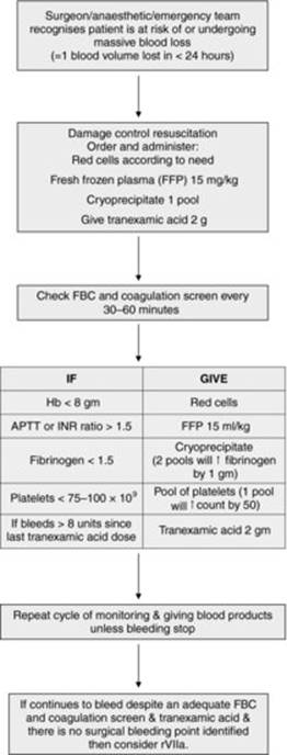
Summary of practical haematological management of a bleeding patient [19] (also see Figure 26.1)
· Send a blood sample to the blood transfusion laboratory for ABO group and RhD group and phone the laboratory indicating the need for blood. If possible, wait for ABO and RhD compatible blood. In emergency cases use group O RhD-negative red cells until patients' ABO and RhD groups are known. Switch to blood of the same ABO and RhD groups as the patient as soon as possible to avoid inappropriate use of group O RhD-negative red cells.
· Send the baseline sample for FBC, coagulation screen, urea and electrolytes.
· When a fast rate of transfusion is required, a vasopressor or infuser or pump and blood warmer should be used.
· Haemostasis. An early coagulation screen and platelet count or thromboelastography will provide a guide to the use of blood components. It is important to appreciate that at least 1.5 blood volumes (i.e. 7–8 litres in adults) must be transfused before the platelet count falls below 50 × 109/L in an average healthy individual, but counts may be lower following massive injury. After initial resuscitation with 1:1:1 or 1:2:2 ratios of plasma, platelets and red cells, transfusion of replacement blood components should be given as necessary according to the results of screening coagulation tests, aiming to keep:
· Platelet count >50 × 109/L;
· PT and APTT ratio less than 1.5 times the control value by giving FFP 15 mL/kg;
· Fibrinogen >1.5 g/L.
· Be mindful of the other possible complications of blood transfusion:
a. Hypocalcemia. Calcium gluconate (2 mL of 10% solution per unit of blood) when calcium concentration is low or there are clinical signs or electrocardiographic changes.
b. Hyperkalaemia may occur due to its high concentration (40 mmol/L) in stored blood. This is only a problem in those with hepatic or renal disease.
c. Acid–base disturbances. Despite the presence of lactic acid in transfused red cells, this usually improves acidosis in shocked patients. Furthermore, transfused citrate produces an alkalosis once it is metabolized.
d. Hypothermia. Warm the patient and the red cell component.
Organization of transfusion of patients with trauma and for major accidents
In a major accident, large numbers of people may be injured within a short space of time. This requires a coordinated approach from the rescue services and the hospital. A ‘major accident procedure’ is a necessity within every hospital and should be tested periodically by holding a ‘major accident exercise’.
The following must be incorporated into the procedure:
· The telephone numbers of those who ‘need to know’ is held by the hospital switchboard.
· Suspend the issue of blood for nonemergency cases.
· Increase the stocks to a predefined level by arranging deliveries from the nearest blood centre and maintain stocks throughout the emergency. The blood transfusion laboratory must have a dedicated telephone line to arrange this, as the main hospital switchboard may be blocked with other calls.
· The risk of clinical clerical errors can be high in this emergency situation so special care must be taken in the identification of casualties and labelling blood samples. In the emergency department, every attempt to maintain good clinical practice should be made. Full identification details of each patient should be given on blood request forms and sample vials, wherever possible, and at least the hospital record number of the patient and their sex.
· The practice of issuing blood in a major disaster is best not changed from routine practice, i.e. compatibility testing should be carried out whenever possible. If this is not possible, every effort should be made to ensure that blood is ABO and RhD group matched. When the recipients blood group is not known group O RhD-negative blood should be given to girls and women in the reproductive age, unless there is life-threatening bleeding and O RhD-negative blood is not available. O RhD-positive blood can be given to males with unknown blood groups.
· Blood components such as FFP and platelets need to be available quickly for those who are receiving massive transfusion.
· Dealing with requests to donate blood. Following a major accident, there may be calls from the public, offering to donate blood. These potential donors should be given the telephone number of the nearest blood centre so that they can attend one of the routine donor clinics.
The future
Resuscitation of massively bleeding patient with initial 1:1:1 ratios of plasma, platelets and red cells is rapidly becoming the norm with demonstrated improved survival and reduced blood use. The efficacy, safety and cost effectiveness of fibrinogen concentrates and 6-factor PCCs need to be assessed to see whether they will replace initial FFP and platelet therapy because of their more rapid availability than thawed plasma, better virological safety and reduced reactions.
Conclusions
The management of bleeding and coagulopathy in massive blood loss has been an area of major research activity in the last five years and is the subject of major ongoing research. The priority of initial treatment is to maintain tissue perfusion while improving haemostasis. Attention to patient characteristics such as hypothermia is critical to good clinical outcomes.
Key points
1. Massive blood transfusion is the administration of one or more blood volumes in 24 hours.
2. The aim of resuscitation is to maintain adequate tissue oxygenation through adequate numbers of red cells and control hemorrhage.
3. Initially use ‘damage control resuscitation’.
4. Check the full blood count and coagulation screen at presentation and then regularly.
5. After initial ‘damage control resuscitation’ carry out the following.
6. Maintain haemoglobin >8 g/dL.
7. Maintain INR and APPT ratio <1.5 with FFP at 15 mL/kg.
8. Maintain platelet counts > 50 × 109/L.
9. Maintain fibrinogen >1.5 g/L.
10. In those bleeding or at risk of bleeding after trauma, give 1 gm tranexamic acid at presentation and then 1 gm in an infusion over 8 hours. Consider tranexamic acid in other major bleeding scenarios.
References
1. Peden M, Scurfield R, Sleet D, Mohan D, Hyder AA, Jarawan E et al. (eds). World Report on road traffic injury prevention. Geneva: World Health Organization; 2004.
2. Dutton RP, Stansbury LG, Leone S, Kramer B, Hess JR & Scalea TM. Trauma mortality in mature trauma systems: are we doing better? An analysis of trauma mortality patterns, 1997–2008. J Trauma 2010; 69: 620–626.
3. World Health Organization. Trends in Maternal Mortality: 1990 to 2008. Geneva: World Health Organization; 2010.
4. Special Issue: Saving Mothers' Lives: Reviewing maternal deaths to make motherhood safer: 2006–2008. The Eighth Report of the Confidential Enquiries into Maternal Deaths in the United Kingdom. Br J Obstet Gynaecol 2011; 118 (Issue Suppl. s1): 1–203.
5. Roullet S, Biais M, Millas E, Revel P, Quinart A & Sztark F. Risk factors for bleeding and transfusion during orthotopic liver transplantation. Ann Fr Anesth Reanim 2011, April; 30(4): 349–352.
6. Fouche Y, Sikorski R & Dutton RP. Changing paradigms in surgical resuscitation. Crit Care Med 2010, September; 38(9 Suppl.): S411–S420.
7. Murthi SB, Stansbury LG, Dutton RP, Edelman BB, Scalea TM & Hess JR. Transfusion medicine in trauma patients: an update. Expert Rev Hematol 2011; 4(5): 527–537.
8. Armand R & Hess JR. Treating coagulopathy in trauma patients. Transfus Med Rev 2003, July; 17: 223–231.
9. Duchesne JC, McSwain Jr NE, Cotton BA, Hunt JP, Dellavolpe J, Lafaro K, Marr AB, Gonzalez EA, Phelan HA, Bilski T, Greiffenstein P, Barbeau JM, Rennie KV, Baker CC, Brohi K, Jenkins DH & Rotondo M. Damage control resuscitation: the new face of damage control. J Trauma 2010, October; 69(4): 976–990.
10. American College of Surgeons Committee on Trauma. Advanced Trauma Life Support Program for Doctors, seventh edn. Chicago, IL: American College of Surgeons; 2004.
11. Koustova E, Stanton K, Gushchin V, Alam HB, Stegalkina S & Rhee PM. Effects of lactated Ringer's solutions on human leukocytes. J Trauma 2002, May; 52(5): 872–878.
12. Jurkovich GJ, Greiser, Luterman A & Curreri PW. Hypothermia in trauma victims: an ominous predictor of survival. J Trauma 1987; 27(9): 1019–1124.
13. Como JJ, Dutton RP, Scalea TJ, Edelman BB & Hess JR. Blood transfusion rates in the care of acute trauma. Transfusion 2004; 44: 809–813.
14. Levi M, Fries D, Gombotz H, van der Linden P, Nascimento B, Callum JL, Bélisle S, Rizoli S, Hardy J-F, Johansson PI, Samama CM, Grottke O, Rossaint R, Henny CP, Goslings JC, Theusinger OM, Spahn DR, Ganter MT, Hess JR, Dutton RP, Scalea TM, Levy JH, Spinella PC, Panzer S & Reesink WH. Prevention and treatment of coagulopathy in patients receiving massive transfusions. Vox Sanguinus 2011; 101: 154–174.
15. Johansson PI, Stensballe J, Rosenberg I, Hilsløv TL, Jørgensen L, Secher NH. Proactive administration of platelets and plasma for patients with a ruptured abdominal aortic aneurysm: evaluating a change in transfusion practice. Transfusion 2007; 47(4): 593–598.
16. McIntyre L, Hebert PC, Wells G, Fergusson D, Marshall J, Yetisir E & Blajchman MJ; Canadian Critical Care Trials Group. Is a restrictive transfusion strategy safe for resuscitated and critically ill trauma patients? J Trauma 2004, September; 57(3): 563–568.
17. Hess JR. Red cell changes during storage. Transfus Apher Sci 2010; 43(1): 51–59.
18. Nienaber U, Innerhofer P, Westermann I et al. The impact of fresh frozen plasma vs coagulation factor concentrates on morbidity and mortality in trauma-associated haemorrhage and massive transfusion. Injury 2011; 42(7): 697–701.
19. Rossaint R, Bouillon B, Cerny V, Coats TJ, Duranteau J, Fernández-Mondéjar E, Hunt BJ, Komadina R, Nardi G, Neugebauer E, Ozier Y, Riddez L, Schultz A, Stahel PF, Vincent JL & Spahn DR; Task Force for Advanced Bleeding Care in Trauma. Management of bleeding following major trauma: an updated European guideline. Crit Care 2010; 14(2): R52.
Further reading
Cotton BA, Reddy N, Hatch QM et al. Damage control resuscitation is associated with a reduction in resuscitation volumes and improvement in survival in 390 damage control laparotomy patients. Ann Surg 2011, October; 254(4): 598–605.
CRASH-2 collaborators, Roberts I, Shakur H, Afolabi A et al. The importance of early treatment with tranexamic acid in bleeding trauma patients: an exploratory analysis of the CRASH-2 randomised controlled trial. Lancet 2011, 26 March; 377(9771): 1096–1101.
Hess JR, Brohi K, Dutton RP et al. The coagulopathy of trauma: a review of mechanisms. J Trauma 2008, October; 65(4): 748–754.
Hess JR, Lindell AL, Stansbury LG, Dutton RP & Scalea TM. The prevalence of abnormal results of conventional coagulation tests on admission to a trauma center. Transfusion 2009; 49: 34–39.
Holcomb JB, Wade CE, Michalek JE et al. Increased plasma and platelet to red blood cell ratios improves outcome in 466 massively transfused civilian trauma patients. Ann Surg 2008; 248(3), 447–458.
Khan KS, Wojdyla D, Say L, Gülmezoglu AM & Van Look PFA. WHO analysis of causes of maternal death: a systematic review. Lancet 2006; 367: 1066–1074.
Klug EG, Sharma GK & Lozano R. The global burden of injuries. Am J Public Health 2000; 90(4): 523–526.
Napolitano LM, Kurek S, Luchette FA et al. Clinical practice guideline: red blood cell transfusion in adult trauma and critical care. Crit Care Med 2009, December; 37(12): 3124–3157.
Rossaint R, Cerny V, Coats TJ, Duranteau J, Fernández-Mondéjar E, Gordini G, Stahel PF, Hunt BJ, Neugebauer E & Spahn DR. Key issues in advanced bleeding care in trauma. Shock 2006, October; 26(4): 322–331.
Winter C, Macfarlane A, Deneux-Tharaux C et al. The European project on haemorrhage reduction: attitudes, trial and early warning system (EUPHRATES). BJOG 2007; 114(7): 845–854.