
David C. Rees
Department of Haematological Medicine, King's College Hospital NHS Foundation Trust, London, UK
Introduction
Haemoglobinopathies are caused by mutations in the globin genes and are probably the commonest single gene disorders in the world. The α-globin and β-globin gene families are found on chromosome 16 and chromosome 11, respectively. Together with haem, they combine to form haemoglobin, which is a tetramer of two α-like and two β-like globins. Developmentally, two different α-globins and four different β-globins are produced, resulting in a variety of haemoglobins (Table 29.1). At birth there is a gradual switch from fetal to adult haemoglobin, which is largely complete in a year [1]. Quantitative defects in globin chain synthesis cause thalassaemia, whereas qualitative defects result in haemoglobin variants; the most important haemoglobin variant is haemoglobin S (HbS, β6 glutamic acid-valine), causing sickle cell disease (SCD). Blood transfusion is important in haemoglobinopathies, allowing correction of anaemia, suppression of abnormal erythropoiesis and replacement of abnormal erythrocytes.
Table 29.1 Normal haemoglobins.
|
Structure |
Name |
Predominant expression |
|
ζ2ɛ2 |
Hb Gower 1 |
0–10th-week gestation |
|
α2ɛ2 |
Hb Gower 2 |
5th–10th-week gestation |
|
ζ2γ2 |
Hb Portland |
5th–10th-week gestation |
|
α2Gγ2 |
HbF |
12th-week gestation – 4th month |
|
α2Aγ2 |
HbF |
12th-week gestation – 4th month |
|
α2β2 |
HbA |
4th month – death |
|
α2δ2 |
HbA2 |
4th month – death |
α-Thalassaemia syndromes
Most people have four α-globin genes and the common types of α-thalassaemia are due to large deletions of one or more of these genes [2]. Deletion of both α-globin genes on a chromosome can occur, although this is only common in Southeast Asia and the eastern Mediterranean. There are three main α-thalassaemia syndromes.
α-Thalassaemia trait
This is usually due to the deletion of one or two α-globin genes. The haemoglobin is normal with mild hypochromia. Blood transfusion is never needed to treat the condition itself.
HbH disease
This occurs when there is only one functional α-globin gene. It is typically a mild condition, with haemoglobin of 7–10 g/dL and HbH (tetramers of β-globin) inclusion bodies in erythrocytes. The spleen is moderately enlarged. Blood transfusion is unusual, but may be necessary following Parvovirus B19 infection.
Hb Bart's hydrops fetalis
A complete or near-complete absence of functional α-globin genes results in progressive fetal anaemia from the 10th week of gestation. Without intervention this results in a hydropic fetus and miscarriage at 30–40 weeks, reflecting the importance of α-globin in forming HbF. Occasionally fetal anaemia has been detected and the pregnancy maintained until term with regular intrauterine transfusions. The resulting babies are transfusion dependent and usually seriously handicapped. This may either result from the effects of fetal anaemia or be caused by large deletions on chromosome 16. If fetal anaemia is found to be due to Hb Bart's hydrops fetalis, the likelihood of serious handicap should be discussed with the parents prior to starting intrauterine transfusions.
β-Thalassaemia syndromes
β-Thalassaemia results from a reduced rate of β-globin synthesis, but much of the pathology arises from the resulting excess of α-globin. β-globin is not part of fetal haemoglobin and there are no adverse fetal or neonatal effects. In contrast to α-thalassaemia, most cases of β-thalassaemia are caused by small mutations or deletions in the β-globin gene. More than 300 different mutations have been identified, and the many different combinations result in a phenotypic continuum from asymptomatic to transfusion dependence.
β-Thalassaemia trait
This results from the inheritance of one mutated β-globin gene. There is minimal anaemia, with hypochromia and microcytosis. Anaemia becomes more marked during pregnancy and occasionally blood transfusion is necessary, although regular or frequent blood transfusions have no role.
β-Thalassaemia intermedia
This is a clinical term referring to a range of conditions characterized by significant anaemia, splenomegaly and increased iron absorption. Patients typically grow and develop normally without the need for regular blood transfusions, at least for the first few years of life. Many different combinations of β-globin mutation cause thalassaemia intermedia [3] (Table 29.2). Anaemia may increase during infection or illness and intermittent transfusions may be necessary. The trigger for blood transfusion is based on clinical signs and symptoms rather than any specific haemoglobin concentration (Hb). Acute symptoms suggesting that transfusion may be beneficial include dyspnoea and fatigue. It can be difficult to decide if someone with severe thalassaemia intermedia would benefit from regular blood transfusions and treatment as for thalassaemia major. In children this is suggested by poor growth, recurrent illness, marked bony expansion, progressive splenomegaly or evidence of organ damage, such as pulmonary hypertension. Sometimes children require regular blood transfusions to progress through puberty; older adults, who previously managed without transfusion, may become transfusion dependent due to reduced cardiorespiratory function. The decision to start regular transfusions is clinical, not based on a particular genotype or Hb. Once regular transfusions start they should be continued long term. Iron overload can be a problem in thalassaemia intermedia, even in the absence of regular transfusions, and iron stores should be monitored regularly and chelation started as necessary [4].
Table 29.2 Causes of β-thalassaemia intermedia.
|
Factors lessening severity of predicted β-thalassaemia major |
|
Mild β-thalassaemia mutations, e.g. HbE/β-thalassaemia |
|
Coinheritance of α-thalassaemia |
|
Coinheritance of increased capacity to make HbF |
|
Unexplained |
|
Factors worsening severity of predicted β-thalassaemia trait |
|
Coinheritance of triplicated α-globin gene |
|
Dominant β-thalassaemia mutation |
|
Unexplained |
β-Thalassaemia major
Thalassaemia major is the term used when a patient with β-thalassaemia is treated with regular blood transfusions from an early age. Without transfusions, the child either dies or is seriously ill, with failure to thrive, bony deformity or a poor quality of life. It is usually due to the coinheritance of severe β-thalassaemia mutations (β° mutations) from both parents, but can be caused by combinations of less severe mutations (β+-thalassaemia) with exacerbation from epigenetic and environmental factors. The clinical problems result from excess α-globin chains, damaging the developing erythroid cells in the marrow such that they fail to mature into circulating red cells (ineffective erythropoiesis). The expanded erythroid component of the marrow produces large amounts of growth differentiation factor 15, which reduces hepcidin production by the liver; hepcidin inhibits gastrointestinal iron absorption and the inappropriately low levels result in increased iron absorption. The main clinical features of thalassaemia major are therefore:
· severe anaemia;
· bone marrow expansion with bony deformity and osteopenia;
· hypersplenism and hypermetabolism;
· iron overload.
Blood transfusion in β-thalassaemia major
The aim of regular, long-term blood transfusions in thalassaemia major is to:
· reduce or eliminate symptoms of anaemia;
· suppress ineffective erythropoiesis to prevent bony deformity;
· prevent the development of significant hypersplenism;
· suppress extramedullary haemopoiesis.
Studies measuring soluble transferrin receptor levels have shown that erythropoiesis is adequately suppressed by maintaining a pretransfusion Hb between 9 and 10 g/dL [5]. The posttransfusion Hb is usually kept below 15 g/dL to avoid problems with high blood viscosity and fluid overload. In practice these parameters are usually achieved by regular, simple transfusions given every 2–5 weeks, the frequency being determined by local resources and pretransfusion symptoms. Occasionally, more intensive transfusion is used to support cardiorespiratory problems or suppress extramedullary haemopoiesis. Automated exchange transfusions are also used, and typically patients have a full-volume exchange every 6 to 8 weeks. Exchange transfusion has the advantages of decreasing iron loading and less frequent hospital attendances, although it involves more donor exposure with increased risk of alloimmunization and infection; good vascular access is also important, and therefore the procedure is more difficult in young children (Table 29.3). It is more expensive than simple transfusion and unavailable in many parts of the world. The insertion of semi-permanent venous access devices is sometimes necessary if venous access is difficult. With adequate blood transfusion and iron chelation from an early age, the expectation is that children will grow and develop normally, with a good quality of life. Life expectancy should approach the normal range, although chelation failure means that median life expectancy is usually shortened; patients in developed countries born in the 1960s had a median survival of 30 years, but this has progressively increased [6].
Table 29.3 Advantages and disadvantages of different types of blood transfusion.
|
Simple transfusion |
Manual exchange |
Automated transfusion |
|
Quick to organize |
Slower to organize |
Slower to organize and not always available |
|
No special skills involved |
Previous experience and training necessary |
Specialist staff and equipment needed |
|
Simple venous access only |
Good venous access |
Very good venous access at two sites |
|
Quick to complete |
Slow to complete |
Quick to complete |
|
Limited scope to reduce HbS percentage |
Significant decrease in HbS percentage |
Very significant decreases in HbS percentage |
|
Fairly predictable final Hb levels and HbS percentage |
Unpredictable final Hb and HbS percentage |
Predictable final Hb and HbS percentage |
|
Limited donor exposure |
Moderate donor exposure |
High donor exposure |
|
Significant increase in iron stores |
Minimal increase in iron stores |
Minimal or no increase in iron stores |
Sickle cell disease (SCD)
SCD refers to a group of conditions in which the patient is either homozygous for the mutated βS allele or coinherits βS with another β-globin mutation (Table 29.4). The primary pathological event is the polymerization of deoxygenated HbS, resulting in damage to the red cell membrane, cellular dehydration and increased cytoplasmic viscosity. These abnormal, rigid sickle cells cause vaso-occlusion, in which precapillary venules are blocked, resulting in both acute and chronic ischaemic damage. Clinical consequences of vaso-occlusion include hyposplenism, acute pain, acute chest syndrome, acute abdominal pain and chronic restrictive lung defects. Vaso-occlusion also leads to a further cascade of interlinked pathological events, including haemolytic anaemia, inflammation, oxidative stress, reperfusion injury, hypercoagulability, nitric oxide deficiency, hypoxaemia and vasculopathy. Vasculopathy and endothelial dysfunction seem to be particularly important for some chronic complications, including cerebrovascular disease and stroke, pulmonaryhypertension, priapism and leg ulcers. Haemolyis is thought to contribute significantly to the vasculopathy by releasing free haemoglobin into the plasma, which inactivates nitric oxide resulting in vascular endothelial dysfunction [7].
Table 29.4 Types of SCD.
|
Severe SCD |
|
HbSS (sickle cell anaemia) |
|
HbS β-thalassaemia |
|
HbS OArab |
|
HbS DPunjab |
|
Mild SCD |
|
HbSC |
|
HbS β+-thalassaemia |
|
HbS Lepore |
|
Very mild SCD |
|
HbSE |
|
HbS/hereditary persistence of fetal haemoglobin |
Blood transfusion plays an important role in combating many of these pathological processes, including increasing Hb, reducing the number of circulating erythrocytes able to cause vaso-occlusion and reducing intravascular haemolysis. Increasing the Hb and haematocrit too much is potentially harmful as it increases blood viscosity, particularly in small blood vessels, and so can reduce oxygen delivery to tissues and precipitate acute ischaemic events, particularly in the brain. When planning a transfusion in SCD, it is important to decide what the target Hb and HbS percentage are, and then plan how best to achieve these. This can be through either a simple top-up transfusion or an exchange transfusion of some sort, in which blood is also removed. In SCD, there is not thought to be any benefit from exchange per se, and it is a way of decreasing the HbS percentage without increasing the haematocrit excessively. Exchange transfusions are performed most efficiently using automated apheresis, although this is not always available as an emergency and requires very good vascular access (Table 29.3).
Indications for transfusions in acute complications of SCD
· Acute anaemia. The need for transfusion is dependent on symptoms rather than on haemoglobin level, but is usually necessary when the Hb falls below 5 g/dL. A single, simple transfusion aiming to increase the Hb to 8–10 g/dL is typically used. Specific causes of acute anaemia include:
· Parvovirus B19 infection – low reticulocyte count, viral symptoms.
· Acute splenic sequestration – high reticulocyte count, enlarging spleen and rapidly falling Hb; potentially fatal without urgent transfusion; it is commonest in children under the age of 5 years and recurrent episodes usually require splenectomy.
· Acute hepatic sequestration – rare complication with enlarging liver and reticulocytosis.
· Acute pain – occasionally the Hb falls significantly (>2 g/dL) during an episode of acute pain and transfusion may be necessary to correct symptomatic anaemia.
· Acute chest syndrome. This is defined as new pulmonary shadowing on a chest X-ray in someone with SCD and is typically accompanied by chest pain, tachypnoea, hypoxia and increasing anaemia. Many cases recover with oxygen and antibiotics, although 5–10% of cases deteriorate and require respiratory support. Blood transfusion has an important role in managing severe cases. Severe cases are usually accompanied by progressive anaemia and observational evidence suggests that early top-up transfusion to increase the Hb to about 10 g/dL can prevent deterioration; this also results in a significant reduction in HbS percentage. If deterioration is rapid or mechanical ventilation necessary, the HbS should be reduced to less than 30% with haemoglobin of about 10 g/dL, which will often involve an urgent exchange transfusion [8].
· Stroke. If acute neurological symptoms occur, urgent blood transfusion should be arranged whilst investigating the cause, which is likely to be cerebrovascular disease. The aim of transfusion is both to reduce HbS to less than 30% and increase the Hb to about 10 g/dL. This will usually require an exchange transfusion and a retrospective study suggested that outcome is better if an exchange transfusion is used initially rather than a top-up [9]. It is probably important to correct significant anaemia rapidly with a top-up transfusion before the exchange, to limit the area of brain ischaemia, if there is going to be a delay of more than a few hours before the exchange transfusion can be performed.
· Multiorgan failure. This can occur following severe sepsis or acute chest syndrome, and usually patients will have been fully exchanged as part of the prodrome to this often agonal event. Generally the HbS is kept at less than 30%.
· There is no evidence to support the use of blood transfusions in the treatment of acute pain, priapism or osteomyelitis. In some cases these may be accompanied by significant anaemia and so benefit from transfusion, or surgery may be necessary and transfusion used preoperatively.
Indications for regular transfusion in SCD
· Secondary stroke prevention. Following a first stroke there is a 20–90% chance of further strokes. Retrospective studies suggest that the risk of recurrence is reduced by up to 90% with regular blood transfusions to keep the HbS less than 30 or 50%. This requires regular exchange or top-up transfusions, with evidence suggesting that the high risk of stroke returns once the transfusions stop [10].
· Primary stroke prevention in children with abnormal transcranial Doppler (TCD) scans. Children with narrowed intracerebral blood vessels, as detected by TCD, are at high risk of acute stroke. A randomized controlled trial showed that keeping HbS less than 30% with regular transfusions reduced stroke risk by 90% [11]. Transfusions are typically continued life-long, although the target HbS is often increased to 50% after about three years without any cerebrovascular deterioration.
· Recurrent acute chest syndrome. Hydroxyurea is effective at stopping recurrent acute chest syndrome in 80% cases; if hydroxyurea fails, regular blood transfusions may help prevent further episodes.
· Progressive organ failure. Hepatic, renal, cardiovascular and pulmonary failure are problems in older SCD patients, and regular transfusions can help support organ function or prevent further deterioration. As survival in SCD improves, there are increasing numbers of older patients and increasing amounts of blood used for this indication.
· Other indications. Regular transfusions may have a role in preventing frequent episodes of acute pain, chronic pain, avascular joint necrosis, leg ulcers, pulmonary hypertension, acute pain in pregnancy and recurrent splenic sequestration, depending on individual circumstances.
Preoperative blood transfusion in SCD
Perioperative complications are increased in SCD, including the development of pain and acute chest syndrome. In high-risk surgery, such as cardiac, brain and long operations, it is accepted that preoperatively the HbS should be less than 30% with minimal anaemia. This will usually involve an exchange transfusion before surgery. For other types of surgery, the need for transfusion is less clear. Studies suggest no advantage of exchange over top-up transfusion, unless the patient has significant organ damage, cerebrovascular disease or had previous severe complications [12]. The recently completed TAPS (transfusion alternatives preoperatively in sickle cell disease) randomized controlled trial suggested that simple preoperative transfusion reduces the risk of complications, such as acute chest syndrome, for some types of operation, particularly abdominal and throat surgery. In practice, most people with sickle cell anaemia and Hb less than 9 g/dL should probably be transfused to a target Hb of about 10 g/dL prior to general anaesthesia for moderate-risk surgery.
Complications of transfusions in haemoglobinopathies
All the routine complications of blood transfusion can occur. Particular problems include:
· Alloimmunization. Rates vary from 10 to 20% depending on the similarity between the ethnicities of donor and recipient populations, and the extent of blood group matching. In many countries, including northern Europe and the USA, the thalassaemic and SCD populations are mostly of a different ethnic origin to the majority of the blood donor population, increasing the risk of alloimmunization. An extended red cell phenotype should be performed before starting transfusion (C, c, D, E, e, K, k, Jka, Jkb, Fya, Fyb, Kpa, Kpb, MNS, Lewis). The risk of alloimmunization can be reduced by choosing blood matched for Rh and Kell groups in both thalassaemia [13] and SCD [14]. Leucocyte reduction reduces the risk of transfusion reactions, prion transmission and possibly alloimmunization.
· Autoantibody formation. This typically accompanies the development of alloantibodies and occurs in up to 25% of thalassaemia major patients and a smaller percentage with SCD. It is associated with non-leucocyte-reduced transfusions and splenectomy. This can result in significant autoimmune haemolysis, in which transfusion fails to increase the Hb significantly. Management includes avoiding unnecessary blood transfusion, with a possible role for corticosteroids, intravenous immunoglobulin and rituximab. Blood transfusion may be unavoidable in severe haemolysis and should be used to treat life-threatening anaemia.
· Infection. The prevalence of transfusion-transmitted infections varies widely but is rare in most developed countries. Prion infection is an increasing concern. Before starting a transfusion programme, children should be vaccinated against hepatitis B.
· Iron overload. This is predominantly due to the iron content of transfused blood, although increased absorption is important in thalassaemia intermedia.
Iron chelation
Regular blood transfusions inevitably cause iron overload. Each unit of transfused blood contains about 200 mg of iron, and typically iron chelation is started after 12 months of regular transfusions or when the ferritin exceeds 1000 μg/L. Without treatment, iron accumulates in and damages the liver, heart and endocrine organs. In thalassaemia, iron-related heart disease is the major cause of death. In SCD marked cardiac iron deposition is unusual, although significant morbidity results from hepatic siderosis.
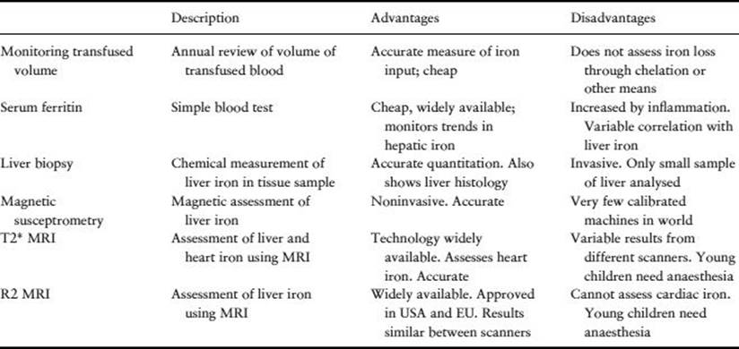
It is important to assess iron stores accurately to monitor chelation therapy (Table 29.5). If there is evidence of progressive iron overload, intensive efforts should be made to improve iron chelation; these will usually focus on improving treatment adherence. Cardiac siderosis is a potentially fatal complication, suggested by the development of arrhythmias, heart failure and increasing iron stores on cardiac MRI, and necessitates intensive chelation therapy. Iron chelation techniques include:
Table 29.5 Assessment of iron overload.
· Venesection rapidly removes excess iron, although clearly this is not possible in transfusion-dependent patients. Venesection is often used in haemoglobinopathy patients post-bone-marrow transplantation.
· Desferrioxamine. This has been used for more than 30 years. Good compliance has been shown to improve survival. Side effects are few but include growth impairment, retinal and cochlear toxicity at higher doses. Side effects become increasingly common as iron stores approach normal. Negative iron balance is typically achieved in a transfusion-dependent patient at a dose of 40 mg/kg 5 nights per week. The main problem is that it has to be given parenterally, usually by overnight subcutaneous infusions. Adherence to desferrioxamine treatment is poor and this results in toxic iron accumulation. In heart failure secondary to iron overload, continuous intravenous desferrioxamine has been shown to be effective [15].
· Deferiprone (L1). This oral iron chelator was developed in the 1980s. Side effects include neutropenia and arthritis. The drug has been licensed as a second-line chelator in thalassaemia in Europe for more than 10 years, but was only licensed for use in North America in 2011 because of concern about toxicity and lack of efficacy. Recent studies suggest that it is particularly effective at removing cardiac iron and various regimes in combination with desferrioxamine have been devised. Because of the risk of agranulocytosis, it is recommended that weekly blood tests are performed on those taking deferiprone [16].
· Deferasirox. This oral iron chelator is licensed as a first-line treatment for transfusional iron overload around the world. It seems to be as effective as desferrioxamine with relatively few side effects. The main toxicity involves increases in serum creatinine, which in general have been nonprogressive and reversible. There is emerging evidence that it also removes cardiac iron [17].
Key points
1. Regular intrauterine transfusions should not be used in fetuses with Hb Bart's hydrops fetalis until the risk of severe handicap has been discussed with the parents.
2. Transfusions should be started in severe β-thalassaemia syndromes on the basis of symptoms rather than a particular Hb or genotype.
3. Blood transfused to haemoglobinopathy patients should be fully matched for Rh and Kell blood groups.
4. Urgent blood transfusion is indicated in SCD in acute anaemia, severe acute chest syndrome, multiorgan failure and acute neurological problems.
5. Regular transfusions are mainly used in SCD for primary and secondary stroke prevention.
6. Iron chelation should be actively considered after 10–12 blood transfusions.
7. Hepatic iron stores should be monitored in SCD and thalassaemia using a combination of transfusion history, serum ferritin and MRI.
8. In thalassaemia major and severe thalassaemia intermedia, cardiac iron should be monitored using MRI and intensive chelation started if there is evidence of significant or progressive cardiac iron overload.
References
1. Weatherall DJ. Pathophysiology of thalassaemia. Bailliere's Clin Haematol 1998; 11(1): 127–146.
2. Higgs DR, Engel JD & Stamatoyannopoulos G. Thalassaemia. Lancet 2012; 379: 373–383.
3. Danjou F, Anni F & Galanello R. Beta-thalassemia: from genotype to phenotype. Haematologica 2011; 96(11): 1573–1575.
4. Taher AT et al. Optimal management of beta thalassaemia intermedia. Br J Haematol 2011; 152(5): 512–523.
5. Cazzola M et al. Relationship between transfusion regimen and suppression of erythropoiesis in beta-thalassaemia major. Br J Haematol 1995; 89(3): 473–478.
6. Telfer P et al. Survival of medically treated thalassemia patients in Cyprus. Trends and risk factors over the period 1980–2004. Haematologica 2006; 91(9): 1187–1192.
7. Rees DC, Williams TN & Gladwin MT. Sickle-cell disease. Lancet 2010; 376(9757): 2018–2031.
8. Vichinsky EP et al. Causes and outcomes of the acute chest syndrome in sickle cell disease. National Acute Chest Syndrome Study Group. N Engl J Med 2000; 342(25): 1855–1865.
9. Hulbert ML et al. Exchange blood transfusion compared with simple transfusion for first overt stroke is associated with a lower risk of subsequent stroke: a retrospective cohort study of 137 children with sickle cell anemia. J Pediat 2006; 149(5): 710–712.
10. Ohene-Frempong K et al. Cerebrovascular accidents in sickle cell disease: rates and risk factors. Blood 1998; 91(1): 288–294.
11. Adams RJ et al. Prevention of a first stroke by transfusions in children with sickle cell anemia and abnormal results on transcranial Doppler ultrasonography. N Engl J Med 1998; 339(1): 5–11.
12. Vichinsky EP et al. A comparison of conservative and aggressive transfusion regimens in the perioperative management of sickle cell disease. The Preoperative Transfusion in Sickle Cell Disease Study Group. N Engl J Med 1995; 333(4): 206–213.
13. Thompson AA et al. Red cell alloimmunization in a diverse population of transfused patients with thalassaemia. Br J Haematol 2011; 153(1): 121–128.
14. Vichinsky EP et al. Alloimmunization in sickle cell anemia and transfusion of racially unmatched blood. N Engl J Med 1990; 322(23): 1617–1621.
15. Davis BA & Porter JB Long-term outcome of continuous 24-hour deferoxamine infusion via indwelling intravenous catheters in high-risk beta-thalassemia. Blood 2000; 95(4): 1229–1236.
16. Tanner MA et al. A randomized, placebo-controlled, double-blind trial of the effect of combined therapy with deferoxamine and deferiprone on myocardial iron in thalassemia major using cardiovascular magnetic resonance. Circulation 2007; 115(14): 1876–1884.
17. Pennell DJ et al. Efficacy of deferasirox in reducing and preventing cardiac iron overload in beta-thalassemia. Blood 2010; 115(12): 2364–2371.
Further reading
Guidelines for the Clinical Management of Thalassaemia, 2nd edn. Thalassaemia International Federation, 2007. Available at: www.thalassaemia.org.cy.
Olivieri NF & Brittenham GM. Iron-chelating therapy and the treatment of thalassemia. Blood 1997; 89: 739–761.
Serjeant GR & Serjeant BE. Sickle Cell Disease, 3rd edn. Oxford, UK: Oxford University Press; 2001.
Weatherall DJ & Clegg JB. The Thalassaemia Syndromes, 4th edn. Oxford, UK: Blackwell Scientific Publications; 2001.