
Anne Zajicek, MD, PharmD, FAAP and Lori D. Karan, MD, FACP, FASAM
CHAPTER OUTLINE
■ BASIC PHARMACOLOGY CONCEPTS
■ SUMMARY
Addiction medicine is based on the application of pharmacologic principles to drugs of abuse, ranging from prescription medications (e.g., narcotics, anxiolytics, stimulants) to Drug Enforcement Administration Class 1 compounds such as heroin, phencyclidine, and hallucinogenic mushrooms. Drugs of abuse are used to produce mood alterations; this requires drug penetration into the brain.
Pharmacologic principles in this chapter will focus on the pharmacokinetics of drug delivery to the brain; pharma-cogenomics, the genetic differences in metabolism, transport, and receptors that effect interindividual differences in drug disposition and response; and pharmacodynamic effects including tolerance and withdrawal.
BASIC PHARMACOLOGY CONCEPTS
Pharmacokinetics
Pharmacokinetics describes the time course of drug concentrations in blood and tissues (e.g., brain). Drug concentrations in blood and other sites are determined by absorption, distribution, metabolism, and elimination. The magnitude of a drug’s pharmacologic effect depends on the free (unbound) drug concentration at its site of action.
Absorption
Absorption is the process of drug movement from the site of drug delivery to the site of action. Psychoactive drugs can be taken orally (ethanol, amphetamines, barbiturates, opiates), intranasally (glue, solvents, amyl nitrate, cocaine, heroin), via smoking (nicotine, marijuana, freebase cocaine), intravenously (heroin, cocaine, methamphetamine), transdermally (fentanyl and nicotine patches), and subcutaneous injection. Figure 6-1 illustrates the differences in drug concentrations over time for the various routes of administration. The more rapidly a psychoactive drug is delivered to its site of action in the central nervous system, the greater is its mood-altering and reinforcing effects. The more rapidly achieved and higher the peak concentrations from intravenous and pulmonary (smoking) routes illustrate this point.

FIGURE 6-1 Venous drug concentrations after different routes of administration.
Bioavailability is defined as the fraction of unchanged drug that reaches the systemic circulation after administration by any route. The bioavailability factor (F) takes into account the portion of the administered dose that is able to enter the circulation unchanged. For intravenously administered drugs, F = 1.0 (100%). Bioavailability depends on a given drug’s site-specific membrane permeability, activity of drug transporters, and its first-pass metabolism. First-pass metabolism is the metabolism that occurs before a drug reaches the systemic circulation and occurs most extensively for lipid-soluble drugs such as morphine, methyl-phenidate, and desipramine and can significantly reduce bioavailability. Morphine, for example, requires nearly twice the dose when administered orally as compared to intravenously. First-pass metabolism is relatively unimportant for drugs administered through the intravenous, sublingual, intramuscular, subcutaneous, and transdermal routes, since drugs administered by these routes enter the general circulation directly.
For orally administered drugs, the rate of absorption is affected by (i) the pharmaceutical properties of the oral dosage form; (ii) the pH of gastric contents (drugs can be destroyed by extreme acid or basic conditions); (iii) gastric emptying time (faster gastric emptying time results in more rapid delivery to the small intestine, the site of absorption, except with dumping where the delivery to the small intestine is too fast to be optimally absorbed); (iv) intestinal transit time (for drugs absorbed in the small intestine, there is an increased rate of absorption with a faster intestinal transit time); (v) integrity of intestinal epithelium; and (vi) the presence of food (which decreases the interaction time between the drug and the intestinal villi) (1).
Efforts have been made to reformulate potentially addictive prescription drugs to reduce the rates of absorption in order to reduce the peak concentrations and reduce this reinforcing effect (2).
Smoked and inhaled drugs bypass the venous system and thus have the most rapid rate of delivery. Absorption of inhaled drug depends on the physical characteristics of the drug, including its volatility, particle size, and lipid solubility (3). Drugs that reach the alveoli of the lungs have rapid access to the bloodstream through closely applied capillary alveolar surfaces on the large pulmonary surface areas. Because a large portion of the cardiac output passes through the pulmonary circulation, the delivery of smoked and freebased drug to the brain is rapid.
Drugs must pass through biologic membranes to be absorbed. With passive diffusion, biologic membranes are more permeable to lipid-soluble and uncharged molecules. Some drugs have diminished absorption because of a reverse transporter associated with P-glycoprotein. This reverse transporter actively pumps drug out of the gut wall cells back into the gut lumen. When P-glycoprotein is inhibited, increased drug absorption results.
Some food and drug interactions alter first-pass metabolism and absorption from the intestinal wall. For example, components of grapefruit juice and other foods that either inhibit (e.g., grapefruit juice) or induce intestinal wall CYP3A4 or P-glycoprotein can lead to altered bioavailability of drugs that are substrates for this cytochrome (4,5). Also, the nonselective monoamine oxidase inhibitors (MAOIs) such as phenelzine and tranylcypromine—and, to a much lesser extent, the MAO B inhibitor selegiline and the reversible MAOI moclobemide—inhibit MAO A in the intestinal wall and liver. This inhibition diminishes the first-pass metabolism of tyramine, which is present in cheeses and various foods (6). When tyramine, an indirect-acting sympathomimetic amine, reaches the systemic circulation, it can produce increased release of norepinephrine from the sympathetic postganglionic neurons; this, in turn, can result in a severe pressor response and hypertensive crisis.
Hastening gastric emptying can help to achieve a more rapid drug effect without altering bioavailability. Gastric emptying can be hastened by taking a drug on an empty stomach with at least 200 mL of water and remaining in an upright position. Food, recumbency, heavy exercise, and drugs that slow gastric emptying (such as narcotics and anticholinergic drugs) can result in later and lower peak concentrations of the index drug.
Upon absorption, when drug concentrations are graphed against time, a peak drug concentration (Cmax) is reached at Tmax. The trough concentration is Cmin. The area under the concentration–time curve (AUC) is a measure of drug exposure that can be calculated and quantified.
Distribution
Once absorbed, a drug is distributed to the various organs and tissues of the body. Distribution is influenced by organ perfusion, organ size, binding of the drug within the blood and tissues, and the permeability of tissue membranes (7).
Most psychoactive drugs enter the brain because they are highly lipid soluble. The blood–brain barrier hinders the ability of non–lipid-soluble drugs to reach the brain tissue by diffusion (8). Unlike the fenestrated capillaries found throughout the body, which allow movement of molecules less than 25,000 Daltons, the endothelial cells lining brain capillaries have tight junctions and do not permit these small molecules to pass through. Without fenestrations, drugs must cross the two membranes of the endothelial cell by passive diffusion in order to enter the brain. The blood–brain barrier limits the admittance of many drugs to the brain and is found throughout the brain and spinal cord at all regions central to the arachnoid membrane, except for the floor of the hypothalamus and the area postrema, including the chemoreceptor trigger zone (where direct-acting chemicals can provoke vomiting).
For some compounds, however, specific active transport systems exist. These active transport systems enable glucose, amino acids, amines, purines, nucleosides, and organic acids to gain access to the brain (9). In contrast, P-glycoprotein is an efflux carrier present in the brain capillary endothelial cell, which bars the drug from translocating across the endothelial cell and actively exports the drug out of the brain (10).
When a drug distributes into all of the body compartments and tissues, it is said to distribute into an apparent volume of distribution (Vd). This volume has no direct physical equivalent because it describes the amount of serum, plasma, or blood that would be required to account for all the drug in the body. Vd can be thought of as the amount of drug in the body (D = dose) divided by the concentration of drug (C) in the plasma, or

Drugs with a small Vd are confined primarily to the intra-vascular space of approximately 5 L. The drugs may be tightly bound to plasma proteins, or they may have a high molecular weight (large proteins, dextrans, and so forth). Drugs can have large Vd values up to 50,000 L if they are highly bound to tissue sites or are lipophilic.
Protein binding affects free (active) drug concentrations. Characteristics of binding proteins are capacity (amount of binding space) and affinity (tightness of binding interaction). Albumin is a high-capacity, low-affinity binding protein, whereas specific transport proteins such as transcortin are low capacity, high affinity. Acidic drugs commonly bind to albumin, the most abundant plasma protein; examples are barbiturates, benzodiazepines, and phenytoin. Basic drugs such as methadone bind to alpha1-acid glycoprotein, and others such as amitriptyline and nortriptyline bind to lipoproteins. Some binding sites are competitive, and a drug with a higher binding site affinity can displace a drug with a lower binding site affinity. Binding can also be stereospecific (specific for one stereoisomer of a compound). Drugs that are greater than 90% bound are considered highly protein bound, and reduced protein binding for these highly protein bound drugs can lead to large increases in drug effect.
The rate of blood flow delivered to specific organs and tissues affects drug distribution. Well-perfused tissues can receive large quantities of drug, provided that the drug can cross the membranes or other barriers present between the plasma and tissue. In contrast, poorly perfused tissues, such as fat, receive and release drug at a slow rate. This action explains why the concentration of drug in fat can be maintained long after the concentration in plasma has begun to decrease; anesthetics are examples of this phenomenon.
Clearance
Elimination refers to disappearance of the parent and/or active molecule from the bloodstream or body, which can occur by metabolism and/or excretion. Excretion is the process of removing a compound from the body without chemically changing that compound. Drugs can be excreted through the urine or feces, exhaled through the lungs, or secreted through sweat or salivary glands. The term clearance (Cl) represents the theoretical volume of blood or plasma that is completely cleared of drug in a given period of time. The factors that determine hepatic clearance are hepatic blood flow, the fraction of drug that is unbound, and the drug’s intrinsic clearance. If the intrinsic clearance of an unbound drug is very small, then this metabolic capacity (i.e., intrinsic clearance) of the liver, rather than hepatic blood flow, becomes the major determinant of hepatic clearance. In this case, functionality of hepatic enzymes determines drug clearance. If the intrinsic clearance of an unbound drug is very large, blood flow to the liver becomes rate limiting. Metabolic capacity determines drug clearance in most cases.
Most drugs display first-order elimination kinetics: the fraction or percentage of the total amount of drug present in the body removed at any one time is constant and independent of dose. Following administration of a drug with first-order kinetics, concentrations show an exponential decline of drug concentrations. The slope of this decay line is the elimination rate constant, kel, which is the percent of drug cleared per unit time (e.g., percent/hour). The half-life (t1/2) of a drug is the amount of time it takes for a drug concentration to decrease by half. One half-life represents a 50% change, and 2, 3, 4, and 5 half-lives represent 75%, 87.5%, 93.7%, and 96.8% changes, respectively. The time to reach steady state depends upon the duration of the half-life, whereas the amount of drug in the body at steady state will depend upon the frequency of drug administration and its dose. With drugs with dose-independent (first-order) disposition and elimination characteristics, five half-lives is a reasonable estimate of the time to reach steady state. For example, if the concentration at 2 hours postdose is 100 μg/mL, and the concentration at 4 hours postdose is 50 μg/mL, the t1/2 is 2 hours.
One means of calculating t1/2 is

Using a more physiologic approach, t1/2 is directly related to the volume of distribution (Vd) and inversely related to the clearance (Cl). This relationship can be written as follows:
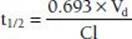
The constant 0.693 in this equation is derived from the natural logarithm of two [ln(2)]. Because drug elimination can be described by an exponential process, the time taken for a twofold decrease can be shown to be proportional to ln(2).
Although it is reasonable to assume that t1/2 and clearance are inversely related (clearance increases, so t1/2 decreases), effects of Vd on t1/2 do occur, which can offset the change in Cl (Vddecreases by the same proportion as clearance decreases, resulting in no change in t1/2).
In contrast, for drugs with zero-order elimination kinetics, the amount of drug removed (rather than the fraction of drug removed) at any one time is constant and dependent on dose. The maximal rate of metabolism and/or elimination is generally due to saturation of a key enzyme. This zero-order process is described by the Michaelis-Menten equation:

where v is the velocity of the reaction, Vmax is the maximum velocity of the reaction, Km is the concentration of the metabolic substrate when the velocity is ½Vmax, and Cpss is the steady-state concentration of drug. In the case of linear kinetics, Km is >>> Cpss, and v = Vmax × Cpss/(Km), or the v is proportional to the drug concentration: the higher the drug concentration, the faster the velocity. When Km <<< Cpss, v = Vmax, which is the case for drugs cleared by zero-order kinetics; the velocity is limited by Vmax. Because the half-life is inversely related to clearance, and clearance changes with drug concentration, the t1/2 is not constant. Therefore, half-life is not a useful descriptor for zero-order drugs. Drug dosing becomes difficult in these cases: a small increase in dose can cause a large increase in concentration, in contrast to drugs with first-order clearance where there is proportionality between dose and concentration. Aspirin, phenytoin, and ethanol are examples of drugs with zero-order elimination (11). See Figure 6-2 for a calculation of the decline in blood alcohol concentrations over time, demonstrating zero-order kinetics.

FIGURE 6-2 Extrapolating the decrease in blood alcohol concentration from two prior readings.

Expect at 4 hours BAL to be 55 mg/dL; 5 hours, BAL to be 35 mg/dL; 6 hours, BAL to be 15 mg/dL; and 7 hours, BAL to have already reached 0.00 mg/dL.
Drug metabolism is the process of chemical modification of drugs and other chemicals by the body, generally into less active and more hydrophilic compounds. These chemical modifications/reactions are generally performed by enzymatic systems, such as the cytochrome P450 enzyme system. Lipophilic drugs are generally transformed to more hydrophilic/polar products that are more readily eliminated by the kidney. Not all metabolites are inactive or nontoxic, and active metabolites need to be considered when assessing a drug’s total activity. In some cases, the administered drug is intentionally designed to be a pharmacologically inactive prodrug that is converted in vivo to a pharmacologically active molecule. Levodopa is one example, which (after crossing the blood–brain barrier) is converted in the basal ganglia to dopamine.
Drugs can be metabolized by Phase I and/or Phase II reactions. Phase I reactions are nonsynthetic reactions in which the drug is chemically altered and oxidized, for example, demethylated. Examples of Phase I reactions include the oxidation of phenobarbital, amphetamine, meperidine, and codeine by microsomal enzymes. Phase II reactions are synthetic reactions in which the drug is conjugated with another moiety, such as glucuronide or sulfate. Examples of synthetic reactions include the glucuronidation of morphine and meprobamate and acetylation of clonazepam and mescaline, which produce a compounds more polar than the parent drugs in order to facilitate elimination. Enzymes are responsible for most drug metabolism. The cytochrome P450 enzymes are most commonly involved and exist in the gut, liver, and brain. The gut and liver enzymes are the best studied. Oxidations can take place by cytochrome P450-dependent and cytochrome P450–independent mechanisms. Cytochrome P450–dependent oxidations include aromatic (phenytoin, amphetamine) and aliphatic (pento-barbital, meprobamate) hydroxylations, epoxidation, and oxidative dealkylation (morphine, caffeine, codeine), deam-ination (amphetamine), desulfurization (thiopental), and dechlorination. Cytochrome P450–independent oxidations include dehydrogenations (ethanol); azo, nitro, and carbonyl reductions (methadone and naloxone); and ester and amide hydrolysis (12).
More than 50 individual CYPs (pronounced “sip” for singular and “sips” for plural) have been identified in humans. As a family of enzymes, CYPs are involved not only in the metabolism of dietary and environmental compounds and medications but in the degradation of bile acids from cholesterol, the metabolism of retinoic and fatty acids including prostaglandins and eicosanoids, and the synthesis of steroids. A large number of specialized CYPs with specific substrate preferences are involved in these latter endogenous functions, in contrast to the relatively few numbers of CYPs that metabolize xenobiotics that have wider ranging and overlapping capabilities. CYPs that metabolize medications not only have a tremendous capacity to oxidize a large number of structurally diverse compounds but can metabolize a single compound at different positions on that molecule. Drug-metabolizing CYPs’ large and fluid substrate–binding sites contribute to their slow catalytic rates. In part, this explains why the half-lives of drugs are much longer than the half-lives of endogenous compounds.
CYPs are named with the root CYP followed by a number designating the family, a letter denoting the subfamily, and another number designating the CYP form. Thus, CYP2B6 is family 2, subfamily B, and gene number 6. Twelve CYPs (1A1, 1A2, 1B1, 2A6, 2B6, 2C8, 2C9, 2C19, 2D6, 2E1, 3A4, and 3A5) are known to metabolize xenobiotics in humans, and CYP3A4 alone is responsible for metabolizing more than 50% of clinically prescribed drugs.
Pharmacogenomics
Pharmacogenomics is the study of the relationship between genetic variations and drug disposition and response. The discipline of pharmacogenetics aims to elucidate cytochrome and other drug-metabolizing enzyme polymorphisms (different enzyme genetic subtypes), the degrees of expression of these polymorphisms, and the functional significance of such expression. Understanding these polymorphisms can help to explain individual differences in drug response.
Genetic variability in drug-metabolizing enzymes can affect drug bioavailability and clearance (13–16). Single nucleotide polymorphisms (SNPs) may alter CYP activity. CYP2D6, for example, which metabolizes codeine to morphine, is the best studied of the drug metabolic enzymes; 48 mutations and 50 alleles have been identified. Genotype and enzyme activity are linked to ethnicity, which varies from no gene/no enzyme activity (6% of whites) to two copies of a fully active gene (33% of Ethiopians). Individuals can be genotyped for 2D6 enzyme function (with classification as poor metabolizers [PMs], intermediate metabolizers [IMs], extensive metabolizers [EMs], and ultrarapid metabolizers [UMs]) (17). Those with PM genotypes generally do not receive adequate analgesia due to the inability to metabo-lize codeine to the active morphine. UMs, on the other hand, metabolize codeine significantly more rapidly and extensively than others, producing rare but life-threatening morphine intoxication. Breast-feeding infants of mothers who are UMs have received morphine overdoses from their mothers who are prescribed codeine for postpartum pain relief (18,19).
CYP2B6 has been less well studied. However, important genotypic influences on enzyme function have been identified, and examples of the importance of the variation in enzyme function on substrate metabolism follow. Several alleles have now been characterized as to enzyme amounts and activities, including CYP2B6 *1 (pronounced sip 2B6 star 1) (wild type), *2, *4, *5, *6, *9, and *18 (20). The *4 allele codes for an enzyme of higher function than the wild type. This allele has been associated with the toxicity of bupropion due to increased rates of conversion of bupro-pion to the active and longer-lasting metabolite, hydroxy-bupropion. Individuals with those genotypes were noted to have less enzyme present and lower enzyme activity as well. The effect of 2B6 genotype on bupropion-facilitated smoking cessation was reported by Zhu et al. (21). Their analysis determined that the hydroxylated metabolite contributed to the pharmacodynamic effect of smoking cessation and that in PMs an upward dosage adjustment could improve the rate of smoking cessation.
CYP3A4 has also had numerous SNPs identified. It appears that prediction of the activity of CYP3A4 is quite complex and not directly related to genotype (13). Several opioids including methadone and buprenorphine are metabolized by CYP3A4. Thus, the role of genetic polymorphisms in metabolism of CYP3A4 substrates is unclear at this time and an area of active research.
Prior to the more routine availability and use of genotyping, phenotyping of CYPs using marker compounds has been, and continues to be, a useful tool. A “cocktail” approach has been used, where subjects are administered microdoses of various compounds, each of which are metabolized by one specific enzyme and would reflect the enzymatic activity for clinically used medications. There are many “cocktail” recipes, one of which is the six-probe cocktail consisting of caffeine (1A2), flurbiprofen (2C9), mephenytoin (2C19), debrisoquine (2D6), chlorzoxazone (2E1), and dapsone (NAT2), where NAT2 is N-acetyltransferase 2 (22,23). A study of a pharmacokinetic interaction with antiretrovirals used midazolam as a marker of CYP3A4, fexof-enadine for P-glycoprotein, and pravastatin for OATP1B1 (organic anion transporter protein 1B1) (24). Many drugs, foods, and environmental chemicals can induce and/or inhibit the activity of the cytochromes, speeding up or slowing down their own metabolism (4,5). Each cytochrome isozyme responds differently to specific exogenous chemicals. For example, CYP1A1 is induced by polycyclic aromatic hydrocarbons, including benzo[α]pyrene contained in cigarette smoke.
Drug interactions at the level of the cytochromes and other metabolizing systems are often clinically significant. Methadone is metabolized primarily by CYP3A4, with contributions from CYP2B6, 2C19, 3A4, and, to a lesser extent, CYP2D6 (5,25). Inhibitors of CYP3A4, including erythro-mycin, diltiazem, ketoconazole, and saquinavir, slow the metabolism of methadone and increase methadone levels. Inducers of CYP3A4 such as carbamazepine, phenobarbital, efavirenz, and St. John’s wort speed the metabolism of methadone and decrease methadone levels. Awareness of potential interactions, clinical observation, and tailoring medication regimens and dosages are needed to optimize therapy and minimize potential toxicities.
Genetically defined differences in drug metabolism may influence risk of addiction, with relative protection for persons who experience adverse drug reactions at lower drug doses. Both the ADH1B2-His47Arg allele of alcohol dehydrogenase 1B and the ALDH-Glu487Lys allele of aldehyde dehydrogenase 2 alone or together can lead to flushing, nausea, and headache, owing to the accumulation of acetaldehyde when alcohol is consumed. Each of these alleles leads to a reduction in the risk of alcoholism, with an additive protective effect when the same person carries both alleles. Persons of South Asian descent are likely to carry both alleles, whereas those with Jewish ancestry often carry the Arg47 allele. Heterozygous carriers of ALDH2 Lys487 have low levels of ALDH2 enzyme activity, whereas ALDH2 Lys487/Lys487 homozygotes are nearly completely protected from alcoholism (26). Tyndale et al. (27) were among the first to postulate the existence of “pharmacogenetic protection factors.” They showed that a sample of whites who inherited two nonfunctional alleles for CYP2D6 were less likely to become dependent on oral opiates (estimated odds ratio >7). Slow nicotine metabolism by CYP2D6 appears to have a protective effect against nicotine addiction (28).
Although drug metabolism occurs largely in the liver, most other tissues and organs, including the lungs, gastrointestinal tract, skin, and kidneys, carry out varying degrees of drug metabolism. Understanding brain metabolism can be especially important in understanding the activity of psychoactive drugs (29). Many P450 cytochromes have been shown to catalyze the metabolism of neuros-teroids as well as psychoactive drugs such as neuroleptics and antidepressants. Alcohol produces a three- to fivefold increase in the level of brain P450 and induces CYP2C, CYP2E1, and CYP4A (30). Brain CYP2D6 can demethylate 3,4-methylenedioxy-N-methylamphetamine (MDMA or “ecstasy”) forming a harmful metabolite, N-methyl-a-methyldopamine. Brain CYP2D6 can O-demethylate para-methoxyamphetamine, a synthetic psychostimulant and hallucinogen, into 4-hydroxyamphetamine, which is also toxic (31). CYP2B6 metabolizes cocaine, phencyclidine, and some amphetamines. Both CYP2B6 and CYP2D6 are inducible by nicotine. P450s in other brain areas are induced by different factors (e.g., levels of CYP2C and CYP4A influence the activity of neurotransmitters such as dopamine, whichuse fatty acid metabolites as intracellular mediators).
Novel brain CYPs, such as 5α-androstane-3β, 17β-diol hydroxylase, CYP7B, and CYP2D4, continue to be discovered (32,33). Because the level of CYPs in the brain is approximately 0.5% to 2% of that in the liver and because brain CYP isoenzymes are of different types than those found in the liver, brain CYPs appear to be locally active but contribute little to overall pharmacokinetics of drugs in the body. The regulation of cytochrome P450 isozyme expression in the brain and elsewhere is being studied.
New areas of research include studies demonstrating novel brain transporter pathways (9,34). Epigenetic factors (35) and use of genome-wide association studies (36) may also yield new insights.
Pharmacodynamics
Pharmacodynamics is the study of the dose–response phenomena, which are the biochemical and physiologic effects of drugs on the body, and the body’s homeostatic response. Most drugs act on specific endogenous targets, or receptors, to modulate the rate and extent of the body’s endogenous functions. Receptors and their associated effector and transducer proteins coordinate signals from multiple ligands with the metabolic activities of the cell to act as integrators of this information.
Potency, Efficacy, and Dose–Response
When drug dose is plotted on a logarithmic scale, a sigmoi-dal curve often results, permitting mathematical manipulation of the results. The maximal efficacy of a drug occurs at Emax, its maximal effect. Efficacy is the extent of functional change imparted to a receptor. Efficacy is determined mainly by the nature of the receptor and its associated effector system. In contrast, potency is primarily determined by the affinity of the receptor for the drug. Potency denotes the amount of drug needed to produce a given effect. The concentration of the drug needed to produce 50% maximal effect occurs at EC50; the more potent the drug, the smaller the dose required to achieve maximal effect. In general, low potency is important only if the drug needs to be administered in undesirably large amounts. Because drug doses are readily adjusted, it is the maximal efficacy that is more often clinically relevant.
A similar sigmoidal curve is attained when the percentage of receptors that bind a drug is plotted against log drug concentration (Fig. 6-3). Here, the concentration at which 50% of the receptors are bound is denoted as Kd, and the maximum number of receptors bound is termed Bmax. Both “dose–response” and “dose–receptor-bound” graphs have a linear or nearly linear middle segment, indicating a first-order process. As the concentration of drug increases, a constant proportion of the drug binds to the receptor, causing a proportionate drug effect.

FIGURE 6-3 Full agonist, partial agonist, and competitive and noncompetitive antagonists. A: No matter how much the dose is increased, a partial agonist always will have a lower maximal efficiency (Emax). A partial agonist may be more potent, less potent, or equally potent as the agonist. In this example, both partial agonists decrease Emax. However, one partial agonist is more potent, and the other partial agonist is equi-potent, when compared to the full agonist. B: If there are no spare receptors, competitive agonists increase the EC50 but do not alter the other Emax. C: Noncompetitive antagonists decrease the Emax. (Modified from Katzung BG, Masters SB. Pharmacodynamics. In: Katzung and trevor’s pharmacology examination and board review. New York, NY: Lange Medical Books/McGraw-Hill, 2002:13.)
A system is said to have spare receptors when the activation of fewer than 50% of the receptors achieves 50% of maximal effect. This determination is made by comparing the concentration for 50% of maximal effect, EC50, with the concentration of 50% of maximal binding, Kd. If the EC50 is less than the Kd, spare receptors are said to be present. The presence of spare receptors does not alter the maximal biologic response, but it does increase the sensitivity to the drug ligand. This relationship occurs because drug–receptor interactions are more likely to appear when there are proportionately more available receptors.
A graded dose–response graph is attained (Fig. 6-4) when the response of a particular receptor–effector system is measured against increasing concentrations of drug. A quantal dose–response graph is achieved when the log dose of a drug is plotted against the cumulative percentage of a population responding to a specified drug response. The results of animal experiments can be plotted in this manner to discern the median effective dose (ED50), median toxic dose (TD50), and median lethal dose (LD50). The therapeutic index is defined as the ratio of the TD50 to the ED50. Because it is unethical to design experiments using a full range of drug doses to determine these indices in humans, the range of therapeutic drug concentrations and the margin of safety are estimated more broadly through extrapolation from animal studies, human drug trials, and clinical experience. In practice, both the risks and benefits of prescribing a medication are taken into account when making therapeutic decisions. Judgment about the clinically acceptable risk of toxicity often is influenced by the severity of the disease being treated.

FIGURE 6-4 Dose-response curves of some opioids. Fentanyl has a lower EC50 than morphine and is more potent than morphine. Both fentanyl and morphine have a higher Emax than codeine and are more efficacious than codeine. Meperidine is less potent than morphine but more efficacious than codeine.
Receptors
The concept of the receptor dates to the turn of the 20th century. Paul Ehrlich introduced the term receptor as he described sites on what he believed to be a single very large molecule of cell protoplasm to which bacterial toxins (and later, drugs) bound to bring about changes in cellular metabolism. John Newport Langley investigated the actions of curare and nicotine on skeletal muscle and put forth the concept that drugs bind to specific binding sites or “receptive substance” to cause their effects. Alfred Joseph Clark then used a simple mathematical model to quantify the relationship between drug concentration and response. These discoveries laid the foundation for experimental pharmacology (37).
Originally, the term receptor was applied generically to all drug targets because there was no clear sense of how binding gave rise to a biologic effect. This chapter focuses on the principles of pharmacology pertinent to psychoactive drugs. In this context, the term receptor will be used to designate controllers of regulatory processes, including transducers for neurotransmitters and hormones that produce endogenous biologic signals.
Receptor Physiology
Receptors contain at least two functional domains: a ligand-binding site and an effector or message propagation (i.e., signaling) area. Receptors can be grouped according to four common types. These are (i) ligand-gated ion channels, (ii) G protein–coupled receptor signaling, (iii) receptors with intrinsic enzymatic activity (guanylate cyclase, serine/threonine kinase, tyrosine kinase activity, tyrosine phosphatases), and (iv) receptors regulating nuclear transcription. The relatively small number of mechanisms for cell signaling is fundamental to how target cells integrate signals from multiple receptors to produce sequential, additive, synergistic, or inhibitory responses. See Figure 6-5 for types of receptor–effector linkages.

FIGURE 6-5 Types of receptor-effector linkage. (From Rang HP, Dale MM, Ritter JM, et al, eds. Pharmacology, 5th ed. Philadelphia, PA: Churchill Livingstone, 2003.)
Ligand-gated Ion Channels
These receptors selectively gate the flow of ions through channels into the cell. Each unit of these multisubunit proteins spans the plasma membrane several times. The association of the subunits allows the formation of a wall or pore. Binding to single or multiple subunits then enables these subunits to rapidly and cooperatively control channel opening and closing. Excitatory neurotransmitters (e.g., acetylcholine and glutamate) result in a net inward current of cations such as Na+, Ca+2, and K+, which depolarize the cell and increase the generation of action potentials. In contrast, inhibitory neurotransmitters (e.g., GABA and glycine) result in the net inward flux of anions, such as Cl−, which hyperpolarize the cell and decrease the generation of action potentials.
Ligand-gated channels enable rapid transformation of information across synapses. They are involved in synaptic plasticity required for learning and memory. The overall function of ligand-gated ion channels can be regulated by multiple mechanisms, including endocytosis and phosphorylation.
G Proteins and Second Messengers
“Serpentine” receptors coupled to G proteins have an extracellular amino (N) terminal and an intracellular carboxyl (C) terminal and commonly transverse the plasma membrane seven times. When agonists approach G protein receptors from the extracellular fluid, they bind to a site surrounded by the transmembrane regions. A change of confirmation occurs that is transmitted to the cytoplasmic loops of the receptor that in turn activates the appropriate G-protein. Several serpentine receptors exist as dimers or larger complexes. Dimerization may influence ligand preference and/or regulate the affinity and specificity of the complex for G protein and for other events important in the termination of action of the receptor (38). Examples of G protein receptors include muscarinic acetylcholinergic receptors, receptors for adrenergic amines, serotonin receptors, and many peptide hormone receptors.
G proteins modify the activity of regulatory proteins and/ or ion channels, which in turn alter the activity of intracellular second messengers that enable signal transduction and amplification. Cells of different tissues may have different G protein–dependent responses to the same initial ligand (e.g., norepinephrine, acetylcholine, serotonin).
Among the well-established second messenger systems are cyclic adenosine monophosphate (cAMP) (by means of Gs and Gi), cyclic guanosine monophosphate (cGMP), and phosphoinositides (by means of Gq). β-Adrenergic amines, glucagon, histamine, serotonin, and numerous other hormones act on Gs to increase adenylyl cyclase and then increase the second messenger, cAMP, whereas 2- adrenergic amines, muscarinic acetylcholine, opioids, serotonin, and others act on Gi1, Gi2, and Gi3 to decrease adenylyl cyclase and then decrease cAMP. cAMP stimulates distinct cAMP-dependent protein kinases that are differentially expressed in varying tissues. When cAMP binds to the regulatory dimer (D) of the kinase, two catalytic (C) chains are released, which diffuse through the cytoplasm and nucleus, transferring phosphate from ATP to other specific enzymes and substrate proteins. When the hormonal stimulation stops, a diverse group of specific and nonspecific phospha-tases quickly reverse the cAMP-induced phosphorylation of enzyme substrates, and cAMP is degraded to 5′AMP by several cyclic nucleotide phosphodiesterases. The cGMP-based signal transduction mechanism closely parallels the cAMP-mediated signaling mechanism, but its presence is limited to a few tissues including intestinal mucosa and vascular smooth muscle. Whereas methylxanthines (e.g., caffeine and theophylline) act by competitively inhibiting cAMP degradation, sildenafil produces vasodilation by inhibiting specific phosphodiesterases, which inhibits cGMP degradation.
Receptors for Gq including muscarinic acetylcholine, bombesin, serotonin (5-HT1c), and others act through G proteins or tyrosine kinases to stimulate phospholipase C in the cell membrane that splits phosphatidylinositol-4,5-bisphosphate (PIP2) into two second messengers, dia-cylglycerol (DAG) and inositol-1,4,5-triphosphate (IPc or InsP3). Confined to the membrane, diacylglycerol activates a phospholipid- and calcium-sensitive protein kinase C, whereas water-soluble IP3 diffuses through the cytoplasm, enabling the release of Ca+2 from internal storage vesicles. Elevated cytoplasmic Ca+2 is bound to calmodulin, which regulates the activities of other enzymes, including calcium-dependent protein kinases. This signaling pathway is inactivated when IP3 is dephosphorylated, and DAG is either phosphorylated to phosphatidic acid and converted back into phospholipids or deacylated to arachidonic acid, and Ca2+ is actively removed by calcium pumps from the cytoplasm. The phosphoinositide signaling pathway is more complex than the cAMP pathway, owing to multiple second messengers and protein kinases. For instance, more than nine structurally distinct types of protein kinase C have been identified. In addition, protein kinases of different cell types may have general or specified substrate targets.
G protein Receptors Undergo Pharmacodynamic Tolerance
G protein receptors acutely attenuate their response by reversible and rapid desensitization. When agonists induce conformational change of this receptor, the receptor binds and activates a family of G protein–coupled receptor kinases (GRKs). The activated GRK then phosphory-lates serine residues in the receptor’s carboxy terminal tail, increasing the affinity for β-arrestin, which in turn diminishes the receptor’s ability to interact with GS, reducing the stimulation of adenylyl cyclase and the agonist response. Cellular phosphatases can terminate GRK activation, facilitate removal of phosphate, and restore initial responsiveness to the agonist.
Desensitization is a Process whereby Response to a Drug is Gradually Decreased
Desensitization may be homologous or heterologous. Homologous desensitization indicates feedback directed to the receptor molecule itself, whereas heterologous desensitization extends to the action of all receptors that share a common signaling pathway. Heterologous desensitization may involve inhibition of one or more downstream proteins that participate in signaling from other receptors as well.
Agonists can also induce endocytosis and membrane trafficking of receptors. β-Arrestin accelerates endocytosis of receptors from the plasma membrane by binding to endocytotic structures in the plasma membrane called coated pits. This endocytosis can either result in the receptor’s recycling through the plasma membrane with continued cellular responsiveness or cause receptor trafficking to lysosomes such that degradation of the receptor causes down- regulation and attenuated cellular responsiveness. Recent work by Kim et al. (39) showed that increased endocytosis and recycling of opiate receptors in the plasma membrane (created by a knock-in mouse expressing mutant MOP-R) was associated with continued morphine analgesia but reduced tolerance and dependence.
Receptors with Intrinsic Enzyme Activity
These polypeptide receptors typically consist of an extracellular growth factor or hormone-binding domain connected to a cytoplasmic enzyme domain by a hydrophobic segment that crosses the plasma membrane’s lipid bilayer. The cytoplasmic enzyme domain may be a tyrosine kinase, a serine/ threonine kinase, or a guanylate cyclase that, when activated, catalyzes the activity of substrate proteins followed by additional downstream signaling proteins. For instance, when epidermal growth factor binds to its receptor, it converts the receptor from its inactive monomeric state to an active dimeric state of two noncovalently bound receptor polypeptides. The enzymatic cytoplasmic domains become activated and catalyze the phosphorylation of substrate proteins. Drugs may target the agonist binding site and/or the enzymatic activity of the receptor.
Some receptors including those for neurotrophic peptides and cytokines lack their own intracellular enzymatic domains. When responding to drug agonists, they bind or activate distinct protein kinases on the cytoplasmic face of the plasma membrane. For instance, cytokine receptors form dimers when activated. This dimerization results in separate activation of mobile Janus kinase molecules that phosphorylate tyrosine residues on the receptor of signal transducers and activation of transcription molecules, which then travel to the nucleus to regulate transcription.
Signaling by insulin and atrial natriuretic peptide, as well as trophic hormones involved in growth and differentiation, begins the processes of invoking enzymatic receptors. Once activated, enzymatic receptors may undergo endocytosis and down-regulation, as occur with platelet-derived growth factor, or translocation in endocytic vesicles from the distal axon to the cell body, as occurs with nerve growth factor.
Receptors Regulating Nuclear Transcription
Receptors that regulate nuclear transcription are soluble DNA-binding proteins that bypass the plasma membrane to reach their intracellular targets. They include (a) the steroid family of androgen, progesterone, glucocorticoid, and mineralocorticoid receptors; (b) the thyroid/retinoid family consisting of thyroid hormone, vitamin D, and retinoic acid; and (c) the orphan receptor family, whose endogenous ligand, if any, remains unknown.
When inactive, these receptors are bound to proteins in the cytoplasm. They are then assembled as homodimers or heterodimers with ligand-binding, DNA-binding, and transcriptional regulation domains. The ligand-binding site confers a negative regulatory role. When hormone binds to the receptor, it relieves an inhibitory constraint, allowing the DNA-binding domain to bind to specific DNA sequences (called response elements) on the genome to activate or inhibit transcription of the nearby gene. For example, when cortisol binds to the glucocorticoid receptor, heat shock protein 90 is released. This allows the DNA-binding and transcription-activating domains of the receptor to fold into their functionally active conformations so that the activated receptor can initiate transcription of target genes. These hormone-mediated gene actions require time for synthesis of new proteins and so have a relatively slower onset and offset of action.
A Mechanistic Classification of Selected Drugs of Abuse
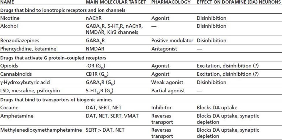
Abused drugs generally activate the mesolimbic system by (a) interacting with ion channel receptors, (b) binding to Gio-coupled receptors, or (c) interfering with monoamine transporters (34). Substances acting through the first two mechanisms tend to inhibit GABA inhibitory interneu-rons, resulting in a net release of dopamine. Drugs acting indirectly or directly upon ion channel receptors can additionally increase dopamine in the nucleus accumbens and ventral tegmental areas. Drugs that interfere with mono-amine transporters block the reuptake or stimulate non-vesicular release of dopamine, causing an accumulation of dopamine in target structures (Table 6-1).
TABLE 6-1 THE MECHANISTIC CLASSIFICATION OF DRUGS OF ABUSE
5-HTxR, serotonin receptor; CB1R, cannabinoid-1; DAT, dopamine transporter; GABA, gamma-aminobutyric acid; Kir3 channels, G protein–coupled inwardly rectifying potassium channels; LSD, lysergic acid diethylamide; -OR, -opioid receptor; nAChR, nicotinic acetylcholine receptor; NET, norepinephrine transporter; NMDAR, N-methyl-D-aspartate receptor; SERT, serotonin transporter; VMAT, vesicular monoamine transporter; ?, data not available. Reprinted from Katzung BG. Basic and clinical pharmacology, 10th ed. New York: McGraw-Hill Companies, Inc., with permission.
Nicotine, benzodiazepines, phencyclidine, and ketamine work through ionotropic receptors. Nicotine activates the nicotinic acetylcholine receptor, and benzodiazepines are modulators of the GABAA receptor. Alcohol alters the function of several ionic receptors including GABAA receptors, K+ inwardly rectifying or G protein activated inwardly rectifying K+ channels (Kir3/GIRK), glycine receptors, N-methyl-D-aspartate (NMDA) receptors, and 5-HT3 receptors. Alcohol also inhibits ENT1, the equilibrative nucleoside transporter for adenosine reuptake, resulting in adenosine accumulation, stimulation of adenosine A2 receptors, and enhanced cAMP response element–binding (CREB) protein signaling. Neither phencyclidine nor ketamine are physically addictive or associated with a physical withdrawal syndrome, but both may lead to a long-lasting psychosis owing to noncompetitive antagonism of the NMDA receptor. Of the inhalants, nitric oxide acts on NMDA receptors, whereas fuel additives enhance GABAA receptor function.
The opioids, cannabinoids, gamma-hydroxybutyric acid, and the hallucinogens all exert their action through Gio. Even though the mu, kappa, and delta opioid receptors all inhibit adenylyl cyclase, their selective neuronal expression results in different effects. For example, mu opioid agonists inhibit GABA inhibition of dopamine, causing a net release of mesolimbic dopamine, reinforcement, and euphoria. In contrast, kappa agonists inhibit dopamine neurons and induce dysphoria.
Cannabinoids cause presynaptic inhibition. The lipid-soluble neurotransmitters 2-arachidonyl glycerol and anan-damide bind to CB1 receptors to induce retrograde signaling from post- to pre-synaptic neurons, where they may inhibit the release of either glutamate or GABA.
The hallucinogens LSD, mescaline, and psilocybin neither stimulate dopamine release nor cause addiction. These drugs act through the 5-HT2A receptor, which couples to Gq proteins and inositol triphosphate (IP3) and leads to intracellular calcium release. Hallucinogens act by enhancing excitatory afferent input from the thalamus and increasing glutamate release in the cortex.
Cocaine, amphetamine, methamphetamine, and ecstasy bind to transporters of biogenic amines. Cocaine inhibits the dopamine transporter, decreasing dopamine clearance from the synaptic cleft and causing an increase in extracellular dopamine. Amphetamine competitively inhibits dopamine transport at the dopamine transporter and interferes with the vesicular monoamine transporter to lessen the storage of dopamine in the synaptic vesicles. As cytoplasmic dopamine increases, there is reversal of the dopamine transporter, increasing nonvesicular release of dopamine and further increasing extracellular dopamine. Ecstasy or MDMA, like amphetamines, causes release of biogenic amines by reversing the serotonin and other transporters.
Allosteric Modulation of G Protein–Coupled Receptors: Activity Based upon the Conformational State
It is now believed that G protein–coupled receptors can exist in multiple conformational states including ones that are active, inactive, partially active, and selectively active and those that produce nonproductive signaling though GTP-binding sites. If the conformational states of a receptor are in equilibrium and the inactive state predominates in the absence of drug, then the basal signal output from the downstream effector will be minimal or absent.
Drugs can bind to receptors at the same site or at a different site than the endogenous compound that physiologically activates that receptor. The relative affinity of the drug for various conformations of the receptor will determine the extent to which the equilibrium is shifted toward the active state. Full agonists have a higher affinity for the active conformation and drive the equilibrium toward the active state. Partial agonists bind to the receptor with only moderately more affinity for the active than for the inactive receptor. Even at saturating concentrations, partial agonists will not enable a full biologic response (see Fig. 6-3).
Buprenorphine is an example of a highly potent mu opioid receptor partial agonist. The drug has a high affinity for mu receptors and displaces morphine, methadone, and other full opiate agonists from these receptors. In contrast to the full agonists, however, increases in buprenorphine dose may result in a longer duration of action but do not result in increased pharmacologic effects. Higher doses of buprenorphine can be given without respiratory depression. The partial agonist properties of buprenorphine may precipitate withdrawal in individuals who have a high level of physical dependence on opioids.
Antagonists have no effect upon response when used alone. They bind with equal affinity to the active and inactive conformations and prevent an agonist from inducing a response (40). Competitive antagonists may be reversed by adding excess agonist, but noncompetitive antagonists cannot be counteracted in this manner. With noncompetitive antagonists, there is a decrease in the agonist-induced Emax(maximal efficiency). The potency of some antagonists, particularly those that act by inhibiting the activity of an enzyme, often are expressed as an I50value, which is the concentration of antagonist needed to elicit a 50% inhibition of enzyme activity.
Inverse agonists have preferential affinity for inactive receptor conformations when otherwise the equilibrium would be shifted toward an active receptor. In this case, inverse agonists produce an effect opposite to those of an agonist. However, if the basal equilibrium lies in the direction of the inactive receptor, then there will be little change in activity, and it will be difficult to distinguish inverse agonism from simple competitive antagonism (38).
Receptors can be constitutively in an active conformation, or they can be activated by physiologic events even in the absence of an agonist. Pharmacologic agents that can induce or stabilize specific receptor conformations may be therapeutic. Inverse agonist drugs may be clinically useful if they can selectively prevent the pathologic aspects of receptor activation (41).
Agents that work at GABA-gated chloride ion channels illustrate this spectrum of drug activity. The endogenous agonist GABA acts at this receptor to produce inhibitory, hyperpolarizing postsynaptic potentials. Depending on variations in receptor structure and location in the central nervous system, this activity produces an assortment of sedative, anxiolytic, and anticonvulsant effects. Both barbiturates and benzodiazepines are pharmaceutical agonists that act at the GABA receptor. Binding of each of these drugs to the GABA receptor complex occurs at distinct sites and facilitates the activity of GABA to open the chloride ion channel. Benzodiazepines increase the frequency of GABA-mediated chloride ion channel opening, whereas barbiturates increase the duration of this opening (42). Bicuculline, a competitive antagonist of GABA, binds selectively to the GABA site, interfering with GABA binding to that site. In contrast, picrotoxin, a noncompetitive antagonist of GABA, binds to the barbiturate site on the receptor, blocking the channel directly. Beta-carboline is an inverse agonist that reduces chloride ion conductance and increases excitability and irritability of the central nervous system; in fact, beta-carboline has no therapeutic use and can precipitate panic attacks. Flumazenil is an antagonist. Flumazenil lacks intrinsic activity but has therapeutic utility in treating benzodiazepine overdose. Flumazenil also blocks beta- carboline activity, although it does not antagonize the actions of ethanol or barbiturates.
Some drugs, depending on their concentrations, act as mixed agonists and antagonists. Mixed opioid agonist–antagonists have been developed in an attempt to produce analgesia with drugs that have less addictive potential and less respiratory depression. For example, nalbuphine and butorphanol are competitive mu receptor antagonists that exert their analgesic actions by acting as agonists at kappa receptors. Although each of these drugs has a place in the therapeutic armamentarium, unfortunately, each is dependence producing and associated with adverse effects, especially at higher doses.
Tolerance, Sensitization, and Physical Dependence
Tolerance and sensitization reflect changes in the way the body responds to a drug when it is used repeatedly. Tolerance is the reduction in response to a drug after its repeated administration. Tolerance shifts the dose–response curve to the right, requiring higher doses than the initial doses to achieve the same effect. Sensitization indicates an increase in drug response after its repeated administration. Sensitization shifts the dose-response curve to the left, so that repeated doses cause a greater effect than that seen with the initial dose.
Tolerance and sensitization develop more readily to some drug effects than to other effects of the same drug. For example, tolerance to the euphoria produced by cocaine occurs much more rapidly than does tolerance to its cardiovascular effects. The discrepancy between tolerance to the “rush” experienced by drug users and tolerance to a drug’s cardiovascular and respiratory effects can be an important cause of mortality in the user who overdoses. Also, chronic opiate users often have constipation and constricted pupils even though they no longer feel “high” after taking their drug (43). Differential toxicity can be explained by the dissimilar rates of drug tolerance that occur in diverse organ systems in individuals with unlike host characteristics.
Rodents experience sensitization, evidenced by an increase in locomotion, after being exposed to intermittent repeated doses of cocaine or amphetamine. In one study (44), this increase in behavioral activity was linked to an increase in dopamine levels in the extracellular fluid of the nucleus accumbens. Rats given repeated daily intraperitoneal cocaine injections (10 mg/kg) for 7 days had higher levels of dopamine detected by microdialysis on the 7th day than on the first day. Sensitization can be a phenomenon underlying chronic stimulant psychosis and/or alcohol withdrawal seizures.
There are several mechanisms by which tolerance can occur. Pharmacokinetic tolerance most often occurs as a consequence of increased metabolism of a drug after its repeated administration, resulting in less drug being available at the receptor for drug activity. For example, the microsomal ethanol-metabolizing system, which usually is not important in metabolizing ethanol, can be induced by prolonged ethanol exposure. Pharmacodynamic tolerance refers to the adaptive changes in receptor density, efficiency of receptor coupling, and/or signal transduction pathways that occur after repeated drug exposure. Homologous and heterologous desensitization were discussed earlier under G protein receptors. Additional discussion about the mechanisms for pharmacologic tolerance is discussed in the chapters that follow.
Learned tolerance refers to a reduction in the effects of a drug because of compensatory mechanisms that are learned. A common example of learned tolerance is the ability for roofers and workers at heights to walk in a straight line despite motor impairment from alcohol intoxication. Conditioned tolerance, which is a subset of learned tolerance, occurs when specific environmental cues such as sights, smells, or circumstances are paired with drug administration so that, when the drug is taken in the presence of the specific environmental cue, a state of expectation occurs. With expectation, the drug effect may be experienced before the drug is taken—and an adaptive response may be learned (45). A powerful example of conditioned tolerance occurred in a study when rats died after being given a dose of opiates to which they previously had been tolerant. The deaths occurred when the rats were put in an unusual environment instead of the home cage where they were used to receiving the drug (46).
Cross-tolerance occurs when tolerance to the repeated use of a specific drug in a given category is generalized to other drugs in that same structural and mechanistic category. The cross-tolerance that occurs between alcohol, barbiturates, and benzodiazepines can be used to facilitate the smooth weaning of a patient from their drug of dependence during detoxification (see Section 6, “Management of Intoxication and Withdrawal.”)
Physical dependence is a state that develops as a result of the adaptation produced by resetting homeostatic mechanisms after repeated drug use and may require medical monitoring during absence of the drug. Physical dependence can arise from many sources, addictive and nonaddictive. One is dependent on a cardiac medication, for example, and could die if it is not taken, but the heart medicine is not addictive. However, drugs such as ethanol and opiates produce withdrawal syndromes that may include increased heart rate and/or blood pressure, sweating, and tremors; more serious withdrawal symptoms such as hallucinations; and medical emergencies such as seizures.
Withdrawal signs and symptoms can occur in a physically dependent person when drug administration is abruptly stopped. Withdrawal symptoms may reflect the interactions of numerous neurocircuits and organ systems. The molecular mechanisms of physical dependence caused by individual drugs are discussed in the chapters that follow.
Patients who take prescribed medications for appropriate medical indications can show tolerance, physical dependence, and withdrawal if the drug is stopped abruptly, even though they do not exhibit the compulsive drug use and negative consequences characteristic of drug addiction (45). The hypertensive rebound that occurs in patients when chronic administration of beta- adrenergic receptor blockers is abruptly discontinued is but one example of this phenomenon. Similarly, patients with pain may develop tolerance to opiates and require increased medication for pain relief. They may also develop physical withdrawal if chronically administered opiates are abruptly discontinued. Pain patients may not be addicted to opiate medication unless they display compulsive behavior with adverse consequences despite being tolerant and/or physically dependent upon this medication (see Section 12, “Pain and Addiction.”)
SUMMARY
This chapter gives an introduction to the pharmacologic principles that underlie the use of drugs for both therapeutic and nontherapeutic purposes. Topics included pharmacokinetics, the study of the time course of drug concentrations as determined by absorption, distribution, metabolism, elimination, and excretion; pharmacodynamics, the study of the biochemical and physiologic effects of drugs and their mechanisms of action; and pharma-cogenomics, the relationship between individual genetic polymorphisms and drug disposition and effect. An introduction to receptor physiology of ligandgated ion channels, G proteins and second messengers, receptors with intrinsic enzyme activity, and receptors regulating nuclear transcription built the foundation for discussing a mechanistic classification of selected drugs of abuse (41). Next, new concepts of allosteric modulation of G protein–coupled receptors formed the bacKdrop for a discussion of full and partial agonists as well as competitive and noncompetitive antagonists. The following chapters in this section elaborate on the pharmacology of individual mood- and mind-altering chemicals and their drug classes, including further information about the topics of tolerance, sensitization, and physical dependence.
ACKNOWLEDGMENTS
The authors wish to express their gratitude to Elinore McCance-Katz, MD, whose contributions to the pharmacogenomics portion of the last version of this chapter set the stage for this fifth edition.
The conclusions in this chapter represent the views of the authors and do not necessarily represent the views of the Eunice Kennedy Shriver National Institute of Child Health and Human Development or the National Institutes of Health.
REFERENCES
1.Stepensky D. Prediction of drug disposition on the basis of its chemical structure. Clin Pharmacokinet 2013;52:415–431.
2.Johnson F, Setnik B. Morphine sulfate and naltrexone hydrochloride extended-release capsules: naltrexone release, pharmacodynamics, and tolerability. Pain Physician 2011;14(4):391–406.
3.Meng Y, Lichtman AH, Bridgen DT, et al. Inhalation studies with drugs of abuse. In: Rapaka RS, Chiang N, Martin BR, eds. Pharmacokinetics, metabolism, and pharmaceutics of drugs of abuse. NIDA Research Monograph 173. Rockville, MD: National Institute on Drug Abuse, 1997:201–224.
4.Won CS, Oberlies NH, Paine MF. Mechanisms underlying food-drug interactions: inhibition of intestinal metabolism and transport. Pharmacol Ther 2012;136:186–201.
5.Hermann R, von Richter O. Clinical evidence of herbal drugs as perpetrators of pharmacokinetic drug interactions. Planta Med 2012;78(13):1458-1477
6.Gardner DM, Shulman KI, Walker SE, et al. The making of a user friendly MAOI diet. J Clin Psychiatry 1996;57(3):99–104.
7.Rowland M, Tozer TN. Clinical pharmacokinetics: concepts and applications, 3rd ed. Baltimore, MD: Lippincott Williams & Wilkins, 1995.
8.Shawahna R, Decleves X, Scherrmann JM. Hurdles with using in vitro models to predict human blood-brain barrier drug permeability: a special focus on transporters and metabolizing enzymes. Curr Drug Metab 2013;14(1):120–136.
9.Geier EG, Schlessinger A, Fan H, et al. Structure-based ligand discovery for the Large-neutral Amino Acid Transporter 1, LAT-1. Proc Natl Acad Sci U S A 2013;110(14):5480–5485.
10.Syvänen S, Eriksson J. Advances in pet imaging of P-glycoprotein function at the blood-brain barrier. ACS Chem Neurosci 2013;4(2):225–237.
11.Atkinson AJ. Clinical pharmacokinetics. In: Atkinson AJ, Abernathy DR, Daniels CE, et al., eds. Principles of clinical pharmacology. New York, NY: Academic Press, 2007:9–20.
12.Ince I, Knibbe CA, Danhof M, et al. Developmental changes in the expression and function of cytochrome P450 3A isoforms: evidence from in vitro and in vivo investigations. Clin Pharmacokinet2013;52(5):333–345.
13.Daly AK. Genetic polymorphisms affecting drug metabolism: recent advances and clinical aspects. Adv Pharmacol 2012;63:137–167.
14.Meyer MR, Maurer HH. Absorption, distribution, metabolism and excretion pharmacogenomics of drugs of abuse. Pharmacogenomics 2011;12(2):215–233.
15.Mroziewicz M, Tyndale RF. Pharmacogenetics: a tool for identifying genetic factors in drug dependence and response to treatment. Addict Sci Clin Pract 2010;5(2):17–29. Review.
16.Sturgess JE, George TP, Kennedy JL, et al. Pharmacogenetics of alcohol, nicotine and drug addiction treatments. Addict Biol 2011;16(3):357–376.
17.Daly AK, Brockmoller J, Broly F, et al. Nomenclature for human CYP2D6 alleles. Pharmacogenomics 1996;6(3):193–201.
18.Kelly LE, Madadi P. Is there a role for therapeutic drug monitoring with codeine? Ther Drug Monit 2012;34(3):249–256.
19.Madadi P, Avard D, Koren G. Pharmacogenetics of opioids for the treatment of acute maternal pain during pregnancy and lactation. Curr Drug Metab 2012;13(6):721–727.
20.Zanger UM, Klein K, Saussele T, et al. Polymorphic CYP2B6: molecular mechanisms and emerging clinical significance. Pharmacogenomics 2007;8(7):743–759.
21.Zhu AZX, Cox LS, Nollen N, et al. CYP2B6 and bupropion’s smoking-cessation pharmacology: the role of hydroxybupropion. Clin Pharmacol Ther 2012;92(6):771–777.
22.Stewart NA, Buch SC, Conrads TP, et al. A UPLC-MS/MS assay of the “Pittsburgh cocktail”: six CYP probe-drug/metabolites from human plasma and urine using stable isotope dilution. Analyst2011;136:605–612.
23.Fuhr U, Jetter A, Kirchheiner J. Appropriate phenotyping procedures for drug metabolizing enzymes and transporters in humans and their simultaneous use in the “cocktail” approach. Clin Pharmacol Ther2007;81(2):270–283.
24.Ieiri I, Tsunemitsu S, Maeda K, et al. Mechanisms of pharmacokinetic enhancement between ritonavir and saquinavir; micro/small dosing tests using midazolam (CYP3A4), fexofenadine (p-Glycoprotein), and pravastatin (OATP1B1) as probe drugs. J Clin Pharmacol 2013;20(20):1–8.
25.Hung CC, Chiou MH, Huang BH, et al. Impact of genetic polymorphisms in ABCB1, CYP2B6, OPRM1, ANKK1 and DRD2 genes on methadone therapy in Han Chinese patients. Pharmacogenomics2011;12(11):1525–1533.
26.Tian JN, Ho IK, Tsou HH, et al. UGT2B7 genetic polymorphisms are associated with the withdrawal symptoms in methadone maintenance patients. Pharmacogenomics 2012;13(8):879–888.
27.Tyndale RF, Droll KP, Sellers EM. Genetically deficient CYP2D6 metabolism provides protection against oral opiate dependence. Pharmacogenetics 1997;7(5):375–379.
28.Chenoweth MJ, O’Loughlin J, Sylvestre MP, et al. CYP2A6 slow nicotine metabolism is associated with increased quitting by adolescent smokers. Pharmacogenet Genomics 2013;23(4): 232–235.
29.Ferguson CS, Tyndale RF. Cytochrome P450 enzymes in the brain: emerging evidence of biological significance. Trends Pharmacol Sci 2011;32(12):708–714.
30.Dutheil F, Beaune P, Loriot MA. Xenobiotic metabolizing enzymes in the central nervous system: contribution of cytochrome P450 enzymes in normal and pathological human brain. Biochimie2008;90(3):426–436.
31.Hedlund E, Gustafsson JA, Warner M. Cytochrome P450 in the brain: a review. Curr Drug Metab 2001;2(3):245–263.
32.Booth Depaz IM, Toselli F, Wilce PA, et al. Differential expression of human cytochrome P450 enzymes from the CYP3A subfamily in the brains of alcoholics and drug free controls. Drug Metab Dispos2013;41(6):1187–1194. doi:10.1124/dmd.113.051359.
33.Ravindranath V, Strobel HW. Cytochrome P450-mediated metabolism in brain: functional roles and their implications. Expert Opin Drug Metab Toxicol 2013;9(5):551–558.
34.Eiden LE, Weihe E. VMAT2: a dynamic regulator of brain monoaminergic neuronal function interacting with drugs of abuse. Ann N Y Acad Sci 2011;1216:86–98.
35.Nielsen DA, Utrankar A, Reyes JA, et al. Epigenetics of drug abuse: predisposition or response. Pharmacogenomics 2012;13(10):1149–1160.
36.Guo X, Liu Z, Wang X, et al. Large scale association analysis for drug addiction: results from SNP to gene. Scientific World Journal 2012;2012:939584.
37.Flower R.Drug receptors: a long engagement. Nature 2002;415(6872):587.
38.Buxton ILO. Pharmacokinetics and pharmacodynamics: the dynamics of drug absorption, distribution, action, and elimination. In: Brunton LL, Lazo JS, Parker KL, eds. Goodman & Gilman’s the pharmacological basis of therapeutics, 11th ed. New York: McGraw Hill Inc., 2006:1–40.
39.Kim JA, Bartlett S, He L, et al. Morphine induced receptor endocytosis in a novel knockin mouse reduces tolerance and dependence. Curr Biol 2008;18(2):129–135.
40.Gilchrist A. Modulating G protein-coupled receptors: from traditional pharmacology to allosterics. Trends Pharmacol Sci 2007;28(8):431–437.
41.Buxton ILO. Rethinking drug action in light of current models of receptor pharmacology. In: Brunton LL, Lazo JS, Parker KL, eds. Goodman & Gilman’s the pharmacological basis of therapeutics, [Supplement to main textbook.]11th ed. New York: McGraw Hill Inc., 2005: http://www.accessmedicine.com
42.Trevor AJ, Way WL. Sedative-hypnotic drugs. In: Katzung BG, ed. Basic and clinical pharmacology, 8th ed. New York: Lange Medical Books/McGraw-Hill, 2001:364–381.
43.Kasai S, Ikeda K. Pharmacogenomics of the human μ-opioid receptor. Pharmacogenomics 2011;12(9):1305–1320.
44.Kalivas PW, Duffy P. Effect of acute and daily cocaine treatment on extracellular dopamine in the nucleus accumbens. Synapse 1990;5(1):48–58.
45.O’Brien CP. Drug addiction and drug abuse. In: Hardman JG, Limbird LE, Gilman AG, eds. Goodman and Gilman’s the pharmacologic basis of therapeutics, 10th ed. New York: McGraw-Hill Inc., 2001:621–642.
46.Siegel S, Hinson RE, Krank MD, et al. Heroin “overdose” death: the contribution of drug-associated environmental cues. Science 1982;216(4544):436–437.