
John J. Woodward, BS, MS, PhD
CHAPTER OUTLINE
■ DEFINITION
■ SUBSTANCES INCLUDED IN THIS CLASS
■ FORMULATIONS AND METHODS OF USE
■ CLINICAL USES
■ BRIEF HISTORICAL FEATURES
■ EPIDEMIOLOGY
■ PHARMACOKINETICS
■ PHARMACODYNAMICS
■ DRUG–DRUG INTERACTIONS
■ NEUROBIOLOGY (MECHANISMS OF ADDICTION)
■ ADDICTION LIABILITY
■ CONCLUSIONS/FUTURE RESEARCH
DEFINITION
Alcohol is a chemical name for a group of related compounds that contain a hydroxyl group (-OH) bound to a carbon atom. The form of alcohol that is voluntarily consumed by humans is ethyl alcohol or ethanol and consists of two carbons and a single hydroxyl group (written as C2H5OH or C2H6O). Unless otherwise noted, the term alcohol is used throughout this chapter to mean ethanol.
SUBSTANCES INCLUDED IN THIS CLASS
In terms of human consumption, all commercially available alcoholic beverages contain ethyl alcohol with concentrations depending upon the type of beverage. Beverages made by fermentation of sugar-containing fruits and grains include beer (3% to 8% ethanol by volume) and wines (11% to 13% ethanol by volume). Spirits are produced after distillation and generally contain at least 30% ethanol. Ethanol can be concentrated by simple distillation up to approximately 95%, while pure ethanol requires addition of benzene or related substances or desiccation using glycerol. Denatured alcohol contains additives or toxins to prevent human consumption. Rubbing alcohol is prepared from denatured alcohol or isopropyl alcohol and is used for topical purposes.
FORMULATIONS AND METHODS OF USE
A bewildering array of alcoholic beverages is available for consumption, and these products contain a wide range of alcohol concentrations. In the United States, a standard alcoholic drink is defined as one that contains 0.6 fluid ounces of alcohol. Thus, this amount of alcohol is typically contained in 12 oz of beer, 5 oz of wine, or 1.5 oz of distilled spirits (40% ethanol by volume) although this can vary depending on the specific type of beverage (Table 7-1). Although most alcohol is consumed orally, there are isolated cases of individuals injecting ethanol intravenously (1). In addition, ethanol vapor can be inhaled, and machines called AWOL (alcohol without liquid; www.awolusa.com) have been introduced into the United States as a novel means of self-administering alcohol. A number of US states have since banned the sale or use of these devices.
TABLE 7-1 AMOUNT OF ALCOHOL IN DIFFERENT BEVERAGES AND RELATIONSHIP TO A STANDARD DRINKa
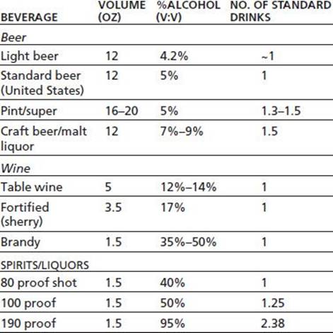
a In the United States, a “standard drink” is one that contains ~0.6 fluid ounces (14 g) of pure alcohol.
CLINICAL USES
In addition to its use as a topical antiseptic, alcohol has several clinical indications including treatment of accidental or voluntary ingestion of methanol or ethylene glycol (2). Ethanol has a higher affinity for alcohol dehydrogenase (ADH) than does methanol and thus reduces the formation of methanol metabolites formaldehyde and formic acid. For both indications, hemodialysis is the recommended first line of treatment. Alcohol has also been used for treatment of various types of cysts (sclerotherapy) and was used historically for treatment of premature labor (2). Safer and more effective medications have largely replaced these uses. Alcohol combined with dextrose is used to increase caloric intake and for replenishing fluids. Alcohol, either given orally or intravenously, is sometimes used by physicians to treat withdrawal, particularly in a hospital setting. For example, a survey of in-patient hospital pharmacies reported that approximately one-half had alcohol available for treatment of withdrawal with surgical services requesting the majority of these uses (3).
BRIEF HISTORICAL FEATURES
Alcohol is one of the oldest used substances. Consumption of alcohol-containing beverages predate recorded human history, while written records of its use are found in Chinese and Middle Eastern texts as far back as 9,000 years ago. Alcoholic beverages have long been consumed as part of the daily diet and are also used for medicinal or symbolic effects. In modern times, alcohol is second only to caffeine in incidence of use, and its manufacture, distribution, and sale are of major economic importance across the world. Alcohol use worldwide is regulated by both societal and religious beliefs. In the United States, consumption of alcohol was legally restricted on the national level with passage of the Volstead Act and the Eighteenth Amendment to the US Constitution in 1919 (4). Amid growing public outcry, prohibition was repealed in 1933 with the ratification of the Twenty-first Amendment that gave states the right to regulate the purchase and sale of alcohol. Today, sales and consumption of alcohol are under control of a wide variety of local and state laws.
EPIDEMIOLOGY
The lifetime exposure to alcohol is high with nearly 88% of the US population reporting using alcohol at least once in their lifetime (5). In 2011, current alcohol use (defined as use in the past 30 days) ranged from 3.9% among 12- to 13-year-olds to nearly 70% of 21- to 25-year-olds. Prevalence decreased among older groups although it was nearly 50% among 60- to 64-year-olds (5). Annual alcohol-related costs in terms of lost productivity and health care are estimated at $223 billion (6).
Clinical studies of alcohol abuse and alcoholism have led to the idea that there may be several types of alcohol use disorders, based on the appearance and severity of certain alcohol-related problems (7). Two particularly well-known examples of these classifications are the Type I and II forms proposed by Cloninger et al. and the Type A and B forms proposed by Babor and Caetano (8). Cloninger's Type I and Babor's Type A share several similarities including (a) later onset of alcohol-related problems (>25 years old), (b) fewer childhood behavior problems, (c) relatively mild alcohol-related issues with fewer hospitalizations, and (d) lower degree of novelty seeking coupled with a preference toward harm avoidance. Type II and B forms show essentially the opposite characteristics as Type I/A and include (a) familial alcoholism, (b) earlier onset of alcohol-related problems, (c) more incidents of alcohol-related problems or violence, and (d) higher preference for risk taking/novelty seeking.
PHARMACOKINETICS
Absorption and Metabolism
Alcohol is a small, water-soluble molecule that is rapidly and efficiently absorbed into the bloodstream from the stomach, small intestine, and colon. The rate of absorption depends on the gastric emptying time and can be delayed by the presence of food in the small intestine. Once in the bloodstream, alcohol is rapidly distributed throughout the body and gains access to all tissues, including the fetus in pregnant women. Although the relationship between alcohol intake and blood levels is body weight dependent, gender is also important. When body weights are equivalent, women show a 20% to 25% higher blood alcohol level than do men following ingestion of the same amount of alcohol (Table 7-2). This appears to be due primarily to less gastric metabolism of alcohol in women as blood levels are not significantly different when alcohol is administered intravenously (9).
TABLE 7-2 ESTIMATED BLOOD ALCOHOL LEVELS FOLLOWING DRINKING

Data represent blood alcohol concentration (gram per 100 mL or gram %) for men (M) and women (F) following consumption of different number of drinks. Subtract 0.01 from each value for each hour of drinking to account for effects of metabolism. 1 drink represents 12 oz beer, 5 oz wine, or 1.5 oz liquor.
Alcohol is metabolized primarily by enzymatic pathways, and only small amounts of alcohol are excreted through the lungs as vapor. The odor of the breath is not a reliable indicator of alcohol consumption, because it is influenced by impurities in the alcoholic beverage. In the liver, alcohol is broken down by ADH and mixed-function oxidases such as P450IIE1 (CYP2E1). Levels of CYP2E1 may be increased in chronic drinkers. ADH converts alcohol to acetaldehyde, which subsequently can be converted to acetate by the actions of acetaldehyde dehydrogenase.
The rate of alcohol metabolism by ADH is relatively constant, as the enzyme is saturated at relatively low blood alcohol levels and thus exhibits zero-order kinetics (constant amount oxidized per unit of time). Alcohol metabolism is proportional to body weight (and probably liver weight) and averages approximately 1 oz of pure alcohol per 3 hours in adults. Thus, the time for an individual to become sober after even moderate intake of alcohol can be substantial. Although stimulants are often used to mask the depressant effects of alcohol, there do not appear to be any truly effective "alcohol antagonists" (amethystic agents) that can quickly reverse the intoxicating effects of alcohol. Naloxone (Narcan), the opiate antagonist, has been tested for its ability to reverse alcohol-induced coma but appears ineffective (10). Several γ-aminobutyric acidA (GABAA) receptor antagonists have also been examined including flumazenil (Anexate) that is used to treat benzodiazepine overdose and the experimental compound RO 15-4513. This compound, which is actually an inverse agonist, has been reported to reverse some of the effects of low to moderate doses of alcohol in animals and may target a very specific subtype of GABAA receptor that shows sensitivity to very low concentration of ethanol (11). In addition to these agents, clinical trials have shown that metadoxine (pyridoxal L-2-pyrrolidone-5-carboxylate) appears to enhance the metabolic clearance of alcohol and speed recovery of intoxicated individuals (12). Although the clinical utility of an antialcohol medication for emergency medicine is undoubtedly high, moral, ethical, and medical concerns must be considered with respect to the development and general use of such a drug since it would not be expected to alter the underlying deleterious effects of alcohol on organ systems.
PHARMACODYNAMICS
Central Nervous System
Acutely, alcohol acts as a central nervous system (CNS) depressant. During the initial phase when blood alcohol levels are rising, a period of disinhibition often occurs, and signs of behavioral arousal are common. These include relief of anxiety, increased talkativeness, feelings of confidence and euphoria, and enhanced assertiveness. As drinking continues, there are impairments in judgment and reaction time, increased emotional outbursts, and ataxia. At higher blood levels, alcohol acts as a sedative and hypnotic, although the quality of sleep often is reduced after alcohol intake. In patients with sleep apnea, alcohol increases the frequency and severity of apneic episodes and the resulting hypoxia. Alcohol potentiates the sedative–hypnotic properties of both benzodiazepines and barbiturates, perhaps reflecting common mechanisms of action for these substances. Acute alcohol intoxication is not always associated with sedation or coma; indeed, some intoxicated individuals display violent behavior that requires administration of other sedative or antipsychotic agents. The use of these agents with a severely intoxicated individual must be approached cautiously to prevent respiratory failure. Chronic abuse of alcohol is associated with several psychoses such as Wernicke's and Korsakoff's that are associated with deficiencies in nutrition and vitamin intake (particularly thiamine).
Other Organ Systems
An in-depth discussion of the actions of alcohol on various organ systems can be found in other chapters of this book. A brief review of the most common effects is given here. Acute alcohol ingestion usually produces a feeling of warmth as cutaneous blood flow is increased, and this is accompanied by a reduction in core body temperature. Gastric secretions are usually increased, although the concentration of alcohol ingested affects this response, with high concentrations (>20%) inhibiting secretions (13). Long-term ingestion of high concentrations of alcohol can lead to a variety of pathologies associated with the gastrointestinal tract including esophageal varices and bleeding, erosive gastritis, and diarrhea and malabsorption of nutrients and vitamins (13). Alcohol consumption is also associated with an increased risk of tumors in the GI system as well as in other tissues including lung and breast (14). Acute and chronic ingestion of alcohol generally decreases sexual performance in both men and women although sexual behavior may be enhanced due to loss of inhibitory control and judgment. Alcohol has manifold effects on the cardiovascular system, and changes in contractility and function can follow both acute and chronic ingestion of alcohol (15). The deleterious effect of alcohol to the heart and other organ systems is countered by protective effects of small amounts of alcohol on cardiovascular tissue (16). Thus, low to moderate alcohol use is associated with a reduced risk of coronary disease, and this may arise from alcohol-induced changes in plasma lipoproteins and alterations in cell protection pathways. Chronic alcohol ingestion is well known to increase fat accumulation in the liver that can progress to severe liver damage and cirrhosis (17). Alcohol-induced liver damage is due in part to the production of acetaldehyde that readily reacts with proteins, lipids, and other compounds leading to impaired mitochondrial function.
DRUG–DRUG INTERACTIONS
Alcohol has depressant actions on the CNS that are similar to other centrally acting drugs such as barbiturates, benzodiazepines, general anesthetics and solvents, and anti-convulsants. Alcohol also enhances the sedative effects of antihistamines that are commonly used in the treatment of nasal congestion. Combining these medications with alcohol can result in significant CNS depression and reduced ability to safely carry out normal functions such as automobile driving. Alcohol can also enhance the hepatotoxic effects of acetaminophen (Tylenol) and the gastric irritating effects of NSAIDs, thus increasing the risk for development of gastritis and upper GI bleeding. Chronic alcohol use can interfere with the metabolism of certain drugs due to enhanced levels of liver enzymes.
NEUROBIOLOGY (MECHANISMS OF ADDICTION)
A widely accepted tenet of addiction research is that addictive substances, by definition, produce pleasurable effects that engender actions promoting further drug seeking and taking. This concept suggests that drugs of abuse, like alcohol, must produce, at least initially, some form of positive reinforcement that provides a strong incentive to reexperience the drug. As drug use proceeds, the degree of acute reinforcement or reward may be attenuated and drinking escalates as negative aspects associated with withdrawal emerge (18). The repetitive nature of drug use also engages mechanisms of learning leading to entrained behaviors that may become highly ritualistic and habit-like. These findings have led to the idea that drug and alcohol addiction is a form of dysfunctional, maladaptive learning that, once established, is difficult to reverse (19). In terms of brain systems that underlie the development of addiction, recent studies suggest an important interaction between midbrain dopamine-based reward systems and cortical mechanisms of brain plasticity and learning that are mediated by the neurotransmitter glutamate (20).
As reviewed in various chapters in Section 1, all drugs of abuse including alcohol affect reward pathways by enhancing the release of dopamine from midbrain dopaminergic projections that regulate neurotransmission within limbic and cortical circuits that regulate motivated behavior (21). The dopamine (DA) neurons involved in this action reside in the midbrain ventral tegmental area (VTA) and project to discrete areas of the brain, including the nucleus accumbens, olfactory tubercle, frontal cortex, amygdala, and septal/hippocampal areas. These regions are thought to be involved in translating emotion and perception into action through the activation of motor pathways; thus, they may be important in initiating and sustaining drug-seeking behavior. Lesions or inactivation of these discrete brain areas in experimental animals can reduce both the acquisition of drug seeking and its reinstatement following long periods of abstinence (22,23).
The initial reinforcing actions of alcohol appear to involve excitation of VTA dopamine neurons. Acutely, alcohol enhances the firing rate of midbrain DA neurons, and animals will self-administer alcohol directly into the posterior but not anterior VTA (24,25). Chronic exposure to alcohol leads to alterations in the excitability of these neurons in the absence of alcohol that may persist for significant periods of time (26,27). Electrophysiologic studies have demonstrated enhanced efficiency of glutamatergic signaling in neurons following exposure to alcohol and other drugs of abuse (28–30). With respect to reward circuits, these findings suggest that enhanced firing of VTA DA neurons during exposure to alcohol facilitates glutamater-gic transmission in limbic, cortical, and striatal areas, thus strengthening the association between behavioral action and outcome. Studies using in vivo microdialysis to monitor levels of dopamine in freely behaving animals report significant dose-dependent increases in extracellular dopamine levels in the nucleus accumbens in rats self-administrating alcohol (31,32). Importantly, in rats genetically selected to consume large amounts of alcohol, significant increases in dopamine were also observed during the 15-minute waiting period that preceded alcohol self-administration (33). This finding suggests that, in animals with drinking experience, the expected reward that ingestion of alcohol provides is itself sufficient to enhance activity in this pathway. The subsequent pharmacologically induced elevation of dopamine that occurs during drinking may further strengthen the motivation to consume alcohol in future sessions. Genetic differences in the responsiveness of this pathway may contribute to the motivational factors that drive greater alcohol-seeking behavior in certain individuals.
Studies with human alcoholics have examined the neurobehavioral aspects of alcohol abuse, using drug discrimination procedures similar to those used in animal studies. In these studies, human alcoholics are asked to rate the effects produced by a variety of drugs in terms of their similarity to those produced by alcohol. For example, ketamine, a dissociative anesthetic that blocks the ethanol-sensitive N-methyl-D-aspartate (NMDA) subtype of glutamate receptors, induces ethanol-like subjective effects in recently detoxified alcoholics (34). These effects were dose dependent: At low doses, they mimicked the effects of one to two standard drinks of alcohol, whereas higher doses produced effects similar to those of eight to nine drinks. Interestingly, the effects of ketamine that were ethanol-like were associated with the descending phase of blood alcohol concentration that is associated with ethanol-induced sedation.
Other human clinical studies using selective pharma-cologic agents have implicated neurotransmitters such as GABA, serotonin, and the opiates in mediating the rewarding and craving aspects of alcohol action. Results from human imaging studies have begun to identify changes in brain activation during exposure to alcohol or alcohol-related cues between control and alcohol-dependent subjects (35,36). Such human studies are important in the context of understanding the underlying causes of alcohol abuse because alcohol, unlike most drugs of abuse, interacts with a wide variety of molecular and cellular processes to produce its pharmacologic, physiologic, and psychological effects.
Molecular Sites of Alcohol Action
Psychostimulants such as cocaine and amphetamine or opiates like heroin and morphine all produce their primary effect by binding to specific protein receptors expressed on brain neurons. In contrast, alcohol is rather promiscuous and interacts with a wide variety of targets including both lipids and proteins. Initial observations made by the German scientists Meyer and Overton over 100 years ago led to a lipid-disordering hypothesis of alcohol and anesthetic action due to the high correlation between lipid solubility of a compound and its potency as an anesthetic. Support for this theory has waned in recent years due to several factors. For example, measurable changes in membrane fluidity require very high concentrations of alcohol, are relatively modest, and are less marked than the effects produced by small changes in temperature that are not associated with behavioral signs of intoxication (37). These findings and the demonstration of effects in lipid-free systems have led to the idea that alcohol's acute actions are likely due to effects on ion channel proteins that regulate the excitability of neurons (38). An important point to consider in these in vitro studies of alcohol action is whether the range of concentrations needed to cause significant effects are similar to those associated with the behavioral actions of alcohol. Blood alcohol concentrations in the range of 40 to 400 mg% are equivalent to 8 to 88 mM and are associated with the full spectrum of alcohol intoxication (described in more detail below). Thus, in vitro studies of alcohol's effect on ion channels are usually conducted at concentrations below 100 mM in order to better approximate levels found in the brain during drinking.
Table 7-3 shows that a variety of ligand-gated ion and voltage-activated ion channels that are expressed by neurons are sensitive to behaviorally relevant concentrations of alcohol.
Alcohol's acute depressant action on neuronal excitability likely results from its ability to enhance the function of inhibitory ion channels while blocking the activity of excit-atory receptors (Fig. 7-1).

FIGURE 7-1 Structure and location of alcohol-sensitive ion channels in the neuronal synapse.
GABAA and Glycine Receptors
As shown in Table 7-3, there are distinct families of subunits that make up GABAA and glycine receptors. Each class of subunits may have multiple members that differ slightly in their sequence and function. Different subunit combinations give rise to GABAA and glycine receptors that can show variable sensitivity to pharmacologic agents, including alcohol. Alcohol generally enhances GABAA and glycine receptor function, although GABAA ρ receptors are inhibited by alcohol. Initial studies with recombinant GABAA receptors suggested that the γ2L subunit that contains an additional eight amino acids including a site for phosphorylation of the receptor by protein kinase C was important for alcohol potentiation of channel function (39,40). Despite these intriguing results, subsequent studies have shown that γ2L expression is not sufficient in itself to confer sensitivity to alcohol (41). In addition, knockout mice lacking this subunit show normal sensitivity to alcohol, although their responsiveness to benzodiazepines is enhanced (42,43). While these results appear to rule out a major role for the γ2 subunit in mediating alcohol action on GABAA receptors, this may be phosphorylation dependent as the alcohol sensitivity of GABAA α1β2γ2 receptors was reported to be markedly enhanced when the kinase PKCε was inhibited (44). Mice lacking PKCε also show an enhanced response to ethanol suggesting that specific subtypes of GABAA receptors may show appreciable alcohol sensitivity under certain conditions (45).
TABLE 7-3 PROPERTIES OF ALCOHOL-SENSITIVE ION CHANNELS
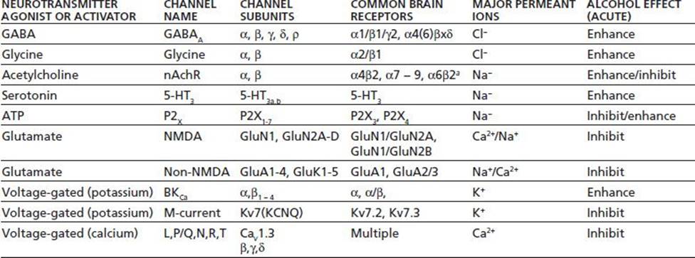
Ion channels are listed according to their natural agonist or mode of activation. Subunit families represent those found in the brain or spinal cord. Brain subtypes are examples of subunit combinations commonly expressed by neurons (aindicates other subunits likely required). Alcohol effect indicates change in ion channel function by alcohol acutely administered to recombinant or native receptor preparations.
In addition to γ-containing GABAA receptors, recent studies suggest that δ-containing GABA receptors possess high sensitivity to relatively low levels of alcohol. These receptors contain the α4(6), β, and δ subunit instead of the γ subunit and are affected by concentrations of ethanol (5 to 10 mM) achieved after only one drink (46). Despite these intriguing findings, not all studies have reported that γ-containing GABAA receptors are sensitive to low concentrations of alcohol (47). While the reasons underlying these conflicting findings are not yet clear, there is substantial evidence to suggest that certain GABAAreceptors may be especially important in mediating low-dose effects of alcohol.
A major approach that has revolutionized the study of alcohol action on ion channels is the use of site-directed mutagenesis to alter putative alcohol-sensitive amino acids within a given receptor. These sites are often located within transmembrane domains of individual receptor subunits, and in GABAA or glycine receptors, replacement of a conserved serine by an isoleucine abolished the potentiating effects of alcohol on receptor currents (48). Subsequent studies have shown that this site may also be involved in mediating the effects of some volatile anesthetics on GABAA and glycine receptor function (49). The identification of these alcohol-sensitive residues combined with the development of ion channel structural models has allowed for detailed molecular modeling of these sites and the development of genetically modified animals that express alcohol-insensitive subunits (50).
Interestingly, GABAA receptor knockout and knock-in mice appear to show relatively subtle changes that are restricted to specific alcohol-induced behaviors with little effect on other alcohol actions (51–53). This may reflect the fact that there are multiple targets for alcohol in the brain and that in the case of knockout animals, changes in expression of related GABA subunits may compensate for the loss of those that are alcohol sensitive (54). In addition, there is now good evidence that some of the effects of alcohol on GABA receptor function arise from changes in the presynaptic release of GABA rather than a direct effect on the GABA receptor itself (55). For example, in certain neurons of the amygdala, a brain structure highly involved in processing emotionally relevant stimuli, alcohol's potentiation of postsynaptic GABAAresponses appears to be due to an increase in the release of GABA rather than a direct effect on the channel (56). The mechanism underlying the sensitivity of GABA release to alcohol is not currently known but could involve specific signaling pathways and proteins that normally regulate vesicle movement and fusion (57).
Glutamate-Activated Ion Channels
Glutamate is the major excitatory neurotransmitter in the brain and activates three major subtypes of ion channels called AMPA, kainate, and NMDA receptors, where AMPA is α-amino-3-hydroxy-5-methyl-4-isoxazolepropionic acid. These channels mediate most of the fast excitatory synaptic transmission in the brain and are critical mediators of most forms of synaptic plasticity thought to underlie learning and memory. NMDA receptors are highly calcium permeable and require both glutamate and glycine for activation, while AMPA and kainate receptors only require glutamate for activation. As reviewed below, NMDA receptors are inhibited by ethanol as well as other abused substances including nitrous oxide, anesthetics, and volatile solvents such as toluene (58–60).
In general, NMDA receptors show appreciable sensitivity to relevant concentrations of alcohol (61), while AMPA and kainate receptors show a more restricted pattern of ethanol sensitivity (62–64). NMDA receptors expressed in most neurons are readily antagonized by alcohol at concentrations (10 to 100 mM) associated with intoxication and sedation although this varies across different brain regions (63–68). Like GABAA and glycine receptors, alcohol inhibition of NMDA receptors may involve an interaction with specific residues within defined transmembrane domains of the receptor that control channel gating (69–71). NMDA responses show a regional and developmental variation in their sensitivity to alcohol, and this may result from differential expression of GluN1 and GluN2 NMDA subunits that regulate some of the alcohol sensitivity of these receptors (72,73). Alcohol's blockade of excitatory NMDA signaling is likely to be important in mediating some of the acute intoxicating and sedative effects of alcohol (74). Inhibition of NMDA receptors in the prefrontal cortex may underlie some of the cognitive deficits and errors in judgment observed during alcohol intoxication (63,75,76). Long-term alcohol consumption may also affect NMDA receptors in medial and orbital cortical areas that are important for adapting behavior to rapidly changing contingencies (77,78).
The antagonism of NMDA receptors by alcohol may also be involved in its rewarding properties because NMDA receptors are thought to be important in regulating the release of dopamine in mesolimbic areas such as the nucleus accumbens. For example, using microdialysis to monitor changes in dopamine in awake animals, NMDA antagonists were shown to increase levels of dopamine in the nucleus accumbens (79). These results suggest that glutamate, by acting on NMDA receptors on interneurons, may exert an inhibitory control over dopamine release by NMDA receptors. Relief of this inhibition during exposure to alcohol may lead to increases in accumbens dopamine. In animal studies designed to evaluate the subjective effects of ethanol on behavior, NMDA antagonists can produce ethanol-appropriate lever response in rats trained to discriminate ethanol from saline (80).
Since NMDA receptor activity is a critical determinant of normal neuronal activity, it is perhaps not surprising that receptor function is also altered following chronic exposure to alcohol. In primary cultures of neurons, chronic exposure to alcohol increases the density and clustering of NMDA but not non-NMDA receptors (81–83). These changes appear to take place specifically within the gluta-matergic synapse as there is no change in NMDA receptors in nonsynaptic locations. These results suggest that NMDA receptors may serve as alcohol sensors and that neurons compensate for inhibition of these receptors by moving more of them to the synapse where they can be activated by glutamate. One important outcome of this adaptive response is an increased susceptibility of animals and humans to seizures that develop during withdrawal from alcohol. Experiments with mice show that NMDA-induced seizure activity is elevated in mice made dependent on alcohol and that NMDA antagonists reduce or prevent seizures during withdrawal (84). Results from studies using isolated cells or intact animals also suggest that the enhancement in NMDA receptor function after chronic alcohol may involve changes in the expression pattern of specific NMDA receptor subunits (77,85–88).
Other Ion Channel Subtypes
5-HT3 Receptors
5-HT3 receptors are ligand-gated ion channels activated by serotonin. They are permeable primarily to monova-lent cations, such as sodium and potassium. In cultured neurons and recombinant expression systems, low doses of alcohol appear to potentiate currents carried by 5-HT3 receptor (89,90). In behavioral studies involving both rats and pigeons, 5-HT3 receptor antagonists blocked the animal's ability to discriminate ethanol from saline, suggesting that alcohol's acute actions on 5-HT3 receptors may underlie some of the subjective effects of alcohol (91).Human studies using the 5-HT3 antagonist ondansetron (Zofran) have generally found that the drug reduces drinking although this effect appears more efficacious in early-onset alcoholics (92).
Acetylcholine Nicotinic Receptors
Acetylcholine activates a family of ligand-gated ion channels expressed in brain neurons that are related to the nicotinic receptor expressed at the neuromuscular junction (93). Alcohol has been shown to either potentiate or inhibit acetylcholine receptors expressed in cultured neurons or oocytes (94,95). In neurons, this biphasic effect appears to result from expression of different subtypes of nicotinic receptors that show a differential response to ethanol. Thus, heteromeric nicotinic receptors composed of just αβ subunits appear to be potentiated by ethanol, whereas homomeric receptors composed of just α subunits (e.g., α7) are inhibited by ethanol (95). It is not yet clear how these different effects of ethanol on neuronal nicotinic receptors are manifested at the behavioral level. However, nicotinic receptors are expressed by neurons in the VTA, nucleus accumbens, and prefrontal cortex where they help shape the excitability of these neurons. Varenicline (Chantix), a medication approved for smoking cessation, is a partial agonist at the α4β2 nicotinic receptor. Results from preclinical studies have reported that varenicline also reduces alcohol seeking in rodents without altering responding for sucrose (96).
ATP-Gated Ion Channels
Adenosine triphosphate (ATP) is released into synapses where it acts on ATP-gated ion channels of the P2X family (97). Alcohol inhibits ATP-gated channels in some neurons (98), while the effect of alcohol on recombinant P2X receptors is subtype specific. P2X2 and P2X4 channels are inhibited by alcohol (98–100), but currents from P2X3 receptors are enhanced (101). This appears to involve differences in a small number of amino acids between subunits at sites near the transmembrane domains that control channel gating (102). The inhibitory action of ethanol on the P2X4 receptor is blocked by the anthelmintic drug ivermectin that is a positive allosteric modulator of P2X4 channel function (103), and in mice, ivermectin reduces ethanol consumption in a variety of drinking models (104).
Potassium- and Calcium-Selective Ion Channels
Potassium channels that are regulated by calcium (SK and BK channels) and those gated by G proteins (GirK) channels are also affected either directly or indirectly by alcohol (105). Potassium channels serve as a brake on excitatory glutamatergic transmission by hyperpolarizing the membrane and thus are critical regulators of neuronal activity. BK channel activity is acutely enhanced by alcohol, and this enhancement may contribute to the inhibition of vasopressin release from neurohypophysial terminals and the resulting diuresis that accompanies alcohol ingestion (105). SK channels do not appear to be inhibited directly by ethanol, but their expression and localization in hippocampal neurons are altered following chronic exposure (106), and these changes may contribute to ethanol withdrawal hyperexcitability. In addition, down-regulation of SK channels in nucleus accumbens following chronic alcohol exposure may contribute to elevated drinking as enhancing SK activity with an allosteric modulator (chlorzoxazone; Parafon Forte) reduced alcohol consumption in rats (107). Most voltage-gated potassium and sodium channels are relatively insensitive to relevant concentrations of alcohol although there are exceptions. For example, recombinant Kv7 (previously termed KCNQ) channels are inhibited by moderate concentrations of ethanol (108), and Kv7.2/Kv7.3 heteromers are thought to underlie the slow, noninactivating M-current that reduces neuronal excitability. In VTA DA neurons, M-current is inhibited by acute ethanol at concentrations that enhance DA neuron firing suggesting another possible mechanism for ethanol-induced increases in DA release (109). Alcohol also has inhibitory actions on certain subtypes of voltage-gated calcium channels (110), and genetically modified mice lacking the N-type calcium channel show reduced ethanol consumption as compared to wild-type animals (111). Alterations in T-type calcium channels in thalamic neurons by alcohol may contribute to disruptions in sleep that are commonly observed in alcohol-dependent individuals (112).
Pharmacologic Studies Implicating Other Neurotransmitter Systems
Much of the working knowledge of alcohol's effects on neuronal function comes from animal studies examining the effects of neurotransmitter-specific agents on alcohol drinking. A brief review of this literature is presented here and is summarized in Table 7-4.
TABLE 7-4 EFFECTS OF ACUTE AND CHRONIC ALCOHOL ON VARIOUS BRAIN ION CHANNELS AND NEUROTRANSMITTER SIGNALING SYSTEMS

aBehaviors that each system/neurotransmitter may contribute to.
Adenosine
Adenosine is a major inhibitory neurotransmitter in the brain and may serve as an endogenous antiepileptic because of its ability to inhibit neuronal function. Alcohol has been shown to inhibit the function of a nucleoside transporter, leading to increased extracellular adenosine levels (113). This leads to activation of A2 adenosine receptors and increases cellular levels of cyclic adenosine monophosphate (cAMP) that can activate protein kinase A (PKA) and stimulate cAMP-dependent changes in gene expression. A2 receptors show cross talk with D2 dopamine receptors, and this interaction may be especially important in regulating the activity of medium spiny neurons in the nucleus accumbens. Block of this sig-naling pathway has been shown to reduce voluntary alcohol drinking in experimental animals (114).
Dopamine
As mentioned earlier in this chapter, increases in the activity of mesolimbic projecting dopamine neurons are thought to be critical steps underlying the reinforcing effects of many drugs of abuse, including alcohol (20). Electrophysiologic studies have demonstrated that alcohol increases the firing of dopamine-containing neurons, located in the VTA leading to enhanced dopamine release in the nucleus accumbens (25). The mechanism underlying this effect of alcohol is not precisely known but may involve both a direct effect on a subtype of potassium channel that regulates the excitability of VTA neurons (26) as well as indirectly via modulation of inputs into the VTA. Interestingly, the sensitivity of VTA neurons to alcohol-induced excitation is lower in mice that show a higher voluntary consumption of alcohol, suggesting that these animals may consume more alcohol to sufficiently activate a dopaminergic reward pathway (25,115). In addition, recent studies using retrograde tracers to identify subpop-ulations of VTA DA neurons reveal that those projecting to nucleus accumbens but not to prefrontal cortex show changes in glutamatergic plasticity following brief exposure to drugs such as cocaine or toluene (29,116). These findings point out that DA neurons within midbrain areas have specialized roles with respect to control of reward-based behaviors.
The role of dopamine in alcohol addiction has also been explored by using pharmacologic agonists and antagonists that directly interact with dopamine receptors. The long-acting dopamine agonist bromocriptine (Parlodel), administered systemically, shifts a rat's preference from alcohol to water, especially in those strains of rats that show alcohol preference (117). Similar findings were demonstrated with another dopamine agonist, apomorphine. These results suggested that administration of direct dopamine agonists reduced the need for alcohol's dopamine-enhancing activity in these animals, such that the "reward state" was achieved at lower alcohol levels. Despite these promising results, human studies with these agents have not found significant effects on alcohol drinking or relapse (92).
Opioids and Other Neuropeptides
The involvement of endogenous opioids (endorphins and enkephalins) and other neuropeptides in alcohol addiction is suggested by several lines of research. One of the first links between alcohol and opioids was suggested by the finding that acetaldehyde could undergo a metabolic reaction with monoamines to form compounds (tetrahydroisoquinolines or TIQs) that were structurally related to morphine (118). TIQs were shown to elicit alcohol-drinking behavior and alcohol preference even after cessation of TIQ administration (119). This hypothesis, although attractive, remains controversial because of the inability to replicate some of these findings and the demonstration that TIQ formation in vivo could be accounted by dietary factors rather than direct effects of alcohol (120). Alcohol does increase the release of certain opioid peptides (such as β-endorphin) from rat pituitary glands and increases blood levels of β-endorphins in humans (121). If alcohol drinking is mediated in part by opioids, then selective opioid antagonists should inhibit alcohol-drinking behavior. Naloxone and naltrexone, two opioid receptor antagonists, reduce alcohol intake in both animals and humans, and various formulations of naltrexone (ReVia) including sustained release and depot forms (Vivitrol, Naltrel, Depotrex) are now approved for treating alcoholism. These agents are thought to work by blocking alcohol-induced increases in β-endorphin, thus reducing the acute rewarding effects of alcohol. As reviewed in a later chapter, a large number of laboratory studies and randomized controlled trials have been conducted, and most of these report that naltrexone, when combined with medical management, is better than placebo in reducing the risk of relapse (122). The results from these human studies are complemented by those using mice genetically modified to lack the μ-opiate receptor. These animals do not self-administer alcohol or respond to the rewarding effects of opiates, nicotine, or cannabinoids (123). Despite these dramatic effects in animal models and positive results in most human trials, the clinical efficacy of opiate antagonists in treating alcohol dependence is rather modest suggesting that other factors may be important (92,124). As discussed in more detail in a later chapter, the variability in response to opiate antagonists may be due to issues related to compliance as well as in differences in opiate receptor polymorphisms and other genetic changes among alcohol-dependent subjects that influence naltrexone efficacy (92,125).
Neuropeptides like neuropeptide Y (NPY) and corticotropin-releasing factor (CRF) are involved in mediating stress and anxiety and have been shown to be important regulators of alcohol drinking. CRF is released following activation of the hypothalamic–pituitary–adrenal axis leading to elevated levels of cortisol that are indicative of a stress response. Alcohol also induces CRF release, and chronic alcohol use is associated with enhanced anxiogenesis that can be blocked by CRF antagonists (126). CRF antagonists appear to be especially effective in reducing the elevated drinking observed in alcohol-dependent rats without altering consumption in nondependent animals (127). Mice engineered to lack CRF1 receptors do not show enhanced drinking following the development of dependence again suggesting an important link between stress and alcohol drinking (128). NPY is a neuropeptide associated with the general regulation of feeding behavior and anxiety. Injection of NPY reduces drinking in rodents, while mice lacking NPY or the NPY Y1 receptor display enhanced ethanol intake (129). These effects may be related to anxiolytic effects of NPY. In addition to CRF and NPY, a variety of studies have shown that the neuropeptide orexin (also called hypocretin) influences drinking. Orexin is synthesized by a small number of neurons in the lateral hypothalamus, and axons from these neurons innervate a wide array of brain structures including those involved in reward and cognition. Studies in rodents show that orexin antagonists can reduce drinking with greatest efficacy in alcohol-preferring individuals (130,131).
Serotonin
As mentioned above, electrophysiologic studies have demonstrated that alcohol enhances cation conductance through 5-HT3 receptors (89). Serotonin also interacts with a large number of non–ion channel G protein–linked receptors that are coupled to various signal transduction pathways. The direct effects of alcohol on these receptor systems are not as well characterized. However, there is a fairly large amount of literature describing the effects on alcohol-drinking behavior of various drugs that modulate serotonergic tone. 5-HT and 5-HT metabolite levels are reduced in the cerebrospinal fluid of many alcohol abusers, suggesting that reduced 5-HT levels or a reduction in 5-HT-mediated neurotransmission may predispose certain people to uncontrollable drinking behavior (132).
It has been suggested that similar deficiencies in 5-HT neurotransmission underlie the development of a variety of other disorders, including bulimia and obsessive–compulsive behavior, disorders that are characterized by a loss of behavioral control. Thus, pharmacologic agents that enhance 5-HT neurotransmission (such as serotonin selective reuptake inhibitors) appear to be therapeutically effective in the treatment of these disorders. However, in terms of treating alcohol dependence, these agents (such as fluoxetine [Prozac] and sertraline [Zoloft]) appear to have limited efficacy (133,134). Of course, one drawback in using a transport inhibitor is the inability to selectively activate specific subtypes of 5-HT receptors. Interestingly, a variety of results from animal studies suggest that the 5HT1b receptor may be especially involved in regulating alcohol intake although the data are conflicting regarding whether receptor activity should be enhanced or blocked to produce these effects (135–137). In addition, it is not completely clear whether these manipulations are selective for alcohol as 5-HT plays a central role in feeding and drinking behaviors.
Endocannabinoids
An extensive series of studies have shown that the endogenous cannabinoid (EC) system is an important modulator of ethanol drinking (138). ECs are natural lipid-derived molecules that activate receptors (CB1, CB2) that also bind THC, the psychoactive constituent of marijuana. ECs regulate both GABAergic and glutamatergic synaptic transmission and are synthesized during periods of intense neuronal depolarization. CB1 agonists including THC are powerful appetite-promoting compounds and antagonists of the CB1 receptor (rimonabant [Acomplia]) were initially marketed in Europe for the treatment of obesity. However, this drug and others in the same class were removed from the market due to significant psychiatric effects (139). In animal studies, CB1 antagonists such as rimonabant reduce ethanol preference in wild-type mice and animals that lack CB1 receptors show reduced alcohol preference (140). This action may reflect the ability of these compounds to block the alcohol-induced increase in dopamine in the nucleus accumbens (141). This effect was also observed for cocaine and nicotine-induced increases in dopamine suggesting a link between the EC system and a variety of drugs of abuse that possess disparate acute mechanisms of action.
ADDICTION LIABILITY
Alcohol is an addictive substance although an individual's susceptibility to developing alcohol use disorders is influenced by a wide range of genetic and environmental factors. The 12-month prevalence for alcohol dependence for males is about 5.4% for men and 2.3% for women (142). Lifetime prevalence of alcohol dependence is approximately 13%, and the risk of developing alcohol dependence shows a strong inverse correlation with the age at which heavy drinking begins (142). Chronic use of alcohol produces several neuroadaptive changes that may be important in the development of alcohol addiction.
Sensitization
Sensitization is defined as an increase in the pharmacologic and physiologic response to a drug after repeated exposures. This phenomenon is best characterized by drugs of abuse such as cocaine or amphetamine where enhanced locomotor activity following repeated drug exposures is associated with changes in glutamatergic signaling in the neurons of the VTA and nucleus accumbens (22). Sensitization to the locomotor effects of alcohol has also been well studied, and the magnitude and duration of these effects depend on genotypic and environmental factors (143,144). Another form of sensitization is characterized by an increase in the severity and intensity of withdrawal signs after multiple episodes of alcohol intoxication and withdrawal (145). This form of sensitization has been suggested to be similar to the kindling phenomena observed after repeated brain seizures and may involve some of the same mechanisms that underlie the adaptation of neurons to impaired neuronal signaling during chronic exposure to alcohol.
Tolerance and Dependence
Tolerance is manifested as a reduced sensitivity to alcohol and is subdivided into several forms, depending on the specific action being measured and the time course over which it develops. For example, concentrations of alcohol required to produce sedation may increase in individuals or animals given alcohol chronically. In human alcoholics, tolerance to the sedative and even lethal effects of alcohol can be profound. For example, while the lethal dose 50% (LD50) in nontolerant humans is approximately 400 to 500 mg%, blood levels exceeding these values are often reported in individuals arrested for drunk driving (146). This form of tolerance is associated with changes in both the capacity to metabolize alcohol and in cellular adaptations that reduce sensitivity to alcohol. A form of short-term acute functional tolerance to alcohol (termed the “Mellanby effect”) is also demonstrated by differences in the degree of impairment produced by the same blood concentration of alcohol achieved during the rising phase of the blood alcohol curve as compared to the falling phase (147). Tolerance development has been extensively studied in rodent models, and the severity of the motor-impairing effects of alcohol is strongly influenced by the context and environment in which they are measured (148). Dependence is defined by the occurrence of symptoms that appear after the cessation of alcohol drinking. These withdrawal symptoms include both physical (tremors, convulsions) and psychological (negative emotions, craving) components. At the cellular level, tolerance and dependence also develop so that the acute effects of alcohol on ion channel or receptor function are diminished in animals chronically exposed to alcohol. As mentioned in the earlier section, this adaptation may involve changes in signaling pathways that regulate subunit expression or the functional state of alcohol-sensitive ion channels as neurons adapt to the chronic presence of alcohol (149,150). Although reward mechanisms are undoubtedly important in the development of heavy alcohol use, processes and brain areas that underlie the development of dependence may be critical for maintaining continued drinking through negative reinforcement (anxiety, stress) generated during withdrawal. In support of this idea, a variety of studies now implicate activation of brain areas such as the amygdala and associated structures (“extended amygdala”) with a relapse to drinking precipitated by alcohol-specific cues or stress (18). These areas in combination with brain stress systems and areas involved in decision-making (prefrontal cortex) may be critically important in driving continued drinking in alcohol-dependent individuals (21).
A main feature of human alcoholism is the strong desire or craving for alcohol in subjects under treatment for alcohol abuse. Bouts of heavy drinking often follow periods of abstinence even when the symptoms of alcohol withdrawal have long subsided. This occurrence suggests that prolonged alcohol abuse may involve long-lasting or permanent changes in brain systems that alter a person's responsiveness to alcohol. Animal models of craving and relapse have been established and involve measuring consumption in alcohol-trained animals after various periods of deprivation (24,151). In most cases, animals display a robust increase in alcohol consumption after deprivation, and this effect is characterized by not only higher rates of drinking but also increased preference for solutions containing higher alcohol concentrations. In addition, the alcohol deprivation effect in animals persists for very long periods of abstinence (up to 9 months; about one-third the lifespan of a rat), suggesting long-lasting or even irreversible changes in mechanisms regulating drinking behavior. Several studies have shown that drugs currently approved for the treatment of alcoholism such as calcium N-acetylhomotaurine (acamprosate [Campral]) and naltrexone (ReVia) can reduce or reverse the increase in ethanol intake produced by periods of forced deprivation (24). These results are important as they validate the utility and predictive value of animal models of craving and relapse and offer hope for the identification of better pharmaco-logic treatments to reduce or prevent relapse to drinking following withdrawal.
Toxicity States
Alcohol produces a well-studied progression of behavioral symptoms that are highly correlated with blood alcohol levels. In nontolerant individuals, low levels (10 to 50 mg%) are anxiolytic and produce a feeling of well-being and increased sociability. As levels increase to 80 to 100 mg%, there is increased release from inhibitions and signs of impaired judgment and motor function. Higher levels (150 to 200 mg%) produce marked ataxia and reduced reaction time, and some individuals may experience blackouts, pos-tintoxication periods where the individual cannot recall events that occurred during intoxication. As levels reach and exceed 300 mg%, an anesthetic level is approached, and individuals may show severe motor impairment and vomiting. As mentioned previously, lethal doses of alcohol in nontolerant individuals are on the order of 400 to 500 mg% although this can vary widely. Alcohol is metabolized under zero-order kinetics such that it is independent of dose and time and blood alcohol levels fall at a rate of about 20 mg/dL/h.
Medical Complications
Alcohol affects nearly all tissue and organ systems studied, and heavy drinkers show skeletal fragility and damage to tissues such as brain, liver, and heart, as well as increased susceptibility to some cancers. Despite these negative effects, beneficial effects of moderate alcohol intake have been demonstrated; these include a reduced risk of coronary heart disease in individuals classified as light to moderate drinkers. Two factors that are important in determining whether alcohol drinking is associated with positive or negative effects are how an individual drinks and for how long. Most beneficial effects of alcohol are associated with light to moderate drinking, consisting of two or fewer drinks per day for men and one or less per day for women. These amounts are well below those of alcohol abusers or alcoholics, who may consume more than 10 to 12 drinks per day. At higher levels, significant toxicity develops in most tissues, including the brain.
A variety of brain imaging techniques have been applied to the study of alcoholism and alcohol abuse. These techniques include computed tomography, magnetic resonance imaging (MRI), single photon emission computed tomography (SPECT), and positron emission tomography (PET). Results from studies of human alcoholics have revealed increases in cortical cerebrospinal fluid in both gray and white matter that is distinct from that found in other neuro-psychiatric disorders such as schizophrenia and Alzheimer disease (152,153). When corrected for age-related changes in these parameters, the frontal lobes and cerebellar gray matter are particularly sensitive to alcohol-induced damage. MRI studies have shown that volume deficits are found in anterior but not poster hippocampus and that these deficits were more severe in patients who displayed symptoms of memory loss and possible Korsakoff syndrome. Prenatal as well as adult exposure to alcohol was shown to disrupt and reduce the area of the corpus callosum. Functional imaging techniques such as magnetic resonance spectroscopy or PET have been used to study alcohol-related changes in brain function (154,155). These techniques monitor the levels of certain metabolites (N-acetylaspartate and myoino-sitol) or glucose metabolism that give useful information as to the integrity and functional status of the brain. These and other studies show reduced brain glucose metabolism in untreated alcoholics as compared with control subjects. Brain glucose metabolism has also been shown to increase 16 to 30 days after withdrawal, consistent with improvements in neuronal integrity measured by MRI. SPECT studies can detect changes in cerebral blood flow and have shown that alcoholics may have low perfusion of frontal lobe areas. Changes in blood flow coupled with structural damage to frontal brain areas may underlie the changes in cognitive and emotional behaviors observed in alcoholics.
Although a lifetime of heavy drinking long has been known to produce substantial changes in brain neuron density, recent data obtained in animal studies suggest that brief episodes of heavy drinking, or binges, also cause neuron loss (156). These findings are particularly relevant to alcohol use during adolescence and young adulthood as these episodes of heavy binge drinking occur during critical periods of brain development (157).
The mechanisms underlying ethanol-induced neuro-toxicity are not completely understood. There is a consensus that some forms of alcohol-induced damage may arise from overactivation of NMDA receptors during alcohol withdrawal. Thus, chronic exposure of neurons to ethanol induces an upregulation in the functional status of the NMDA receptor that is revealed during withdrawal (158,159). Enhanced receptor activation by glutamate may lead to above-normal production of cellular signals that contribute to cell death.
In other cases of ethanol-induced neurotoxicity, it appears that a non–NMDA-mediated mechanism is at work. In an acute binge model of alcohol intoxication, rats given large doses of alcohol over a 3- to 4-day period show pronounced loss of neurons in specific brain areas, including the entorhinal cortex and dentate gyrus. The toxic actions of ethanol were not blocked by NMDA antagonists but were attenuated by the diuretic furosemide (Lasix), suggesting other pathways for ethanol-induced brain damage (160). Chronic alcohol drinking may also induce activation of microglia and astrocytes in brain tissue and promote aberrant signaling of the neuroimmune system (161). This may lead to overexpression of proinflammatory molecules such as cytokines, oxidases, and proteases that contribute to dysfunctional frontal circuits observed in alcoholics.
Heavy alcohol use during pregnancy can lead to a variety of birth defects and alterations in normal growth and development of the newborn (162). Fetal alcohol spectrum disorder (FASD) consists of a variety of characteristic symptoms in newborns exposed to alcohol in utero, and one in three infants born to alcoholic mothers displays symptoms of FASD. These include CNS dysfunction, such as low IQ and microcephaly; delayed growth; and facial abnormalities, among others. FASD generally is associated with heavy drinking, especially early in pregnancy, although it is not known if there is any safe lower limit for alcohol consumption.
CONCLUSIONS/FUTURE RESEARCH
It is clear that a great deal of progress has been made in recent years in understanding the sites and mechanisms of alcohol's effects on the brain. There is a growing appreciation that alcohol and other drugs of abuse initially target brain circuits involved in reward and learning and with repeated use cause long-lasting changes in areas involved in habit formation, stress, and cognitive control of behavior. With regard to sites of action, a consensus has emerged that specific ligand-gated and voltage-gated ion channels represent a likely site for many of the acute effects of alcohol on neuronal function although exactly how alcohol produces these effects remains unclear. Compensatory mechanisms of neuroplasticity are likely engaged during repeated episodes of alcohol drinking and withdrawal as neurons and neuronal circuits attempt to adapt to the periodic presence of alcohol. These effects may involve changes in the expression and distribution of ion channel subunits and their downstream signaling processes that are normally involved in allowing an organism to learn and adapt to its environment. Chronic abuse of alcohol may usurp these mechanisms and result in a near permanent altered neuropsychological state that promotes continued alcohol consumption. More work is needed with transgenic or knock-in animals that express proteins with altered alcohol sensitivity, so as to better understand the correlation between these targets and alcohol's behavioral effects. For example, if ion channels gated by glutamate, GABA, acetylcholine, ATP, and serotonin represent primary targets of alcohol action, how does perturbation of these channels lead to behaviors such as reward, craving, and reinforcement that appear to involve complex neurocircuitry and multiple neurotransmitters such as dopamine, opioids, and neuropeptides?
Other areas that need attention include elucidating the normal physiologic processes that operate in response to food and other natural reinforcers as well as what genetic and environmental factors contribute to an individual's risk for developing an alcohol use disorder. Better use of brain imaging techniques in conjunction with electrophysiologic recording and network modeling would also improve our understanding of the neural regions that are involved in mediating the various behaviors associated with alcohol abuse and alcoholism. As outlined in later chapters, more effort is needed to develop better pharmacologic treatments for alcohol dependence including those that can target the different subtypes of alcoholism that may respond differentially to currently available compounds. Lastly, there needs to be enhanced emphasis and support for more research into the causes and treatments of alcoholism and better public awareness that alcoholism is a chronic relapsing disorder that, like other chronic diseases, can be treated.
ACKNOWLEDGMENTS
Development of this chapter was supported by grant R37 AA009986 from the National Institute on Alcohol Abuse and Alcoholism.
REFERENCES
1.Mahdi AS, McBride AJ. Intravenous injection of alcohol by drug injectors: report of three cases. Alcohol Alcohol 1999;34:918.
2.Ethanol. Micromedex 2.0. Greenwood Village, CO: Thomson Healthcare, 2013. http://www.thomsonhc.com/micromedex2
3.Rosenbaum M, McCarty T. Alcohol prescription by surgeons in the prevention and treatment of delirium tremens: historic and current practice. Gen Hosp Psychiatry 2002;24:257.
4.Blocker JS Jr. Did prohibition really work? Alcohol prohibition as a public health innovation. Am J Public Health 2006;96:233.
5.Substance Abuse and Mental Health Administration. Results from the 2011 National Survey on Drug Use and Health. 2012. NSDUH Series H-44, HHS Publication No. SMA 12-4713.
6.Bouchery EE, Harwood HJ, Sacks JJ, et al. Economic costs of excessive alcohol consumption in the U.S., 2006. Am J Prev Med 2011;41:516.
7.Leggio L, Kenna GA, Fenton M, et al. Typologies of alcohol dependence. From Jellinek to genetics and beyond. Neuropsychol Rev 2009;19:115.
8.Babor TF, Caetano R. Subtypes of substance dependence and abuse: implications for diagnostic classification and empirical research. Addiction 2006;101(Suppl 1):104.
9.Baraona E, Abittan CS, Dohmen K, et al. Gender differences in pharmacokinetics of alcohol. Alcohol Clin Exp Res 2001;25:502.
10.Garces JM, de la Torre R, Gutierrez J, et al. Clinical effectiveness of naloxone in acute ethanol intoxication. Rev Clin Esp 1993;193:431.
11.Paul SM. Alcohol-sensitive GABA receptors and alcohol antagonists. Proc Natl Acad Sci U S A 2006;103:8307.
12.Vonghia L, Leggio L, Ferrulli A, et al. Acute alcohol intoxication. Eur J Intern Med 2008;19:561.
13.Bujanda L. The effects of alcohol consumption upon the gastrointestinal tract. Am J Gastroenterol 2000;95:3374.
14.Boffetta P, Hashibe M. Alcohol and cancer. Lancet Oncol 2006;7:149.
15.Lucas DL, Brown RA, Wassef M, et al. Alcohol and the cardiovascular system: research challenges and opportunities. J Am Coll Cardiol 2005;45:1916.
16.O'Keefe JH, Bybee KA, Lavie CJ. Alcohol and cardiovascular health: the razor-sharp double-edged sword. J Am Coll Cardiol 2007;50:1009.
17.Lieber CS. Alcoholic fatty liver: its pathogenesis and mechanism of progression to inflammation and fibrosis. Alcohol 2004;34:9.
18.Koob G, Kreek MJ. Stress, dysregulation of drug reward pathways, and the transition to drug dependence. Am J Psychiatry 2007;164:1149.
19.Berke JD, Hyman SE. Addiction, dopamine, and the molecular mechanisms of memory. Neuron 2000;25:515.
20.Luscher C, Malenka RC. Drug-evoked synaptic plasticity in addiction: from molecular changes to circuit remodeling. Neuron 2011;69:650.
21.Koob GF, Volkow ND. Neurocircuitry of addiction. Neuropsychopharmacology 2009;35:217.
22.Kalivas PW. Glutamate systems in cocaine addiction. Curr Opin Pharmacol 2004;4:23.
23.Stefanik MT, Moussawi K, Kupchik YM, et al. Optogenetic inhibition of cocaine seeking in rats. Addict Biol 2013;18:50.
24.Rodd ZA, Bell RL, Sable HJ, et al. Recent advances in animal models of alcohol craving and relapse. Pharmacol Biochem Behav 2004;79:439.
25.Brodie MS, Pesold C, Appel SB. Ethanol directly excites dopaminergic ventral tegmental area reward neurons. Alcohol Clin Exp Res 1999;23:1848.
26.Okamoto T, Harnett MT, Morikawa H. Hyperpolarization-activated cation current (Ih) is an ethanol target in midbrain dopamine neurons of mice. J Neurophysiol 2006;95:619.
27.Hopf FW, Martin M, Chen BT, et al. Withdrawal from intermittent ethanol exposure increases probability of burst firing in VTA neurons in vitro. J Neurophysiol 2007;98:2297.
28.Saal D, Dong Y, Bonci A, et al. Drugs of abuse and stress trigger a common synaptic adaptation in dopamine neurons. Neuron 2003;37:577.
29.Beckley JT, Evins CE, Fedarovich H, et al. Medial prefrontal cortex inversely regulates toluene-induced changes in markers of synaptic plasticity of mesolimbic dopamine neurons. J Neurosci 2013;33:804.
30.Wang J, Lanfranco MF, Gibb SL, et al. Long-lasting adaptations of the NR2B-containing NMDA receptors in the dorsomedial striatum play a crucial role in alcohol consumption and relapse. J Neurosci2010;30:10187.
31.Weiss F, Hurd YL, Ungerstedt U, et al. Neurochemical correlates of cocaine and ethanol self-administration. Ann N Y Acad Sci 1992;654:220.
32.Doyon WM, York JL, Diaz LM, et al. Dopamine activity in the nucleus accumbens during consummatory phases of oral ethanol self-administration. Alcohol Clin Exp Res 2003;27:1573.
33.Weiss F, Lorang MT, Bloom FE, et al. Oral alcohol self-administration stimulates dopamine release in the rat nucleus accumbens: genetic and motivational determinants. J Pharmacol Exp Ther1993;267:250.
34.Krystal JH, Petrakis IL, Webb E, et al. Dose-related ethanol-like effects of the NMDA antagonist, ketamine, in recently detoxified alcoholics. Arch Gen Psychiatry 1998;55:354.
35.Sinha R, Li CS. Imaging stress- and cue-induced drug and alcohol craving: association with relapse and clinical implications. Drug Alcohol Rev 2007;26:25.
36.Myrick H, Anton RF, Li X, et al. Differential brain activity in alcoholics and social drinkers to alcohol cues: relationship to craving. Neuropsychopharmacology 2004;29:393.
37.Forman SA, Miller KW. Molecular sites of anesthetic action in postsynaptic nicotinic membranes. Trends Pharmacol Sci 1989;10:447.
38.Franks NP, Lieb WR. Seeing the light: protein theories of general anesthesia. 1984. Anesthesiology 2004;101:235.
39.Wafford KA, Burnett DM, Leidenheimer NJ, et al. Ethanol sensitivity of the GABAA receptor expressed in xenopus oocytes requires 8 amino acids contained in the gamma2L subunit. Neuron 1991;7:27.
40.Wafford KA, Whiting PJ. Ethanol potentiation of GABAA receptors requires phosphorylation of the alternatively spliced variant of the gamma2 subunit. FEBS Lett 1992;313:113.
41.Mori T, Aistrup GL, Nishikawa K, et al. Basis of variable sensitivities of GABA(A) receptors to ethanol. Alcohol Clin Exp Res 2000;24:965.
42.Homanics GE, Harrison NL, Quinlan JJ, et al. Normal electrophysiological and behavioral responses to ethanol in mice lacking the long splice variant of the gamma2 subunit of the gamma-aminobutyrate type A receptor. Neuropharmacology 1999;38:253.
43.Quinlan JJ, Firestone LL, Homanics GE. Mice lacking the long splice variant of the gamma 2 subunit of the GABA(A) receptor are more sensitive to benzodiazepines. Pharmacol Biochem Behav2000;66:371.
44.Qi ZH, Song M, Wallace MJ, et al. Protein kinase C epsilon regulates gamma-aminobutyrate type A receptor sensitivity to ethanol and benzodiazepines through phosphorylation of gamma2 subunits. J Biol Chem 2007;282:33052.
45.Hodge CW, Mehmert KK, Kelley SP, et al. Supersensitivity to allosteric GABA(A) receptor modulators and alcohol in mice lacking PKCepsilon. Nat Neurosci 1999;2:997.
46.Wallner M, Hanchar HJ, Olsen RW. Ethanol enhances alpha 4 beta 3 delta and alpha 6 beta 3 delta gamma-aminobutyric acid type A receptors at low concentrations known to affect humans. Proc Natl Acad Sci U S A 2003;100:15218.
47.Borghese CM, Storustovu S, Ebert B, et al. The delta subunit of gamma-aminobutyric acid type A receptors does not confer sensitivity to low concentrations of ethanol. J Pharmacol Exp Ther2006;316:1360.
48.Mihic SJ, Ye Q, Wick MJ, et al. Sites of alcohol and volatile anaesthetic action on GABAA and glycine receptors. Nature 1997;389:385.
49.Mascia MP, Trudell JR, Harris RA. Specific binding sites for alcohols and anesthetics on ligand-gated ion channels. Proc Natl Acad Sci U S A 2000;97:9305.
50.Howard RJ, Slesinger PA, Davies DL, et al. Alcohol-binding sites in distinct brain proteins: the quest for atomic level resolution. Alcohol Clin Exp Res 2011;35:1561.
51.Werner DF, Blednov YA, Ariwodola OJ, et al. Knockin mice with ethanol-insensitive alpha1-containing gamma-aminobutyric acid type A receptors display selective alterations in behavioral responses to ethanol. J Pharmacol Exp Ther 2006;319:219.
52.Mihalek RM, Bowers BJ, Wehner JM, et al. GABA(A)-receptor delta subunit knockout mice have multiple defects in behavioral responses to ethanol. Alcohol Clin Exp Res 2001;25:1708.
53.Blednov YA, Borghese CM, McCracken ML, et al. Loss of ethanol conditioned taste aversion and motor stimulation in knockin mice with ethanol-insensitive alpha2-containing GABA(A) receptors. J Pharmacol Exp Ther 2011;336:145.
54.Woodward JJ. GABAA alpha 4 receptor subunits and ethanol: a knockout punch?. Alcohol Clin Exp Res 2008;32:8.
55.Weiner JL, Valenzuela CF. Ethanol modulation of GABAergic transmission: the view from the slice. Pharmacol Ther 2006;111:533.
56.Roberto M, Madamba SG, Moore SD, et al. Ethanol increases GABAergic transmission at both pre- and postsynaptic sites in rat central amygdala neurons. Proc Natl Acad Sci U S A2003;100:2053.
57.Bajo M, Cruz MT, Siggins GR, et al. Protein kinase C epsilon mediation of CRF- and ethanol-induced GABA release in central amygdala. Proc Natl Acad Sci U S A 2008;105:8410.
58.Ogata J, Shiraishi M, Namba T, et al. Effects of anesthetics on mutant N-methyl-D-aspartate receptors expressed in Xenopus oocytes. J Pharmacol Exp Ther 2006;318:434.
59.Cruz SL, Balster RL, Woodward JJ. Effects of volatile solvents on recombinant N-methyl-D-aspartate receptors expressed in Xenopus oocytes. Br J Pharmacol 2000;131:1303.
60.Beckley JT, Woodward JJ. The abused inhalant toluene differentially modulates excitatory and inhibitory synaptic transmission in deep-layer neurons of the medial prefrontal cortex. Neuropsychopharmacology 2011;36:1531.
61.Lovinger DM. Alcohols and neurotransmitter gated ion channels: past, present and future. Naunyn-Schmiedebergs Arch Pharmacol 1997;356:267.
62.Mameli M, Zamudio PA, Carta M, et al. Developmentally regulated actions of alcohol on hippocampal glutamatergic transmission. J Neurosci 2005;25:8027.
63.Weitlauf C, Woodward JJ. Ethanol selectively attenuates NMDAR-mediated synaptic transmission in the prefrontal cortex. Alcohol Clin Exp Res 2008;32:690.
64.Badanich KA, Mulholland P, Beckley JT, et al. Ethanol Reduces Neuronal Excitability of Lateral Orbitofrontal Cortex Neurons Via a Glycine Receptor Dependent Mechanism. Neuropsychopharmacology2013.;38(7):1176-1188.
65.Lovinger DM, White G, Weight FF. Ethanol inhibits NMDA-activated ion current in hippocampal neurons. Science 1989;243:1721.
66.Hoffman PL, Moses F, Tabakoff B. Selective inhibition by ethanol of glutamate-stimulated cyclic GMP production in primary cultures of cerebellar granule cells. Neuropharmacology1989;28:1239.
67.Woodward JJ, Gonzales RA. Ethanol inhibition of N-methyl-D-aspartate-stimulated endogenous dopamine release from rat striatal slices: Reversal by glycine. J Neurochem 1990;54:712.
68.Gonzales RA, Woodward JJ. Ethanol inhibits N-methyl-D-aspartate-stimulated [3H]norepinephrine release from rat cortical slices. J Pharmacol Exp Ther 1990;252:1138.
69.Ronald KM, Mirshahi T, Woodward JJ. Ethanol inhibition of N-methyl-D-aspartate receptors is reduced by site-directed mutagenesis of a transmembrane domain phenylalanine residue. J Biol Chem2001;276:44729.
70.Honse Y, Ren H, Lipsky RH, et al. Sites in the fourth membrane-associated domain regulate alcohol sensitivity of the NMDA receptor. Neuropharmacology 2004;46:647.
71.Xu M, Smothers CT, Trudell J, et al. Ethanol inhibition of constitutively open N-methyl-D-aspartate receptors. J Pharmacol Exp Ther 2012;340:218.
72.Jin C, Smothers CT, Woodward JJ. Enhanced ethanol inhibition of recombinant N-methyl-D-aspartate receptors by magnesium: role of NR3A subunits. Alcohol Clin Exp Res2008;32:1059.
73.Jin C, Woodward JJ. Effects of 8 different NR1 splice variants on the ethanol inhibition of recombinant NMDA receptors. Alcohol Clin Exp Res 2006;30:673.
74.Palachick B, Chen Y-C, Enoch AJ, et al. Role of major NMDA or AMPA receptor subunits in MK-801 potentiation of ethanol intoxication. Alcohol Clin Exp Res 2008;32:1.
75.Tu Y, Kroener S, Abernathy K, et al. Ethanol inhibits persistent activity in prefrontal cortical neurons. J Neurosci 2007;27:4765.
76.Abernathy K, Chandler LJ, Woodward JJ. Alcohol and the prefrontal cortex. Int Rev Neurobiol 2010;91:289.
77.Kroener S, Mulholland PJ, New NN, et al. Chronic alcohol exposure alters behavioral and synaptic plasticity of the rodent prefrontal cortex. PLoS One 2012;7:e37541.
78.Badanich KA, Becker HC, Woodward JJ. Effects of chronic intermittent ethanol exposure on orbitofrontal and medial prefrontal cortex-dependent behaviors in mice. Behav Neurosci2011;125:879.
79.Jentsch JD, Tran A, Taylor JR, et al. Prefrontal cortical involvement in phencyclidine-induced activation of the mesolimbic dopamine system: behavioral and neurochemical evidence. Psychopharmacology (Berl) 1998;138:89.
80.Colombo G, Grant KA. NMDA receptor complex antagonists have ethanol-like discriminative stimulus effects. Ann N Y Acad Sci 1992;654:421.
81.Iorio KR, Reinlib L, Tabakoff B, et al. Chronic exposure of cerebellar granule cells to ethanol results in increased N-methyl-D-aspartate receptor function. Mol Pharmacol 1992;41:1142.
82.Carpenter-Hyland EP, Woodward JJ, Chandler LJ. Chronic ethanol induces synaptic but not extrasynaptic targeting of NMDA receptors. J Neurosci 2004;24:7859.
83.Clapp P, Gibson ES, Dell'acqua ML, et al. Phosphorylation regulates removal of synaptic N-methyl-D-aspartate receptors after withdrawal from chronic ethanol exposure. J Pharmacol Exp Ther2010;332:720.
84.Grant KA, Valverius P, Hudspith M, et al. Ethanol withdrawal seizures and the NMDA receptor complex. Eur J Pharmacol 1990;176:289.
85.Blevins T, Mirshahi T, Woodward JJ. Increased agonist and antagonist sensitivity of N-methyl-D-aspartate stimulated calcium flux in cultured neurons following chronic ethanol exposure. Neurosci Lett1995;200:214.
86.Follesa P, Ticku MK. Chronic ethanol treatment differentially regulates NMDA receptor subunit mRNA expression in rat brain. Mol Brain Res 1995;29:99.
87.Snell LD, Nunley KR, Lickteig RL, et al. Regional and subunit specific changes in NMDA receptor mRNA and immunoreactivity in mouse brain following chronic ethanol ingestion. Brain Res Mol Brain Res1996;40:71.
88.Szumlinski KK, Ary AW, Lominac KD, et al. Accumbens Homer2 overexpression facilitates alcohol-induced neuroplasticity in C57BL/6J mice. Neuropsychopharmacology 2008;33:1365.
89.Lovinger DM, White G. Ethanol potentiation of 5-hydroxytryptamine3 receptor-mediated ion current in neuroblastoma cells and isolated adult mammalian neurons. Mol Pharmacol1991;40:263.
90.Hu XQ, Hayrapetyan V, Gadhiya JJ, et al. Mutations of L293 in transmembrane two of the mouse 5-hydroxytryptamine3A receptor alter gating and alcohol modulatory actions. Br J Pharmacol 2006;148:88.
91.Grant KA, Barrett JE. Blockade of the discriminative stimulus effects of ethanol with 5-HT3 receptor antagonists. Psychopharmacology (Berl) 1991;104:451.
92.Johnson BA. Update on neuropharmacological treatments for alcoholism: Scientific basis and clinical findings. Biochem Pharmacol 2008;75:34.
93.Dani JA, Bertrand D. Nicotinic acetylcholine receptors and nicotinic cholinergic mechanisms of the central nervous system. Annu Rev Pharmacol Toxicol 2007;47:699.
94.Aistrup GL, Marszalec W, Narahashi T. Ethanol modulation of nicotinic acetylcholine receptor currents in cultured cortical neurons. Mol Pharmacol 1999;55:39.
95.Cardoso RA, Brozowski SJ, Chavez-Noriega LE, et al. Effects of ethanol on recombinant human neuronal nicotinic acetylcholine receptors expressed in Xenopus oocytes. J Pharmacol ExpTher1999;289:774.
96.Steensland P, Simms JA, Holgate J, et al. Varenicline, an alpha4beta2 nicotinic acetylcholine receptor partial agonist, selectively decreases ethanol consumption and seeking. Proc Natl Acad Sci U S A2007;104:12518.
97.Khakh BS, North RA. Neuromodulation by extracellular ATP and P2X receptors in the CNS. Neuron 2012;76:51.
98.Li C, Aguayo L, Peoples RW, et al. Ethanol inhibits a neuronal ATP-gated ion channel. Mol Pharmacol 1993;44:871.
99.Xiong K, Li C, Weight FF. Inhibition by ethanol of rat P2X(4) receptors expressed in Xenopus oocytes. Br J Pharmacol 2000;130:1394.
100.Koles L, Wirkner K, Furst S, et al. Trichloroethanol inhibits ATP-induced membrane currents in cultured HEK 293-hP2X3 cells. Eur J Pharmacol 2000;409:R3.
101.Davies DL, Kochegarov AA, Kuo ST, et al. Ethanol differentially affects ATP-gated P2X(3) and P2X(4) receptor subtypes expressed in Xenopus oocytes. Neuropharmacology2005;49:243.
102.Asatryan L, Popova M, Woodward JJ, et al. Roles of ectodomain and transmembrane regions in ethanol and agonist action in purinergic P2X2 and P2X3 receptors. Neuropharmacology2008;55:835.
103.Asatryan L, Popova M, Perkins D, et al. Ivermectin antagonizes ethanol inhibition in purinergic P2X4 receptors. J Pharmacol Exp Ther 2010;334:720.
104.Yardley MM, Wyatt L, Khoja S, et al. Ivermectin reduces alcohol intake and preference in mice. Neuropharmacology 2012;63:190.
105.Mulholland PJ, Hopf FW, Bukiya AN, et al. Sizing up ethanol-induced plasticity: the role of small and large conductance calcium-activated potassium channels. Alcohol Clin Exp Res2009;33:1125.
106.Mulholland PJ, Becker HC, Woodward JJ, et al. Small conductance calcium-activated potassium type 2 channels regulate alcohol-associated plasticity of glutamatergic synapses. Biol Psychiatry2010;69:625.
107.Hopf FW, Simms JA, Chang SJ, et al. Chlorzoxazone, an SK-Type potassium channel activator used in humans, reduces excessive alcohol intake in rats. Biol Psychiatry 2010;69:618.
108.Cavaliere S, Gillespie JM, Hodge JJ. KCNQ channels show conserved ethanol block and function in ethanol behaviour. PLoS One 2012;7:e50279.
109.Koyama S, Brodie MS, Appel SB. Ethanol inhibition of M-current and ethanol-induced direct excitation of ventral tegmental area dopamine neurons. J Neurophysiol 2006;97:1977.
110.Walter HJ, Messing RO. Regulation of neuronal voltage-gated calcium channels by ethanol. Neurochem Int 1999;35:95.
111.Newton PM, Orr CJ, Wallace MJ, et al. Deletion of N-type calcium channels alters ethanol reward and reduces ethanol consumption in mice. J Neurosci 2004;24:9862.
112.Graef JD, Huitt TW, Nordskog BK, et al. Disrupted thalamic T-type Ca2+ channel expression and function during ethanol exposure and withdrawal. J Neurophysiol 2011;105:528.
113.Choi DS, Cascini MG, Mailliard W, et al. The type 1 equilibrative nucleoside transporter regulates ethanol intoxication and preference. Nat Neurosci 2004;7:855.
114.Ruby CL, Adams CA, Knight EJ, et al. An essential role for adenosine signaling in alcohol abuse. Curr Drug Abuse Rev 2010;3:163.
115.Brodie MS, Appel SB. Dopaminergic neurons in the ventral tegmental area of C57BL/6J and DBA/2J mice differ in sensitivity to ethanol excitation. Alcohol Clin Exp Res 2000;24:1120.
116.Lammel S, Hetzel A, Hackel O, et al. Unique properties of mesoprefrontal neurons within a dual mesocorticolimbic dopamine system. Neuron 2008;57:760.
117.Weiss F, Mitchiner M, Bloom FE, et al. Free-choice responding for ethanol versus water in alcohol preferring (P) and unselected Wistar rats is differentially modified by naloxone, bromocriptine, and methysergide. Psychopharmacology (Berl) 1990;101:178.
118.Davis VE, Walsh MJ. Alcohol, amines, and alkaloids: a possible biochemical basis for alcohol addiction. Science 1970;167:1005.
119.Myers RD. Isoquinolines, beta-carbolines and alcohol drinking: involvement of opioid and dopaminergic mechanisms. Experientia 1989;45:436.
120.Collins MA. Acetaldehyde and its condensation products as markers in alcoholism. Recent Dev Alcohol 1988;6:387.
121.Oswald LM, Wand GS. Opioids and alcoholism. Physiol Behav 2004;81:339.
122.Anton RF, O'Malley SS, Ciraulo DA, et al. Combined pharmacotherapies and behavioral interventions for alcohol dependence: the COMBINE study: a randomized controlled trial. JAMA2006;295:2003.
123.Koob GF. The neurobiology of addiction: a neuroadaptational view relevant for diagnosis. Addiction 2006;101(Suppl 1):23.
124.Mann K, Lemenager T, Hoffmann S, et al. Results of a double-blind, placebo-controlled pharmacotherapy trial in alcoholism conducted in Germany and comparison with the US COMBINE study. Addict Biol2012. doi: 10.1111/adb.12012. [Epub ahead of print].
125.Oroszi G, Anton RF, O'Malley S, et al. OPRM1 Asn40Asp predicts response to naltrexone treatment: a haplotype-based approach. Alcohol Clin Exp Res 2009;33:383.
126.Valdez GR, Roberts AJ, Chan K, et al. Increased ethanol self-administration and anxiety-like behavior during acute ethanol withdrawal and protracted abstinence: regulation by corticotropin-releasing factor. Alcohol Clin Exp Res 2002;26:1494.
127.Funk CK, Zorrilla EP, Lee MJ, et al. Corticotropin-releasing factor 1 antagonists selectively reduce ethanol self-administration in ethanol-dependent rats. Biol Psychiatry 2007;61:78.
128.Chu K, Koob GF, Cole M, et al. Dependence-induced increases in ethanol self-administration in mice are blocked by the CRF1 receptor antagonist antalarmin and by CRF1 receptor knockout. Pharmacol Biochem Behav 2007;86:813.
129.Thiele TE, Koh MT, Pedrazzini T. Voluntary alcohol consumption is controlled via the neuropeptide Y Y1 receptor. J Neurosci 2002;22:RC208.
130.Moorman DE, Aston-Jones G. Orexin-1 receptor antagonism decreases ethanol consumption and preference selectively in high-ethanol-preferring Sprague-Dawley rats. Alcohol2009;43:379.
131.Lawrence AJ, Cowen MS, Yang HJ, et al. The orexin system regulates alcohol-seeking in rats. Br J Pharmacol 2006;148:752.
132.Sellers EM, Higgins GA, Sobell MB. 5-HT and alcohol abuse. Trends Pharmacol Sci 1992;13:69.
133.Nunes EV, Levin FR. Treatment of depression in patients with alcohol or other drug dependence: a meta-analysis. JAMA 2004;291:1887.
134.Kranzler HR, Mueller T, Cornelius J, et al. Sertraline treatment of co-occurring alcohol dependence and major depression. J Clin Psychopharmacol 2006;26:13.
135.Crabbe JC, Phillips TJ, Feller DJ, et al. Elevated alcohol consumption in null mutant mice lacking 5-HT1B serotonin receptors. Nat Genet 1996;14:98.
136.Yan QS, Zheng SZ, Feng MJ, et al. Involvement of 5-HT1B receptors within the ventral tegmental area in ethanol-induced increases in mesolimbic dopaminergic transmission. Brain Res 2005;1060:126.
137.Hoplight BJ, Sandygren NA, Neumaier JF. Increased expression of 5-HT1B receptors in rat nucleus accumbens via virally mediated gene transfer increases voluntary alcohol consumption. Alcohol2006;38:73.
138.Pava MJ, Woodward JJ. A review of the interactions between alcohol and the endocannabinoid system: implications for alcohol dependence and future directions for research. Alcohol2012;46:185.
139.Janero DR, Makriyannis A. Cannabinoid receptor antagonists: pharmacological opportunities, clinical experience, and translational prognosis. Expert Opin Emerg Drugs 2009;14:43.
140.Wang L, Liu J, Harvey-White J, et al. Endocannabinoid signaling via cannabinoid receptor 1 is involved in ethanol preference and its age-dependent decline in mice. Proc Natl Acad Sci U S A2003;100:1393.
141.Cheer JF, Wassum KM, Sombers LA, et al. Phasic dopamine release evoked by abused substances requires cannabinoid receptor activation. J Neurosci 2007;27:791.
142.Grant BF, Dawson DA, Stinson FS, et al. The 12-month prevalence and trends in DSM-IV alcohol abuse and dependence: United States, 1991-1992 and 2001-2002. Drug Alcohol Depend 2004;74:223.
143.Crabbe JC, Phillips TJ, Harris RA, et al. Alcohol-related genes: contributions from studies with genetically engineered mice. Addict Biol 2006;11:195.
144.Phillips TJ, Huson M, Gwiazdon C, et al. Effects of acute and repeated ethanol exposures on the locomotor activity of BXD recombinant inbred mice. Alcohol Clin Exp Res 1995;19:269.
145.Becker HC, Hale RL. Repeated episodes of ethanol withdrawal potentiate the severity of subsequent withdrawal seizures: an animal model of alcohol withdrawal "kindling". Alcohol Clin Exp Res1993;17:94.
146.Jones AW. The drunkest drinking driver in Sweden: blood alcohol concentration 0.545% w/v. J Stud Alcohol 1999;60:400.
147.Wu PH, Tabakoff B, Szabo G, et al. Chronic ethanol exposure results in increased acute functional tolerance in selected lines of HAFT and LAFT mice. Psychopharmacology (Berl)2001;155:405.
148.Kalant H. Research on tolerance: what can we learn from history? Alcohol Clin Exp Res 1998;22:67.
149.Ron D, Messing RO. Signaling pathways mediating alcohol effects. Curr Top Behav Neurosci 2013;13:87.
150.Wu PH, Coultrap S, Browning MD, et al. Correlated changes in NMDA receptor phosphorylation, functional activity, and sedation by chronic ethanol consumption. J Neurochem2010;115:1112.
151.Koob GF, Weiss F, Tiffany ST, et al. Animal models of craving: a roundtable discussion. Alcohol Res Health 1999;23:233.
152.Sullivan EV, Pfefferbaum A. Neurocircuitry in alcoholism: a substrate of disruption and repair. Psychopharmacology (Berl) 2005;180:583.
153.Harper C. The neurotoxicity of alcohol. Hum Exp Toxicol 2007;26:251.
154.Martinez D, Kim JH, Krystal J, et al. Imaging the neurochemistry of alcohol and substance abuse. Neuroimaging Clin N Am 2007;17:539.
155.Volkow ND, Fowler JS, Wang GJ. The addicted human brain viewed in the light of imaging studies: brain circuits and treatment strategies. Neuropharmacology 2004;47(Suppl 1):3.
156.Obernier JA, White AM, Swartzwelder HS, et al. Cognitive deficits and CNS damage after a 4-day binge ethanol exposure in rats. Pharmacol Biochem Behav 2002;72:521.
157.Crews F, He J, Hodge C. Adolescent cortical development: a critical period of vulnerability for addiction. Pharmacology, Biochemistry and Behavior 2007;86:189.
158.Chandler LJ, Newsom H, Sumners C, et al. Chronic ethanol exposure potentiates NMDA excitotoxicity in cerebral cortical neurons. J Neurochem 1993;60:1578.
159.Mulholland PJ, Chandler LJ. The thorny side of addiction: adaptive plasticity and dendritic spines. Scientific World Journal 2007;7:9.
160.Collins MA, Zou JY, Neafsey EJ. Brain damage due to episodic alcohol exposure in vivo and in vitro: furosemide neuroprotection implicates edema-based mechanism. FASEB J1998;12:221.
161.Crews FT. Immune function genes, genetics, and the neurobiology of addiction. Alcohol Res 2012;34:355.
162.Spadoni AD, McGee CL, Fryer SL, et al. Neuroimaging and fetal alcohol spectrum disorders. Neurosci Biobehav Rev 2007;31:239.