
Steven Pfau, MD and Howard S. Friedman, MD
CHAPTER OUTLINE
■ ALCOHOL
■ NICOTINE
■ COCAINE
■ OPIOIDS
■ AMPHETAMINES
■ CANNABIS
Diseases of the heart and blood vessels account for substantial medical morbidity and mortality worldwide and affect a staggering proportion of the population. Lifetime risk for coronary heart disease alone at age 40 approaches 50% for men and 33% for women in the United States (1). While some addictions may rarely precipitate rare cardiovascular conditions—cocaine use causing aortic dissection, for example—much more importantly, drugs of abuse are critical to the pathogenesis of very common cardiovascular conditions (Table 73-1). The most common heart diseases in Western society—hypertension, coronary artery disease (CAD), atrial fibrillation, heart failure, and stroke—have important associations with the use of both alcohol and nicotine (through cigarette smoking). As a general practitioner, a specialist in addiction medicine, or a cardiovascular specialist, it is important to understand the implications of these relationships, as it is likely that treatment of a common cardiac condition (e.g., hypertension) will require recognition and potential treatment for a common addiction (e.g., alcohol). Similarly, the treatment of common addictions will require recognition of risk for common cardiac diseases. Here, we consider these very frequent associations and the relationship of drugs of abuse to the development and clinical manifestations of heart disease.
TABLE 73-1 SALIENT CARDIOVASCULAR EFFECTS OF ALCOHOL AND OTHER DRUGS
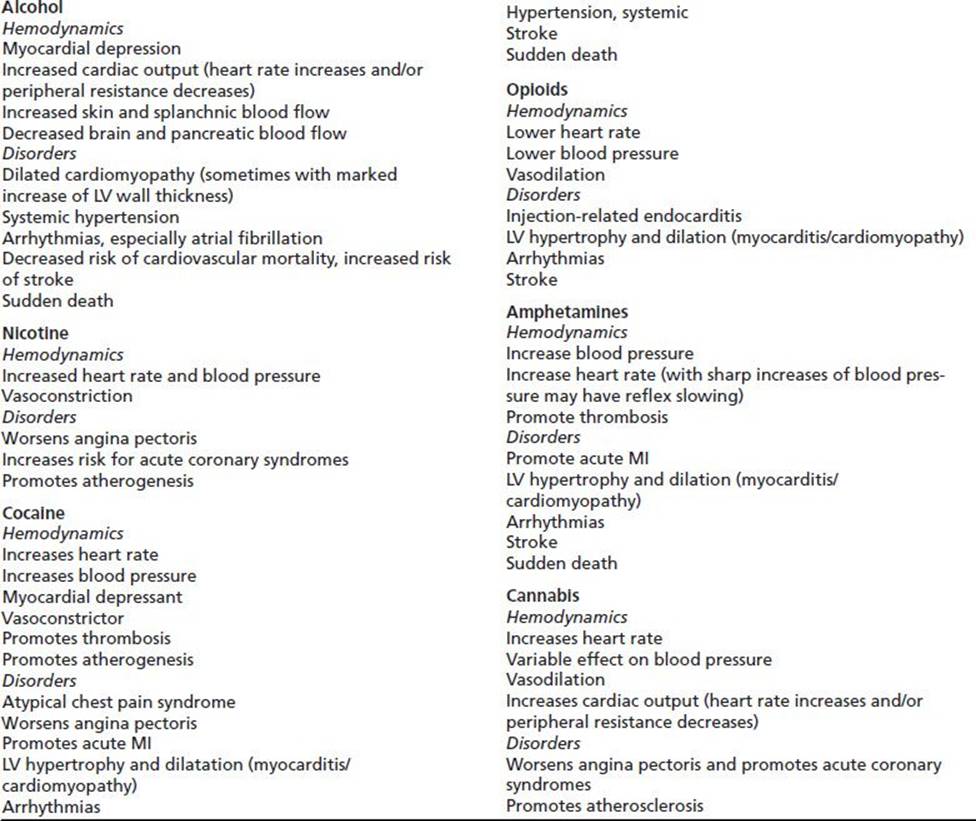
ALCOHOL
Clinical heart disease related to heavy alcohol use is an old observation, familiar to physicians for at least two centuries. The term “alcoholic heart disease” was first used by William MacKenzie in 1902 (2). The association with so-called moderate, better labeled as low, or lower-risk alcohol consumption and protection from cardiovascular diseases is a more recent observation (3,4), leading to a complicated relationship between alcohol and heart disease. The manifestations of alcohol-related heart disease depend in large part on the amount of alcohol ingested and the time period of exposure. Alcohol use and related disorders are a group of conditions that may be defined by a variety of parameters and consequences of alcohol consumption (5), and specific threshold amounts may be relatively crude descriptors. However, for the purpose of definitions used in this discussion, drinking no more than 3 drinks in a day or 7 drinks a week for women (and those over 65 years old) and drinking no more than 4 drinks in a day or 14 drinks a week for men is considered lower risk drinking.
Direct Effects
Ingestion of even a single small-dose alcohol will have acute effects on the circulatory system in adults. Because ethanol and its metabolites, acetaldehyde and acetate, also have adrenergic (6) and vasodilatory (7) effects, the direct myocardial depressant actions of ethanol may be obscured when cardiac “pump” function is assessed after ethanol administration. Cardiac output usually increases after alcohol ingestion in healthy subjects, reflecting the changes in heart rate and peripheral resistance that ensue (8,9); by contrast, a more sensitive index of cardiac function, such as left ventricular (LV) ejection fraction, generally worsens (6,10).
Previously nutritional factors were considered to be primary in the development of cardiac dysfunction in patients with chronic heavy alcohol ingestion. It is now generally accepted that the development of cardiomyopathy is independent of nutritional status and that direct effects of alcohol on the myocardium are responsible for the clinical disease (11). Acute depression of myocardial contractility is observed in the presence of alcohol, and a number of metabolic abnormalities are associated with the presence of alcohol that may contribute. Ethanol has been shown to reduce the amplitude, duration, and the rate of rise of the myocardial transmembrane action potential, an effect that relates directly to the reduction in contractile force (12). Such changes on myocardial contractility may be related in part to an intracellular acidosis resulting from an ethanol concentration–related inhibition of the acid extruder, Na+/H+ exchange (22). The total calcium available to the con tractile proteins, measured as light-indicator calcium transients, is also reduced by ethanol (13,14). These changes do not, however, appear to be related to a reduction in calcium entry (15). Even at concentrations lower than those required to affect calcium transients, ethanol depresses contractility, suggesting that ethanol has effects at the level of the myofilament (16). Acetaldehyde, a metabolite of alcohol produced by alcohol dehydrogenase in the liver, also has direct myocardial depressant effects (17). Alcohol has additionally been associated with abnormal contractile protein and heat shock protein turnover (18). Finally, there are likely genetic factors that predispose to the development of cardiac dysfunction with alcohol use. For example, polymorphisms of the gene for angiotensin-converting enzyme have been associated with the development of cardiomyopathy in men with alcohol use disorders (19). Chronic activation of the renin–angiotensin system is present in people with alcohol use disorders (20), and animal models indicate that this may contribute to the development of cardiomyopathy (21). While all of these metabolic abnormalities have been associated with alcohol use, the exact mechanism by which alcohol results in the development of clinical cardiomyopathy remains less clear.
Alcoholic Cardiomyopathy
Chronic heavy alcohol consumption can result in a clinical picture characterized by four-chamber cardiac enlargement, loss of LV systolic function, and the development of typical symptoms of congestive heart failure: dyspnea on exertion, orthopnea, and nocturnal dyspnea. In Western societies, alcoholic myopathy is second only to ischemia as a cause of secondary dilated cardiomyopathies (22,23). Worldwide, as many as one-third to one-half of cases of dilated cardiomyopathy may be attributable to alcohol use (24). Alcohol use disorders remain very common in the United States, affecting as much as 17% of the population (25).
The likelihood of developing clinical cardiomyopathy correlates with the mean daily alcohol intake and the duration of drinking (26), but the proportion of heavy drinkers who develop overt cardiomyopathy is difficult to estimate. Asymptomatic LV dysfunction is present in as many as one-third of alcoholics (27). Men develop alcoholic cardiomyopathy more often than women, primarily because men drink alcohol more frequently than women and in greater amounts (25). Differences in alcohol metabolism between women and men, however, may explain the observation that women develop cardiomyopathy at a lower total alcohol dose when compared to men (27,28). The relationship between alcoholic cardiomyopathy and alcoholic liver disease is debated: Some studies suggest that the likelihood of cardiomyopathy is higher in patients with alcoholic cirrhosis than without (29), and the coexistence of both entities carries a worse prognosis than when cardiomyopathy is present alone (30). Others suggest that overt alcoholic liver disease and alcoholic cardiomyopathy rarely coexist, but rather a separate entity of cirrhotic cardiomyopathy may be present (22). Cirrhotic cardiomyopathy is less well defined but is characterized by high cardiac output at rest and a blunted cardiac response to stress (31).
While acute alcohol ingestion may depress myocardial contractility by mechanisms discussed above, patients with chronic heavy alcohol use begin to manifest abnormalities of cardiac function even before clinical symptoms of heart failure are apparent. A variety of echocardiographic abnormalities have been described in people with alcohol use disorders without apparent heart disease (32). People with alcohol use disorders may have increased wall thickness, left ventricular (LV) mass, and LV volumes even before there is a decrease in ejection fraction (33). Similarly, cardiac performance with exercise can be impaired in people with moderate to severe alcohol use disorders compared to controls, even when resting ejection fraction is normal (34). Autopsy studies also confirm a high incidence of findings of cardiomyopathy in people with moderate to severe alcohol use disorders who have no clinical heart disease (35).
The signs and symptoms of alcoholic cardiomyopathy are not unique, and so the history is primary in establishing the diagnosis. A history of chronic and prolonged alcohol use, usually 80 to 90 g of alcohol daily for at least 5 years, is of primary importance (36). In the United States, ischemic cardiomyopathy is the most common form of car-diomyopathy (37), and so ischemia must be excluded. This can be difficult, given adult patients who present with car-diomyopathy may also have important risk factors for atherosclerosis, such as hypertension, elevated cholesterol, and cigarette use. Additionally, heavy alcohol use is also a risk factor for ischemic heart disease (38,39). A history of prior myocardial infarction (MI), chest discomfort, or evidence of ischemia and or infarction on the electrocardiogram must move the clinician away from a diagnosis of alcoholic myopathy.
Ancillary testing in new-presentation cardiomyopathy is most helpful in separating alcoholic myopathy from other diagnoses, and the echocardiogram is the standard first step in the evaluation. Four-chamber enlargement is the typical echocardiographic appearance of an alcoholic myopathy, with either normal or increased wall thickness. The presence of significant valvular heart disease or regional wall motion abnormalities suggests that either valve disease or CAD is the primary etiology of the myopathy, even in the presence of alcohol use. Either a functional study, such as a stress test, or an anatomic study, such as a coronary angiogram or CT angiogram, may be necessary to exclude a diagnosis of ischemic disease. The separation of ischemic disease is critical because timely coronary revascularization is the cornerstone of therapy in patients with this diagnosis.
Other causes of dilated cardiomyopathy must be considered, including toxin exposure (especially chemotherapy), trace element exposure (lead, mercury), familial/genetic myopathies, prolonged arrhythmias (such as tachycardia-induced myopathy), and viral infections. Although endomyocardial biopsy may be helpful in establishing the diagnosis of alcoholic myopathy if performed early in the disease and before significant fibrosis, in general, difficulties with sampling error and risk have limited its utility in routine clinical evaluation of cardiomyopathy (40). Cardiac magnetic resonance imaging (MRI) is a useful technique in structural heart disease, with increasing utility in the evaluation of dilated cardiomyopathy (41). Cardiac MR is particularly useful in suspected acute myocarditis, hypertrophic myopathy, amyloidosis, hemochromatosis, and fibrosis, but abnormalities of cardiac MR specific to alcoholic cardio-myopathy have not been defined (42,43). Ultimately, the diagnosis of alcoholic cardiomyopathy is one of exclusion, established only after more common conditions are no longer likely.
The prognosis in alcoholic cardiomyopathy is correlated strongly with continued use of alcohol. When detected early and treated aggressively with alcohol abstinence and standard medical therapy for dilated myopathies and congestive symptoms, the prognosis may be better than in other dilated myopathies. This may be explained in large part by improvement in LV function with abstinence (44–46). However, with continued alcohol use, the prognosis is as poor or worse than in other nonischemic dilated cardiomyopathies (47). As many of 19% of patients who experience sudden cardiac death in the absence of ischemic heart disease have alcoholic cardiomyopathy (48), second only to obesity-related myopathy. Without complete abstinence, the 4-year mortality approaches 50% (49). Treatment, therefore, centers on abstinence, with other standard medical therapies for heart failure: loop diuretics, angiotensin-converting enzyme inhibitors, beta blockers, and spironolactone (50). Advanced therapies, such as implanted defibrillators, LV assist devices, and cardiac transplantation, imply a high level of patient compliance and social support and necessarily demand complex decision making (51); these therapies are unlikely to be applicable in alcoholic cardiomyopathy unless the patient is able to demonstrate abstinence. Although there are no data available specific to heart transplantation, in liver transplantation, posttransplant alcohol use relapse rates are high, even for those with more than 6 months of abstinence prior to transplant (52).
Hypertension
Alcohol use, especially in moderate to heavy amounts, is associated with hypertension. Alcohol may be responsible for as much as 16% of the global burden of hypertensive disease (53). While the most recent data have suggested cardiovascular benefit from light or low-risk alcohol intake, it is important to remember that the relationship between alcohol use and disease relies in large part on self-reporting of amounts of alcohol intake, which may be inaccurate (54).
The mechanisms by which alcohol raises blood pressure are through direct sympathetic vasomotor effects and through secondary mechanisms, such as worsened sleep apnea (55). Alcohol and its metabolites, acetaldehyde and acetate, have both direct vasodilatory effects (7) and sympathetic vasoconstrictive actions. When alcohol is imbibed (56) or administered intravenously (57), muscle sympathetic nerve traffic (of the peroneal nerve) increases. Initially, blood pressure does not increase, but as blood levels of ethanol decrease (and the vasodilator actions abate) and as sympathetic nerve traffic increases, blood pressure elevates (58). The α-adrenergic blocker phentolamine blocks this vasopressor response, and both the sympathetic neural response and the blood pressure increase are blocked by dexamethasone. The acute hypertensive effects of alcohol are, therefore, mediated centrally, with participation of corticotropin-releasing hormone, and act directly through α-adrenergic mechanisms (57). Alcohol ingestion also impairs the baroreceptor reflex (59). When alcohol is administered to normotensive subjects, slowing of heart rate to a sudden elevation in blood pressure is attenuated, and the reflex is reset to a higher blood pressure; this effect is directly related to blood ethanol concentrations (59). Alcohol is also a potent activator of the renin–angiotensin system, with elevation of angiotensin-converting enzyme persisting even after 4 weeks of abstinence (20).
Regular consumption of more than 2 drinks a day is associated with increased blood pressure (60), and the degree of blood pressure increase is dose dependent (54). The elevation in blood pressure is accompanied by the expected rise in hypertension-related cardiovascular morbidity and mortality. Although there is some debate regarding the possible benefits of red wine, the hypertensive effects of alcohol are consistent regardless of source (61). In hypertensive individuals, the sympathetic and hemodynamic responses to alcohol are accentuated when compared to normotensive individuals (62). Alcohol can be an important factor in hypertension that is refractory to standard therapy (55), which may be 10% to 15% of the general hypertension population (63,64) and has an increased risk of cardiovascular complications (65,66) when compared to medication-responsive hypertension. Reduction of alcohol consumption is associated with significant decreases in blood pressure (67,68) in hypertensive patients. In patients being treated for hypertension, alcohol consumption should be limited to 2 drinks per day or less (69).
Atrial Fibrillation
Atrial fibrillation is the most common clinical arrhythmia in adults, with a lifetime risk of 1 in 4 for men and women over the age of 40 (70). Prevalence is highly age dependent, with the arrhythmia being much more common with increasing age. The prevalence varies from 1% in patients less than 60 years of age to 17.8% in patients older than 85 (71). In addition to age, the next two most important risk factors are hypertensive heart disease and coronary heart disease. A myriad of other conditions are associated with the development of atrial fibrillation and include heart failure, congenital heart disease, valvular heart disease, hypertrophic cardiomyopathy, family history, obesity, chronic kidney disease, diabetes, hyperthyroidism, sleep apnea, increased birth weight, pericardial fat, and autonomic dysfunction (70,72).
Alcohol use has also been associated with an increased risk for the development of atrial fibrillation. “Holiday Heart” is a term applied to atrial fibrillation that occurs after binge drinking and typically implies the absence of other heart disease. However, alcohol use has a complex relationship to atrial fibrillation. Light or low-risk alcohol use is of debatable risk, with some authors suggesting that these levels are benign (73,74), and others more recently suggesting a linear increase in risk for atrial fibrillation with any level of alcohol use (75). There is little debate that heavy alcohol use is associated with a significant risk of atrial fibrillation (76,77). Because of early structural abnormalities that occur in the heart with chronic heavy alcohol use, alterations in cardiac function with acute ingestion (78), and associations between alcohol and coronary heart disease, hypertension, and cardiomyopathy, the occurrence of atrial fibrillation in those patients with a history of low-risk to heavy alcohol use must be considered as a potential harbinger of significant heart disease. Atrial fibrillation in the setting of alcohol use must be evaluated in the same manner as new-onset atrial fibrillation occurring in any other context: a full history and physical exam, 12-lead electrocardiogram, echocardiogram, and blood tests for renal, thyroid, and liver function (79).
Coronary Artery Disease and Stroke
The relationship between alcohol use and the incidence of coronary artery atherosclerosis is complex. In examining incidence of CAD by noninvasive or invasive testing, or incident MI, or mortality from coronary disease, low to moderate alcohol consumption appears to be largely protective when compared to those patients who abstain from alcohol. In patients with established coronary heart disease, both cardiovascular mortality and all-cause mortality are lower with low to moderate alcohol intake, with the dose response curve indicating benefit between 5 and 26 g/d (4). Similarly, there is a suggestion that low-risk amounts of alcohol intake may be associated with a lower likelihood of developing congestive heart failure (80) and lower risk of MI and all-cause mortality in patients with established LV dysfunction (80).
The evidence for the beneficial effect of alcohol at low consumption levels has been debated, particularly since the epidemiologic data suffer the potential bias of self-reporting of alcohol intake and lack any randomized controlled data (81). Recent US studies continue to support this point of view (82), but studies from China (83) and India (84) indicate there may be different relationships in different genetic or ethnic groups. There are reasonable biologic mechanisms that would support a protective effect of alcohol though these generally do not account for the magnitude of benefit observed in observational studies (85,86). In addition to favorably affecting lipid profiles (87), low to moderate alcohol consumption has been associated with improved insulin sensitivity (88) and decreased incidence of type 2 diabetes (89). There may also be particular advantages to red wine over other forms of alcohol because of the presence of polyphenols, tannins, and flavonoids, which have been associated with favorable effects on lipids, platelet function, endothelial function, and inflammatory parameters (90).
The epidemiologic evidence for increasing CAD risk with heavy alcohol use has also been consistent (4,91–93), leading to the so-called J-shaped curve of alcohol-related cardiovascular risk (see Fig. 1 in Ref. (93); http://archinte.jamanetwork.com/article.aspx?articleid=769554). The prevalence of coronary artery calcium on CT is a marker for coronary atherosclerosis in asymptomatic patients and is predictive of risk of incident MI and cardiovascular mortality. Studies have consistently failed to demonstrate a J-shaped relationship between coronary artery calcification and alcohol intake, but heavy alcohol use is associated with the highest risk (94–97). Progression of coronary calcification over a 2- to 4-year period is increased with heavy alcohol consumption (96) across all ethnic groups. Over the period of follow-up in this study, incident coronary events occurred most often in the group with the highest alcohol intake, independent of coronary calcification. These data suggest that protection from coronary events is not through prevention of atherosclerosis but through those mechanisms that promote the clinical manifestations of atherosclerosis such as thrombosis. The mechanism of higher clinical events in the heaviest drinkers is less clear.
Consumption of low-risk amounts of alcohol is also associated with a lower risk of stroke, although the data are less robust than that for CAD. At any level of consumption, alcohol is associated with a lower risk of ischemic stroke and a higher risk of hemorrhagic stroke (98) and may be explained by those antithrombotic properties of alcohol that may be protective from CAD (99). The increased risk of stroke with heavy alcohol use is consistent across many studies, with an increased relative risk of total, ischemic, and hemorrhagic stroke of approximately 1.6 (99). Similar to coronary disease, some authors have suggested a J-shaped curve to the risk relationship. The potential explanations include the well-recognized risks of alcohol-related hypertension, atrial fibrillation, and cardiomyopathy.
NICOTINE
Nicotine use is a common clinical entity, with the most common method of ingestion being through cigarette smoking. It is estimated that 19.3% of the US adult population were active smokers in 2010 (>45 million people) (100). While nicotine has direct and immediate effects on the cardiovascular system, the specific role of nicotine in cigarette-related heart disease is debated.
Within minutes of a nicotine dose, either by smoking (101) or by direct administration of the drug (102), both heart rate and blood pressure rise. These hemodynamic changes are accompanied by increases in plasma epinephrine and norepinephrine (101,103). Nicotine exerts its adrenergic actions mainly by release of norepinephrine from nerve terminals (102) and by release of epinephrine from the adrenal medulla (104). A central nervous system stimulatory effect by nicotine is disputed by the observation that muscle sympathetic nerve traffic (of the peroneal nerve) assessed by microneurography decreases as blood pressure increases (103,105). The complex adrenergic response to smoking may account for the lack of elevation of plasma norepinephrine in some studies (104,105). Smoking also attenuates baroreceptor responses to blood pressure elevations (103,105) and the heart rate fluctuations that occur with normal respiration (105). In spite of measurable acute changes, nicotine use is characterized by a rapid onset of tolerance to these physiologic effects, so that in chronic use, these are likely important to overall cardiovascular risk (106).
Nicotine use, both acute and chronic, has also been implicated as a risk for cardiac arrhythmias, especially atrial fibrillation (107,108). In both animal models (109,110) and cell culture (111), nicotine can alter atrial structure, with increased fibrosis and increased likelihood of atrial fibrillation. However, in human studies of stable cardiac disease (112) or those hospitalized with acute coronary syndrome (113), there is no evidence for any increase in cardiac arrhythmias attributable to nicotine replacement therapy. It is likely that while nicotine administration to otherwise healthy hearts may be arrhythmogenic, there is net benefit and overall decreased events when nicotine administration replaces cigarette smoking, which is also highly associated with both supraventricular and ventricular arrhythmias (114).
The development and progression of atherosclerosis requires arterial (especially endothelial) injury, recruitment of leukocytes, and adherence and activation of platelets (115,116). Clinical manifestations of atherosclerosis often are the result of alterations in the arterial microenvironment that favor thrombosis at the site of plaque (117,118). In patients who smoke cigarettes, it appears that the role of nicotine in both atherogenesis and atherothrombosis is small (119,120) and other components of cigarette smoke play more important roles. Cigarette smoke is a complex mixture of chemicals, many of which have been more closely associated with a direct role in coronary atherosclerosis and its clinical manifestations (121). Endothelial injury and dysfunction occur early in smokers, with impaired nitric oxide production (122) and endothelium-dependent vasodilatation (123). Markers of endothelial injury are apparent within 20 minutes of smoking two cigarettes (124). People who smoke have increased markers of endothelial cell activation (125) and evidence for increased adhesion and migration of leukocytes into the artery wall (126). Chronic smoking alters normal platelet function, with evidence of increase aggregation when stimulated compared to nonsmokers (127). Finally, soluble plasma proteins such as fibrinogen are increased in smokers (128); elevated fibrinogen is among the strongest acquired risk factors for cardiovascular risk (129). Particulate matter inhalation, through either air pollution or cigarette smoke, has also been demonstrated to be proinflammatory and contribute to the mechanisms of atherogenesis (121).
The importance of cigarette smoking as a risk for atherosclerotic vascular disease cannot be overemphasized. Cigarette smoking has been identified as an important risk factor for virtually every clinical manifestation of atherosclerosis, including the presence (130) and progression (131) of CAD, carotid disease (132), peripheral vascular disease (133) including abdominal aneurysm (134), thoracic aortic plaque burden (135), ischemic stroke (136), and particularly the incidence of acute MI (137–141), Significantly, mandatory smoking prohibition programs have repeatedly been related to decreased hospitalization rates for acute MI (142–145). In a given individual, smoking cessation is associated with a rapid decline in the risk for acute MI (140), approaching the level of a nonsmoker after 2 to 3 years (146). Every individual addicted to nicotine through smoking should be considered as high risk for cardiovascular disease.
COCAINE
Cocaine is a local anesthetic with potent sympathomimetic actions. The adrenergic effects are the result of blocking pre-synaptic norepinephrine and dopamine reuptake, thereby making more catecholamines available at postsynaptic receptors (147,148), stimulating the sympathetic nervous system (149), and enhancing the effects of endogenous catecholamines (150,151). As a local anesthetic, cocaine inhibits transmembrane sodium flux during electrical excitation, producing a delay in the upstroke and amplitude of the myocardial action potential; this action diminishes intracellular calcium indirectly (making less sodium available for sodium–calcium exchange) (152). Cocaine may also directly block (L-type channel) calcium entry into the myocyte (153,154), inhibit the release of calcium from the sarcoplasmic reticulum (155), and make the contractile proteins less responsive to available calcium (155). Thus, depending on the dosage and the clinical or experimental conditions, cocaine may produce seemingly contradictory effects, reflecting whether the sympathomimetic or the local anesthetic actions are predominant (155).
The so-called “recreational” use of cocaine has been associated with a wide variety of cardiovascular complications. Cocaine is the illicit drug most likely to precipitate an emergency room visit, responsible for 57% of recreational drug-related emergency room visits (156). Many of these visits are for complaints of chest pain; this is particularly important as cocaine is the most common illicit drug used by individuals over 50 years of age (63%) (157). In addition to chest pain, other conditions such as acute MI, aortic dissection, stroke, heart failure, and sudden cardiac death have all been associated with acute and chronic cocaine abuse (158). A high proportion of asymptomatic cocaine users have MRI evidence of ischemia-related fibrosis on cardiac MRI or LV dysfunction by echocardiography, especially over the age of 40 (159,160). Cocaine can alter hemodynamics, arterial muscular tone, and both cellular and humoral parameters of coagulation—all factors that conspire to produce the observed cardiovascular pathology.
Acute hemodynamic effects of cocaine administration are elevation in heart rate, systolic blood pressure, diastolic blood pressure, and mean arterial pressure (161). Repeated use results in the development of tolerance, with limited hemodynamic responses to increasing doses and serum concentrations (162). Animal data indicate that inhibition of norepinephrine and dopamine reabsorption raises serum concentrations, leading to increased sympathetic stimulation (163); this is presumed to be the primary mechanism of hemodynamic alterations in humans. In the coronary arteries of human subjects, cocaine use results in vasoconstriction (164,165), an effect that may be accentuated by the presence of atherosclerosis (166) and cigarette smoking (167).
Cocaine affects all three components of the normal system of hemostasis: blood cells, soluble plasma proteins, and the vessel wall (particularly the endothelium). Cocaine shifts the balance of this tightly regulated system toward arterial thrombus formation, particularly in the presence of atherosclerosis. Platelets are of primary importance in both physiologic and pathologic arterial thrombus formation (118), and activation of platelets is apparent in both chronic users (168,169) and after acute ingestion (170). Monocyte–platelet aggregates, soluble CD40 ligand, neutrophilactivating peptide-2, and regulated on activation, normal T cells expressed and secreted (RANTES)—all indicators of platelet activation—are higher at baseline in chronic cocaine users when compared to controls (168). These effects are completely reversed following 4 weeks of abstinence, indicating specific effects of the drug on the platelets rather than an effect of some other cocaine-induced vascular process. However, at least some measures of platelet reactivity are not altered by direct exposure of platelets in vitro to doses of cocaine similar to plasma levels obtained with recreational use, indicating an indirect mechanism such as catecholamines (170). Evidence for acute endothelial dysfunction related to direct exposure to cocaine exists in cultured human aortic endothelial cells, with increases in endothelin-1 and decreases in nitric oxide production and endothelial nitric oxide synthase expression (171). Finally, levels of soluble plasma proteins such as fibrinogen and von Willebrand factor are increased in chronic cocaine users when compared to drug-free users (172). These markers are associated with increased long-term risk for cardiovascular disease (173) and predict short-term risk in those with acute coronary syndromes (174).
Although some have suggested that cocaine use accelerates the atherosclerotic process (175), it is difficult to separate cocaine from other major environmental, genetic, and behavioral factors that confound this relationship in those who use cocaine, for example, cigarettes, alcohol, and hypertension. The development and progression of atherosclerosis necessarily proceed through three related steps: arterial injury, inflammatory cell recruitment, and platelet adhesion and activation (115). Cocaine directly (in the case of elevation of blood pressure) or indirectly (by increased cigarette use while using cocaine) impacts all three of these pathologic processes.
Cocaine levels peak in the bloodstream at different times depending upon method of ingestion, ranging from 3 minutes with smoking to 40 to 60 minutes with intranasal administration (156). Cocaine metabolites are hemodynamically active and can persist at detectable levels for 48 hours after a single dose. Concomitant use of alcohol can slow the clearance of metabolites as well as produce additional active metabolites (176). In general, presentation with cardiovascular symptoms occurs early after cocaine use, with a markedly elevated risk of cocaine-related MI within the first 24 hours (177).
Cocaine-related chest pain is the most frequent cardiovascular presentation associated with cocaine use. The incidence of acute MI in this group of patients is relatively low, however, with reports ranging from 0.7% to 6% (178) (179). The most recent ACC/AHA guidelines for the evaluation of unstable angina indicate that, in the absence of other high-risk features, a history of cocaine use is associated with a lower risk (180). Still, as the demographic of cocaine shifts to an older population with a high proportion of other CAD risk factors, the diagnosis must be considered carefully. Compared to non–cocaine users, risk stratification tools such as the TIMI risk score are not clinically useful, as more than half of cardiovascular events occur in patients with a TIMI risk score of less than 1 (181). Similarly, stress testing and coronary CT angiography rarely lead to identification of cocaine users with significant CAD and do not affect length of stay (182). Similar to conventional (non–cocaine-related) MI, measurement of troponin maintains a high level of sensitivity and specificity in cocaine-related chest pain (183). The high index of suspicion with poor discriminatory power of conventional risk stratification tools leads to frequent utilization of observation units and hospital admission in patients with cocaine-associated chest pain (184). It appears that observation with serial measurement of troponin is the best compromise of cost and sensitivity when evaluating this group of patients (185). During the observation period, both benzodiazepines and nitroglycerin have been shown in small studies to alleviate symptoms (186,187).
When MI in the context of cocaine abuse does occur, the treatment approach differs from non–cocaine- associated infarction in several important aspects. Antiplatelet therapy with aspirin and an inhibitor of the P2Y12 receptor on platelets is a cornerstone of current infarction management (188). Given the evidence for platelet activation by cocaine, it would seem reasonable that antiplatelet therapy with aspirin or P2Y12 inhibitors would be beneficial, and most experts agree with following standard treatment algorithms for antiplatelet therapy in these patients (189). In contrast to conventional infarction, however, there are no specific data with regard to cocaine-related MI on which to base these recommendations.
Beta-adrenergic receptor blockers are a class of medications that have demonstrated mortality benefit in a large number of clinical trials when administered in the setting of acute MI (180). In the setting of recent cocaine use, the possibility of precipitating arterial vasoconstriction by unopposed alpha-receptor stimulation raises concern about administration of beta blockers in cocaine-related infarction. Arterial vasoconstriction in response to beta blockers in the context of cocaine appears to be corroborated by two small studies in humans without acute infarction (165,190). More recent data suggest that this may not be as concerning as previously thought. In two studies (191,192), each including over 300 subjects with cocaine-associated chest pain, the effect of beta blocker administration to patients who were subsequently found to be cocaine positive was examined with regard to blood pressure and clinical outcome. In fact, subjects who had received beta blockers had lower blood pressure and lower rate of MI, and in one study (192), there was a trend toward decreased postdischarge mortality when compared to subjects who did not receive them. One randomized study of a combined alpha and beta blocker (labetalol) showed similar blood pressure reduction compared to diltiazem with no in-hospital adverse events (193). Still, the most recent practice guidelines to address cocaine-related chest pain/acute coronary syndrome do not endorse beta blockers in early management and give a class II recommendation to the use of labetalol only for the treatment of associated hypertension (189).
Reperfusion therapy, by either systemic thrombolytics or primary angioplasty with stent placement, is the standard of care of ST-segment elevation MI because of demonstrated mortality benefit in large randomized clinical trials (194). Thrombolytic therapy achieves the best results in younger patients, especially those who smoke (195), and this would seem to favor the use of thrombolytics in cocaine-related ST-segment elevation MI. Unfortunately, this has never been tested formally, and the existing literature consists of case reports and small series (196). Similarly, primary angioplasty with stenting has improved MI outcomes across many demographic groups (197), but stent placement in patients who use cocaine has been associated with an increased risk for stent thrombosis (198), a platelet-mediated event. Presumably, this observation can be attributed to both the activation of platelets by ongoing cocaine use and lack of adherence to prescribed dual antiplatelet therapy.
Other important presentations of cocaine-related cardiovascular diseases include aortic dissection and stroke. The proportion of aortic dissection that is related to cocaine varies depending upon the population studied, ranging from 0.5% to 37% (199–201). Cocaine-related dissection tends to occur more frequently in young, hypertensive subjects (202). Cocaine use may accelerate the progression of atherosclerosis, and the presence of other traditional atherosclerosis risk factors in chronic cocaine users such as hypertension and cigarette smoking (203) contributes to the arterial pathology that predisposes to dissection.
Cocaine-related stroke was first described in 1977. Both ischemic stroke and intracerebral hemorrhage have been reported, with approximately equal frequency. Hemorrhage appears to be more frequent in current users, while ischemic stroke and transient ischemic attack are more common in patients who are remote cocaine users. The mechanisms of stroke in cocaine use are not entirely clear, but severe vasospasm, in either normal or atherosclerotic arteries, is implicated from animal studies and observations in patients. Decrease in cerebral blood flow is observed in human subjects after a single intravenous dose of cocaine (204). Consistent with the effects of cocaine on platelet activation, platelet-rich thrombi have been observed after fatal cerebral infarcts in chronic cocaine users (205). Large artery atherosclerotic disease is the most common finding in ischemic stroke, consistent with the hypothesis that cocaine use accelerates atherosclerotic vascular disease.
In summary, cocaine has direct and indirect effects on the cardiovascular system that are associated with an increased risk for clinical manifestations of atherosclerotic vascular disease. Cocaine-related chest pain and alterations in heart rate and blood pressure are the most commonly encountered problems in clinical practice.
OPIOIDS
From a strictly cardiovascular perspective, opiates have significant medicinal value. The hemodynamic effects of lowering heart rate and blood pressure, as well as decreasing preload, combined with their analgesic and anxiolytic properties, have made morphine a cornerstone of the treatment of MI and acute pulmonary edema. Opiates have not been associated with any direct toxic effects on myocytes or other components of the heart, a fact underscored by the general lack of abnormalities in cardiac dimension and structure in heroin addicts (206).
The primary risks of opioid use from a cardiovascular point of view are those associated with method of administration, namely, intravenous injection. Although rare when compared to, for example, cocaine-related chest pain, infectious endocarditis is important because of the extremely high morbidity and mortality and associated costs of treatment. It is estimated that as many as 21 million people worldwide injected drugs in 2007 (207), indicating a very large pool of subjects at risk. It is difficult to estimate the incidence of injection-related endocarditis, but the number of hospitalizations for this diagnosis is rising in the United States (208).
Infectious endocarditis in someone who has used injection drug is characterized by several features that distinguish this entity from other cases of endocarditis. First, it is much more common for the right heart valves, especially the tricuspid valve, to be involved (209). While less than 10% of endocarditis in nonusers is right sided, as much as 75% of endocarditis in drug users involves the right heart valves (210). The mechanisms may be related to direct toxicity to the right-sided valve structures by the injected drugs or contaminants (209). Left-sided endocarditis in those who use drugs may be associated with a worse prognosis (211). Second, polymicrobial infections and endocarditis due to fungal pathogens are much more common in those who use injection drugs (212). Polymicrobial endocarditis is much more likely to require surgery and carries a higher mortality than single-organism infections (212). Fungal endocarditis mortality ranges from 37% to 80%, even with aggressive surgical and medical therapy (213,214). Finally, because patients with opioid dependence have a high likelihood of continued use, recurrent endocarditis is common. Recurrence of endocarditis after valve replacement surgery carries a particularly ominous prognosis (215).
AMPHETAMINES
Amphetamines (a contraction of alpha-methylphenethyl-amine) are a group of synthetic compounds used both medicinally and recreationally. Amphetamines have a common β-phenylethylamine chemical structure, which they share with catecholamines; the differences in their chemical composition determine their modes of action, relative potency, and sympathomimetic properties (216). Though these substances differ in their central nervous system and peripheral effects, as a group, they act by stimulating the sympathetic nervous system, by displacing catecholamines or interfering with reuptake from their storage sites, by blocking the actions of monoamine oxidase inhibition, and/ or by direct adrenergic actions (216). Amphetamines and their derivatives such as methamphetamine include medications used in the treatment of attention deficit hyperactivity disorder and traumatic brain injury. Phenylpropanolamine is an amphetamine frequently used as an appetite suppressant and nasal decongestant. Other derivatives are used only recreationally and include 3,4-methylenedioxymeth-amphetamine (MDMA or “ecstasy”) and methyldiethanol-amine (MDEA or “eve”).
Amphetamines produce a dose-dependent elevation of blood pressure and an increase of heart rate (217,218). The magnitude of the changes reflects their relative α1-adrenergic (elevate blood pressure with reflex slowing of heart rate) and β1-adrenergic (enhance cardiac contractility and increase heart rate) effects. When amphetamines are taken parenterally, the peak vasopressor effects are evident within a half hour (217), whereas when taken by mouth, the peak changes are observed within 1 to 2 hours (218). Under experimental conditions, amphetamines have been shown to increase systolic and diastolic blood pressure in healthy subjects by 30 and 20 mm Hg, respectively (217,218). The blood pressure elevations dissipate over 3 to 4 hours (217,218). Though for MDMA and methylphenidate an increase in heart rate (20 beats/min) parallels their blood pressure changes, for amphetamine and methamphetamine, only modest heart rate changes are initially observed (the magnitude of the heart rate changes actually having an inverse relation to the blood pressure changes) (217,218). Three to four hours after the administration of amphetamine, methamphetamine, or methylphenidate, as the blood pressure elevations dissipate and the reflex baroreceptor response is attenuated, further heart rate increases ensue (a total change of as many as 20 to 30 beats/min) that may persist above baseline for 10 hours (217,218).
Given the sympathomimetic properties of amphetamines, one would expect cardiovascular complications to be similar to those of cocaine. The most common presentation in patients seen in the emergency room after recent amphetamine use is tachycardia and hypertension (219). The presence of early CAD may be related to chronic amphetamine use (220,221), although the evidence is not as compelling as for cocaine. Amphetamine-related stimulants are reported to be associated with acute MI (222) and may be associated with a worse prognosis in those with acute coronary syndrome (223). Amphetamines may cause myocardial ischemia by several mechanisms, including focal or diffuse vasospasm, increased myocardial demand on preexisting coronary disease, and increased platelet reactivity (223). Similarly, ischemic and hemorrhagic stroke have been associated with amphetamine use (224). Phenylpropanolamine, a common ingredient in appetite suppressants, was removed from over-the-counter sale by the FDA in the United States because of a strong association with an increased risk for hemorrhagic stroke, especially in young women (225). Some amphetamine derivatives developed as appetite suppressants (such as fenfluramine) are strong serotonin receptor (especially the 5-HT2B receptor) agonists and were removed from the market after evidence linking them to the development of a specific form of valvular heart disease previously associated with serotonin-secreting carcinoid tumors; similar lesions have been described in recreational amphetamine use (226–228).
Cardiomyopathy is perhaps the most commonly associated heart disease in chronic amphetamine use (229–232). As many as 5% of all patients presenting to emergency rooms in the United States with decompensated heart failure may be chronic stimulant users (233). Concentric hypertrophy is a common finding in methamphetamine users (220,234) and may precede the development of heart failure. In general, patients tend to be young and have more severe depression of LV function when compared to patients presenting with other forms of nonischemic myopathy (229). Both animal models and human pathologic data suggest that direct toxicity to myocytes can occur with chronic amphetamine exposure (220,235,236). Contraction band necrosis, a myocyte injury pattern seen in exposure to high levels of catecholamines, may be present; this finding can also be seen in stress-related cardiomyopathy and in the cardiomyopathy related to pheochromocytoma (237).
CANNABIS
After oral ingestion, intravenous administration, or by smoking, cannabis increases heart rate and cardiac output (238). The effects on the cardiovascular system are likely mediated through sympathetic activation (239). With continued use, tolerance develops, blunting the cardiovascular changes observed (240).
Use of cannabis may be through direct ingestion, but more commonly, it is through smoking the dried plant. At least two types of cannabis receptors have been identified (CB1 and CB2) and exist on a variety of tissues, including those that play a role in the development of atherosclerosis: endothelial cells (241), leukocytes (242), vascular smooth muscle cells (243), and platelets (244). However, stimulation of these receptors is associated with both changes that would be protective (245) from atherosclerosis or promote (241) atherosclerosis. Effects on lipid oxidation (246), lipid accumulation in macrophages (247), and vascular smooth muscle (248) are similarly divergent, with most data suggesting proatherogenic effects from CB1 agonists and antiatherogenic effects from CB2 agonists (249).
Based upon the body of preclinical scientific data, three clinical trials have sought to demonstrate benefit of antagonism of the CB1 receptor in studies of coronary atheroma volume (250), carotid artery intima media thickness (251), and overall vascular events (252). The negative findings of these studies suggest that CB1 receptors are not likely to be involved in the development of atherosclerosis or its clinical manifestations.
Observational data suggest a relationship between smoking of marijuana and the onset of acute MI (253) and worsened outcome of MI (254). Most summaries of the association of marijuana and MI rely heavily on case reports (255). However, population-based studies have found no increased risk of mortality associated with marijuana use (256,257). Similar to cigarette smoking, components of marijuana smoke would likely predispose to atherosclerosis, as inhaled particulates are associated with vascular inflammation and the development of atherosclerosis (258). The most accurate statement regarding the cardiovascular risk from cannabis is that the subject is limited by a striking absence of published data.
REFERENCES
1.Lloyd-Jones DM, Larson MG, Beiser A, et al. Lifetime risk of developing coronary heart disease. Lancet 1999;353(9147): 89–92.
2.George A, Figueredo VM. Alcoholic cardiomyopathy: a review. J Card Fail 2011;17(10):844–849.
3.Malinski Mk, Sesso HD, Lopez-Jimenez F, et al. Alcohol consumption and cardiovascular disease mortality in hypertensive men. Arch Intern Med 2004;164(6):623–628.
4.Costanzo S, Di Castelnuovo A, Donati MB, et al. Alcohol consumption and mortality in patients with cardiovascular disease: a meta-analysis. J Am Coll Cardiol 2010;55(13):1339–1347.
5.Reid M, Fiellin DA, O’Connor PG. Hazardous and harmful alcohol consumption in primary care. Arch Intern Med 1999;159(15):1681–1689.
6.Kelbaek H, Gjorup T, Hartling OJ, et al. Left ventricular function during alcohol intoxication and autonomic nervous blockade. Am J Cardiol 1987;59(6):685–688.
7.Altura BM, Altura BT. Microvascular and vascular smooth muscle actions of ethanol, acetaldehyde, and acetate. Fed Proc 1982;41(8):2447–2451.
8.Riff DP, Jain AC, Doyle JT. Acute hemodynamic effects of ethanol on normal human volunteers. Am Heart J 1969;78(5):592–597.
9.Blomqvist G, Saltin B, Mitchell JH. Acute effects of ethanol ingestion on the response to submaximal and maximal exercise in man. Circulation 1970;42(3):463–470.
10.Delgado CE, Gortuin NJ, Ross RS. Acute effects of low doses of alcohol on left ventricular function by echocardiography. Circulation 1975;51(3):535–540.
11.Patel VB, Why HJ, Richardson PJ, et al. The effects of alcohol on the heart. Adverse Drug React Toxicol Rev 1997;16(1):15–43.
12.Williams ES, Mirro MJ, Bailey JC. Electrophysiological effects of ethanol, acetaldehyde, and acetate on cardiac tissues from dog and guinea pig. Circ Res 1980;47(3):473–478.
13.Thomas AP, Sass EJ, Tun-Kirchmann TT, et al. Ethanol inhibits electrically-induced calcium transients in isolated rat cardiac myocytes. J Mol Cell Cardiol 1989;21(6):555–565.
14.Danziger RS, Sakai M, Capogrossi MC, et al. Ethanol acutely and reversibly suppresses excitation-contraction coupling in cardiac myocytes. Circ Res 1991;68(6):1660–1668.
15.Mongo KG, Vassort G. Inhibition by alcohols, halothane and chloroform of the Ca current in single frog ventricular cells. J Mol Cell Cardiol 1990;22(9):939–953.
16.Guarnieri T, Lakatta EG. Mechanism of myocardial contractile depression by clinical concentrations of ethanol. A study in ferret papillary muscles. J Clin Invest 1990;85(5):1462–1467.
17.Lange LG, Sobel BE. Myocardial metabolites of ethanol. Circ Res 1983;52(4):479–482.
18.Preedy VR, Atkinson LM, Richardson PJ, et al. Mechanisms of ethanol-induced cardiac damage. Br Heart J 1993;69(3):197–200.
19.Fernandez-Sola J, Nicolas JM, Oriola J, et al. Angiotensin-converting enzyme gene polymorphism is associated with vulnerability to alcoholic cardiomyopathy. Ann Intern Med2002;137(5 Part 1): 321–326.
20.Okuno F, Arai M, Ishii H, et al. Mild but prolonged elevation of serum angiotensin converting enzyme (ACE) activity in alcoholics. Alcohol 1986;3(6):357–359.
21.Cheng CP, Cheng HJ, Cunningham C, et al. Angiotensin II type 1 receptor blockade prevents alcoholic cardiomyopathy. Circulation 2006;114(3):226–236.
22.Ripoll C, Yotti R, Bermejo J, et al. The heart in liver transplantation. J Hepatol 2011;54(4):810–822.
23.Piano MR. Alcoholic cardiomyopathy: incidence, clinical characteristics, and pathophysiology. Chest 2002;121(5): 1638–1650.
24.Regan TJ. Alcoholic cardiomyopathy. Prog Cardiovasc Dis 1984;27(3):141–152.
25.Hasin DS, Stinson FS, Ogburn E, et al. Prevalence, correlates, disability, and comorbidity of DSM-IV alcohol abuse and dependence in the United States: results from the National Epidemiologic Survey on Alcohol and Related Conditions. Arch Gen Psychiatry 2007;64(7):830–842.
26.Kajander OA, Kupari M, Laippala P, et al. Dose dependent but non-linear effects of alcohol on the left and right ventricle. Heart 2001;86(4):417–423.
27.Urbano-Marquez A, Estruch R, Fernandez-Sola J, et al. The greater risk of alcoholic cardiomyopathy and myopathy in women compared with men. JAMA 1995;274(2):149–154.
28.Fernandez-Sola J, Estruch R, Nicolas JM, et al. Comparison of alcoholic cardiomyopathy in women versus men. Am J Cardiol 1997;80(4):481–485.
29.Estruch R, Fernandez-Sola J, Sacanella E, et al. Relationship between cardiomyopathy and liver disease in chronic alcoholism. Hepatology 1995;22(2):532–538.
30.Henriksen JH, Moller S. Cardiac and systemic haemodynamic complications of liver cirrhosis. Scand Cardiovasc J 2009; 43(4):218–225.
31.Moller S, Henriksen JH. Cirrhotic cardiomyopathy. J Hepatol 2010;53(1):179–190.
32.Mathews EC, Jr, Gardin JM, Henry WL, et al. Echocardiographic abnormalities in chronic alcoholics with and without overt congestive heart failure. Am J Cardiol 1981;47(3):570–578.
33.Lazarevic AM, Nakatani S, Neskovic AN, et al. Early changes in left ventricular function in chronic asymptomatic alcoholics: relation to the duration of heavy drinking. J Am Coll Cardiol2000;35(6):1599–1606.
34.Kelbaek H, Eriksen J, Brynjolf I, et al. Cardiac performance in patients with asymptomatic alcoholic cirrhosis of the liver. Am J Cardiol 1984;54(7):852–855.
35.Davidson DM. Cardiovascular effects of alcohol. West J Med 1989;151(4):430–439.
36.McKenna CJ, Codd MB, McCann HA, et al. Alcohol consumption and idiopathic dilated cardiomyopathy: a case control study. Am Heart J 1998;135(5 Pt 1):833–837.
37.Ho KK, Anderson KM, Kannel WB, et al. Survival After the Onset of Congestive Heart Failure in Framingham Heart Study Subjects. Circulation 1993;88(1):107–115.
38.Corrao G, Rubbiati L, Bagnardi V, et al. Alcohol and coronary heart disease: a meta-analysis. Addiction 2000;95(10):1505–1523.
39.Britton KA, Gaziano JM, Sesso HD, et al. Relation of alcohol consumption and coronary heart disease in hypertensive male physicians (from the Physicians’ Health Study). Am J Cardiol2009;104(7):932–935.
40.Hunt SA, Abraham WT, Chin MH, et al. 2009 focused update incorporated into the ACC/AHA 2005 Guidelines for the Diagnosis and Management of Heart Failure in Adults: a report of the American College of Cardiology Foundation/American Heart Association Task Force on Practice Guidelines: developed in collaboration with the International Society for Heart and Lung Transplantation. Circulation2009;119(14):e391–e479.
41.Dickerson JA, Raman SV, Baker PM, et al. Relationship of cardiac magnetic resonance imaging and myocardial biopsy in the evaluation of nonischemic cardiomyopathy. Congest Heart Fail 2013;19(1):29–38.
42.Bluemke DA. MRI of nonischemic cardiomyopathy. AJR Am J Roentgenol 2010;195(4):935–940.
43.Shehata ML, Turkbey EB, Vogel-Claussen J, et al. Role of cardiac magnetic resonance imaging in assessment of nonischemic cardiomyopathies. Top Magn Reson Imaging2008;19(1):43–57.
44.La Vecchia LL, Bedogni F, Bozzola L, et al. Prediction of recovery after abstinence in alcoholic cardiomyopathy: role of hemodynamic and morphometric parameters. Clin Cardiol1996;19(1):45–50.
45.Guillo P, Mansourati J, Maheu B, et al. Long-term prognosis in patients with alcoholic cardiomyopathy and severe heart failure after total abstinence. Am J Cardiol 1997;79(9):1276–1278.
46.Nicolas JM, Fernandez-Sola J, Estruch R, et al. The effect of controlled drinking in alcoholic cardiomyopathy. Ann Intern Med 2002;136(3):192–200.
47.Fauchier L, Babuty D, Poret P, et al. Comparison of long-term outcome of alcoholic and idiopathic dilated cardiomyopathy. Eur Heart J 2000;21(4):306–314.
48.Hookana E, Junttila MJ, Puurunen VP, et al. Causes of nonischemic sudden cardiac death in the current era. Heart Rhythm 2011;8(10):1570–1575.
49.Laonigro I, Correale M, Di Biase M, et al. Alcohol abuse and heart failure. Eur J Heart Fail 2009;11(5):453–462.
50.Hunt SA, Abraham WT, Chin MH, et al. ACC/AHA 2005 Guideline Update for the Diagnosis and Management of Chronic Heart Failure in the Adult: a report of the American College of Cardiology/American Heart Association Task Force on Practice Guidelines (Writing Committee to Update the 2001 Guidelines for the Evaluation and Management of Heart Failure): developed in collaboration with the American College of Chest Physicians and the International Society for Heart and Lung Transplantation: endorsed by the Heart Rhythm Society. Circulation 2005;112(12):e154–e235.
51.Allen LA, Stevenson LW, Grady KL, et al. Decision making in advanced heart failure: a scientific statement from the American Heart Association. Circulation 2012;125(15):1928–1952.
52.Dew MA, DiMartini AF, Steel J, et al. Meta-analysis of risk for relapse to substance use after transplantation of the liver or other solid organs. Liver Transpl 2008;14(2):159–172.
53.Rehm J, Room R, Monteiro M, et al. Alcohol as a risk factor for global burden of disease. Eur Addict Res 2003;9(4):157–164.
54.Beilin LJ, Puddey IB. Alcohol and hypertension: an update. Hypertension 2006;47(6):1035–1038.
55.Sarafidis PA, Bakris GL. Resistant hypertension: an overview of evaluation and treatment. J Am Coll Cardiol 2008;52(22): 1749–1757.
56.Grassi GM, Sommers VK, Rek WS, et al. Effects of alcohol intake on blood pressure and sympathetic nerve activity in normotensive humans: a preliminary report. J Hypertens Suppl1989;7(Suppl 6): S20–S21.
57.Randin D, Vollenweider P, Tappy L, et al. Suppression of alcohol-induced hypertension by dexamethasone. N Engl J Med 1995;332(26):1733–1737.
58.van de Borne P, Mark AL, Montano N, et al. Effects of alcohol on sympathetic activity, hemodynamics, and chemoreflex sensitivity. Hypertension 1997;29(6):1278–1283.
59.Abdel-Rahman AR, Merrill RH, Wooles WR. Effect of acute ethanol administration on the baroreceptor reflex control of heart rate in normotensive human volunteers. Clin Sci (Lond)1987;72(1):113–122.
60.Klatsky AL, Friedman GD, Siegelaub AB, et al. Alcohol consumption and blood pressure Kaiser-Permanente Multiphasic Health Examination data. N Engl J Med 1977;296(21):1194–1200.
61.Zilkens RR, Burke V, Hodgson JM, et al. Red wine and beer elevate blood pressure in normotensive men. Hypertension 2005;45(5):874–879.
62.Hering D, Kucharska W, Kara T, et al. Potentiated sympathetic and hemodynamic responses to alcohol in hypertensive vs. normotensive individuals. J Hypertens 2011;29(3):537–541.
63.McAdam-Marx C, Ye X, Sung JC, et al. Results of a retrospective, observational pilot study using electronic medical records to assess the prevalence and characteristics of patients with resistant hypertension in an ambulatory care setting. Clin Ther 2009;31(5):1116–1123.
64.Persell SD. Prevalence of resistant hypertension in the United States, 2003–2008. Hypertension 2011;57(6):1076–1080.
65.Pierdomenico SD, Lapenna D, Bucci A, et al. Cardiovascular outcome in treated hypertensive patients with responder, masked, false resistant, and true resistant hypertension. Am J Hypertens2005;18(11):1422–1428.
66.Daugherty SL, Powers JD, Magid DJ, et al. Incidence and prognosis of resistant hypertension in hypertensive patients. Circulation 2012;125(13):1635–1642.
67.Xin X, He J, Frontini MG, et al. Effects of alcohol reduction on blood pressure: a meta-analysis of randomized controlled trials. Hypertension 2001;38(5):1112–1117.
68.McFadden CB, Brensinger CM, Berlin JA, et al. Systematic review of the effect of daily alcohol intake on blood pressure. Am J Hypertens 2005;18(2 Pt 1):276–286.
69.Chobanian AV, Bakris GL, Black HR, et al. Seventh report of the Joint National Committee on Prevention, Detection, Evaluation, and Treatment of High Blood Pressure. Hypertension2003;42(6):1206–1252.
70.Magnani JW, Rienstra M, Lin H, et al. Atrial fibrillation: current knowledge and future directions in epidemiology and genomics. Circulation 2011;124(18):1982–1993.
71.Heeringa J, van der Kuip DA, Hofman A, et al. Prevalence, incidence and lifetime risk of atrial fibrillation: the Rotterdam study. Eur Heart J 2006;27(8):949–953.
72.Schoonderwoerd BA, Smit MD, Pen L, et al. New risk factors for atrial fibrillation: causes of “not-so-lone atrial fibrillation.” Europace 2008;10(6):668–673.
73.Djousse L, Levy D, Benjamin EJ, et al. Long-term alcohol consumption and the risk of atrial fibrillation in the Framingham Study. Am J Cardiol 2004;93(6):710–713.
74.Mukamal KJ, Psaty BM, Rautaharju PM, et al. Alcohol consumption and risk and prognosis of atrial fibrillation among older adults: the Cardiovascular Health Study. Am Heart J2007;153(2):260–266.
75.Kodama S, Saito K, Tanaka S, et al. Alcohol consumption and risk of atrial fibrillation: a meta-analysis. J Am Coll Cardiol 2011;57(4):427–436.
76.Frost L, Vestergaard P. Alcohol and risk of atrial fibrillation or flutter: a cohort study. Arch Intern Med 2004;164(18):1993–1998.
77.Mukamal KJ, Tolstrup JS, Friberg J, et al. Alcohol consumption and risk of atrial fibrillation in men and women: the Copenhagen City Heart Study. Circulation 2005;112(12):1736–1742.
78.Kelbaek H, Gjorup T, Brynjolf I, et al. Acute effects of alcohol on left ventricular function in healthy subjects at rest and during upright exercise. Am J Cardiol 1985;55(1):164–167.
79.Fuster V, Ryden LE, Cannom DS, et al. ACC/AHA/ESC 2006 Guidelines for the Management of Patients with Atrial Fibrillation: a report of the American College of Cardiology/American Heart Association Task Force on Practice Guidelines and the European Society of Cardiology Committee for Practice Guidelines (Writing Committee to Revise the 2001 Guidelines for the Management of Patients With Atrial Fibrillation): developed in collaboration with the European Heart Rhythm Association and the Heart Rhythm Society. Circulation 2006;114(7):e257–e354.
80.Walsh CR, Larson MG, Evans JC, et al. Alcohol consumption and risk for congestive heart failure in the Framingham Heart Study. Ann Intern Med 2002;136(3):181–191.
81.Klatsky AL. Alcohol and cardiovascular mortality: common sense and scientific truth. J Am Coll Cardiol 2010;55(13):1336–1338.
82.Mukamal KJ, Chen CM, Rao SR, et al. Alcohol consumption and cardiovascular mortality among U.S. adults, 1987 to 2002. J Am Coll Cardiol 2010;55(13):1328–1335.
83.Zhou X, Li C, Xu W, et al. Relation of alcohol consumption to angiographically proved coronary artery disease in chinese men. Am J Cardiol 2010;106(8):1101–1103.
84.Roy A, Prabhakaran D, Jeemon P, et al. Impact of alcohol on coronary heart disease in Indian men. Atherosclerosis 2010;210(2):531–535.
85.Zakhari S. Molecular mechanisms underlying alcohol-induced cardioprotection: contribution of hemostatic components. Introduction to the symposium. Alcohol Clin Exp Res1999;23(6):1108–1110.
86.Booyse FM, Parks DA. Moderate wine and alcohol consumption: beneficial effects on cardiovascular disease. Thromb Haemost 2001;86(2):517–528.
87.Gaziano JM, Buring JE, Breslow JL, et al. Moderate alcohol intake, increased levels of high-density lipoprotein and its subfractions, and decreased risk of myocardial infarction. N Engl J Med1993;329(25):1829–1834.
88.Davies MJ, Baer DJ, Judd JT, et al. Effects of moderate alcohol intake on fasting insulin and glucose concentrations and insulin sensitivity in postmenopausal women: a randomized controlled trial. JAMA2002;287(19):2559–2562.
89.Baliunas DO, Taylor BJ, Irving H, et al. Alcohol as a risk factor for type 2 diabetes: A systematic review and meta-analysis. Diabetes Care 2009;32(11):2123–2132.
90.Di Minno MN, Franchini M, Russolillo A, et al. Alcohol dosing and the heart: updating clinical evidence. Semin Thromb Hemost 2011;37(8):875–884.
91.Di Castelnuovo A, Rotondo S, Iacoviello L, et al. Meta-analysis of wine and beer consumption in relation to vascular risk. Circulation 2002;105(24):2836–2844.
92.White IR, Altmann DR, Nanchahal K. Alcohol consumption and mortality: modelling risks for men and women at different ages. BMJ 2002;325(7357):191.
93.Di Castelnuovo A, Costanzo S, Bagnardi V, et al. Alcohol dosing and total mortality in men and women: an updated meta-analysis of 34 prospective studies. Arch Intern Med2006;166(22):2437–2445. http://archinte.jamanetwork.com/article.aspx?articleid=769554
94.Yang T, Doherty TM, Wong ND, et al. Alcohol consumption, coronary calcium, and coronary heart disease events. Am J Cardiol 1999;84(7):802–806.
95.Pletcher MJ, Varosy P, Kiefe CI, et al. Alcohol consumption, binge drinking, and early coronary calcification: findings from the Coronary Artery Risk Development in Young Adults (CARDIA) Study. Am J Epidemiol 2005;161(5):423–433.
96.McClelland RL, Bild DE, Burke GL, et al. Alcohol and coronary artery calcium prevalence, incidence, and progression: results from the Multi-Ethnic Study of Atherosclerosis (MESA). Am J Clin Nutr2008;88(6):1593–1601.
97.Vliegenthart R, Oei HH, van den Elzen AP, et al. Alcohol consumption and coronary calcification in a general population. Arch Intern Med 2004;164(21):2355–2360.
98.Ronksley PE, Brien SE, Turner BJ, et al. Association of alcohol consumption with selected cardiovascular disease outcomes: a systematic review and meta-analysis. BMJ2011;342:d671.
99.Reynolds K, Lewis B, Nolen JD, et al. Alcohol consumption and risk of stroke: a meta-analysis. JAMA 2003;289(5):579–588.
100.Vital signs: current cigarette smoking among adults aged ≥18 years—United States, 2005–2010. MMWR Morb Mortal Wkly Rep 2011;60(35):1207–1212.
101.Cryer PE, Haymond MW, Santiago JV, et al. Norepinephrine and epinephrine release and adrenergic mediation of smoking-associated hemodynamic and metabolic events. N Engl J Med1976;295(11):573–577.
102.Benowitz NL, Jacob P III, Jones RT, et al. Interindividual variability in the metabolism and cardiovascular effects of nicotine in man. J Pharmacol Exp Ther 1982;221(2):368–372.
103.Grassi G, Seravalle G, Calhoun DA, et al. Mechanisms responsible for sympathetic activation by cigarette smoking in humans. Circulation 1994;90(1):248–253.
104.Benowitz NL, Gourlay SG. Cardiovascular toxicity of nicotine: implications for nicotine replacement therapy. J Am Coll Cardiol 1997;29(7):1422–1431.
105.Niedermaier ON, Smith ML, Beightol LA, et al. Influence of cigarette smoking on human autonomic function. Circulation 1993;88(2):562–571.
106.Adamopoulos D, van de Borne P, Argacha JF. New insights into the sympathetic, endothelial and coronary effects of nicotine. Clin Exp Pharmacol Physiol 2008;35(4):458–463.
107.Stewart PM, Catterall JR. Chronic nicotine ingestion and atrial fibrillation. Br Heart J 1985;54(2):222–223.
108.Rigotti NA, Eagle KA. Atrial fibrillation while chewing nicotine gum. JAMA 1986;255(8):1018.
109.Mehta MC, Jain AC, Mehta A, et al. Cardiac arrhythmias following intravenous nicotine: experimental study in dogs. J Cardiovasc Pharmacol Ther 1997;2(4):291–298.
110.Miyauchi M, Qu Z, Miyauchi Y, et al. Chronic nicotine in hearts with healed ventricular myocardial infarction promotes atrial flutter that resembles typical human atrial flutter. Am J Physiol Heart Circ Physiol2005;288(6):H2878–H2886.
111.Wang H, Shi H, Wang Z. Nicotine depresses the functions of multiple cardiac potassium channels. Life Sci 1999;65(12):PL143–PL149.
112.Joseph AM, Norman SM, Ferry LH, et al. The safety of transdermal nicotine as an aid to smoking cessation in patients with cardiac disease. N Engl J Med 1996;335(24):1792–1798.
113.Woolf KJ, Zabad MN, Post JM, et al. Effect of nicotine replacement therapy on cardiovascular outcomes after acute coronary syndromes. Am J Cardiol 2012;110(7):968–970.
114.D’Alessandro A, Boeckelmann I, Hammwhoner M, et al. Nicotine, cigarette smoking and cardiac arrhythmia: an overview. Eur J Prev Cardiol 2012;19(3):297–305.
115.Ross R. Atherosclerosis—an inflammatory disease. N Engl J Med 1999;340(2):115–126.
116.Ross R. The pathogenesis of atherosclerosis: a perspective for the 1990s. Nature 1993;362(6423):801–809.
117.Ruberg FL, Loscalzo J. Prothrombotic determinants of coronary atherothrombosis. Vasc Med 2002;7(4):289–299.
118.Lippi G, Franchini M, Targher G. Arterial thrombus formation in cardiovascular disease. Nat Rev Cardiol 2011;8(9):502–512.
119.Ambrose JA, Barua RS. The pathophysiology of cigarette smoking and cardiovascular disease: an update. J Am Coll Cardiol 2004;43(10):1731–1737.
120.Blann AD, Steele C, McCollum CN. The influence of smoking and of oral and transdermal nicotine on blood pressure, and haematology and coagulation indices. Thromb Haemost1997;78(3):1093–1096.
121.Talhout R, Schulz T, Florek E, et al. Hazardous compounds in tobacco smoke. Int J Environ Res Public Health 2011;8(2):613–628.
122.Kugiyama K, Yasue H, Ohgushi M, et al. Deficiency in nitric oxide bioactivity in epicardial coronary arteries of cigarette smokers. J Am Coll Cardiol 1996;28(5):1161–1167.
123.Barua RS, Ambrose JA, Eales-Reynolds LJ, et al. Dysfunctional endothelial nitric oxide biosynthesis in healthy smokers with impaired endothelium-dependent vasodilatation. Circulation2001;104(16):1905–1910.
124.Blann AD, Kirkpatrick U, Devine C, et al. The influence of acute smoking on leucocytes, platelets and the endothelium. Atherosclerosis 1998;141(1):133–139.
125.Bermudez EA, Rifai N, Buring JE, et al. Relation between markers of systemic vascular inflammation and smoking in women. Am J Cardiol 2002;89(9):1117–1119.
126.Adams MR, Jessup W, Celermajer DS. Cigarette smoking is associated with increased human monocyte adhesion to endothelial cells: reversibility with oral L-arginine but not vitamin C. J Am Coll Cardiol1997;29(3):491–497.
127.Rival J, Riddle JM, Stein PD. Effects of chronic smoking on platelet function. Thromb Res 1987;45(1):75–85.
128.Kannel WB, D’Agostino RB, Belanger AJ. Fibrinogen, cigarette smoking, and risk of cardiovascular disease: insights from the Framingham Study. Am Heart J 1987;113(4):1006–1010.
129.Danesh J, Lewington S, Thompson SG, et al. Plasma fibrinogen level and the risk of major cardiovascular diseases and nonvascular mortality: an individual participant meta-analysis. JAMA2005;294(14):1799–1809.
130.Ramsdale DR, Faragher EB, Bray CL, et al. Smoking and coronary artery disease assessed by routine coronary arteriography. Br Med J (Clin Res Ed) 1985;290(6463):197–200.
131.Waters D, Lesperance J, Gladstone P, et al. Effects of cigarette smoking on the angiographic evolution of coronary atherosclerosis. A Canadian Coronary Atherosclerosis Intervention Trial (CCAIT) Substudy. CCAIT Study Group. Circulation 1996;94(4):614–621.
132.Howard G, Burke GL, Szklo M, et al. Active and passive smoking are associated with increased carotid wall thickness. The Atherosclerosis Risk in Communities Study. Arch Intern Med1994;154(11):1277–1282.
133.Joosten MM, Pai JK, Bertoia ML, et al. Associations between conventional cardiovascular risk factors and risk of peripheral artery disease in men. JAMA 2012;308(16):1660–1667.
134.Sakalihasan N, Limet R, Defawe OD. Abdominal aortic aneurysm. Lancet 2005;365(9470):1577–1589.
135.Inoue T, Oku K, Kimoto K, et al. Relationship of cigarette smoking to the severity of coronary and thoracic aortic atherosclerosis. Cardiology 1995;86(5):374–379.
136.Wolf PA, D’Agostino RB, Kannel WB, et al. Cigarette smoking as a risk factor for stroke. The Framingham Study. JAMA 1988;259(7):1025–1029.
137.Rogot E, Murray JL. Smoking and causes of death among U.S. veterans: 16 years of observation. Public Health Rep 1980;95(3):213–222.
138.Prescott E, Hippe M, Schnohr P, et al. Smoking and risk of myocardial infarction in women and men: longitudinal population study. BMJ 1998;316(7137):1043–1047.
139.Wilhelmsson C, Vedin JA, Elmfeldt D, et al. Smoking and myocardial infarction. Lancet 1975;1(7904):415–420.
140.Gordon T, Kannel WB, McGee D, et al. Death and coronary attacks in men after giving up cigarette smoking. A report from the Framingham study. Lancet 1974;2(7893):1345–1348.
141.Doll R, Peto R. Mortality in relation to smoking: 20 years’ observations on male British doctors. Br Med J 1976;2(6051):1525–1536.
142.Pell JP, Haw S, Cobbe S, et al. Smoke-free legislation and hospitalizations for acute coronary syndrome. N Engl J Med 2008;359(5):482–491.
143.Juster HR, Loomis BR, Hinman TM, et al. Declines in hospital admissions for acute myocardial infarction in New York state after implementation of a comprehensive smoking ban. Am J Public Health2007;97(11):2035–2039.
144.Bartecchi C, Alsever RN, Nevin-Woods C, et al. Reduction in the incidence of acute myocardial infarction associated with a citywide smoking ordinance. Circulation 2006;114(14):1490–1496.
145.Bruintjes G, Bartelson BB, Hurst P, et al. Reduction in acute myocardial infarction hospitalization after implementation of a smoking ordinance. Am J Med 2011;124(7):647–654.
146.Rosenberg L, Palmer JR, Shapiro S. Decline in the risk of myocardial infarction among women who stop smoking. N Engl J Med 1990;322(4):213–217.
147.Kloner RA, Hale S, Alker K, et al. The effects of acute and chronic cocaine use on the heart. Circulation 1992;85(2):407–419.
148.Pitts WR, Lange RA, Cigarroa JE, et al. Cocaine-induced myocardial ischemia and infarction: pathophysiology, recognition, and management. Prog Cardiovasc Dis 1997;40(1):65–76.
149.Shannon RP, Mathier MA, Shen Y. Role of cardiac nerves in the cardiovascular response to cocaine in conscious dogs. Circulation 2001;103(12):1674–1680.
150.Trendelenburg U. The effect of cocaine on the pacemaker of isolated guinea-pig atria. J Pharmacol Exp Ther 1968;161(2):222–231.
151.Greenberg R, Innes IR. The role of bound calcium in supersensitivity induced by cocaine. Br J Pharmacol 1976;57(3):329–334.
152.Richie JM, Rall TW, Nies AS, et al. The pharmacologic basis of therapeutics, 7th ed. New York, NY: Pergamon Press, 1990.
153.Josephson I, Sperelakis N. Local anesthetic blockade of Ca2+− mediated action potentials in cardiac muscle. Eur J Pharmacol 1976;40(2):201–208.
154.Kimura S, Bassett AL, Xi H, Myerburg RJ. Early afterdepolarizations and triggered activity induced by cocaine. A possible mechanism of cocaine arrhythmogenesis. Circulation1992;85(6):2227–2235.
155.Egashira K, Morgan KG, Morgan JP. Effects of cocaine on excitation-contraction coupling of aortic smooth muscle from the ferret. J Clin Invest 1991;87(4):1322–1328.
156.Finkel JB, Marhefka GD. Rethinking cocaine-associated chest pain and acute coronary syndromes. Mayo Clin Proc 2011;86(12):1198–1207.
157.The DAWN Report: Emergency Department Visits Involving Illicit Drug Use by Older Adults: 2008. [Online Website]. September 9, 2010:http://oas.samhsa.gov/2k10/DAWN015/IllicitAbuseHTML.pdf
158.Basso C, Marra MP, Thiene G. Cocaine and the heart: more than just coronary disease. Heart 2011;97(24):1995–1996.
159.Paraschin K, Guerra De Andrade A, et al. Assessment of myocardial infarction by CT angiography and cardiovascular MRI in patients with cocaine-associated chest pain: a pilot study. Br J Radiol2012;85(1015):e274–278.
160.Aquaro GD, Gabutti A, Meini M, et al. Silent myocardial damage in cocaine addicts. Heart 2011;97(24):2056–2062.
161.Resnick RB, Kestenbaum RS, Schwartz LK. Acute systemic effects of cocaine in man: a controlled study by intranasal and intravenous routes. Science 1977;195(4279):696–698.
162.Fischman MW, Schuster CR, Javaid J, et al. Acute tolerance development to the cardiovascular and subjective effects of cocaine. J Pharmacol Exp Ther 1985;235(3):677–682.
163.Whitby LG, Hertting G, Axelrod J. Effect of cocaine on the disposition of noradrenaline labelled with tritium. Nature 1960;187:604–605.
164.Brogan WC III, Lange RA, Kim AS, et al. Alleviation of cocaine-induced coronary vasoconstriction by nitroglycerin. J Am Coll Cardiol 1991;18(2):581–586.
165.Boehrer JD, Moliterno DJ, Willard JE, et al. Influence of labetalol on cocaine-induced coronary vasoconstriction in humans. Am J Med 1993;94(6):608–610.
166.Flores ED, Lange RA, Cigarroa RG, et al. Effect of cocaine on coronary artery dimensions in atherosclerotic coronary artery disease: enhanced vasoconstriction at sites of significant stenoses. J Am Coll Cardiol 1990;16(1):74–79.
167.Moliterno DJ, Willard JE, Lange RA, et al. Coronary-artery vasoconstriction induced by cocaine, cigarette smoking, or both. N Engl J Med 1994;330(7):454–459.
168.Pereira J, Saez CG, Pallavicini J, et al. Platelet activation in chronic cocaine users: effect of short term abstinence. Platelets 2011;22(8):596–601.
169.Heesch CM, Wilhelm CR, Ristich J, et al. Cocaine activates platelets and increases the formation of circulating platelet containing microaggregates in humans. Heart 2000;83(6):688–695.
170.Rinder HM, Ault KA, Jatlow PI, et al. Platelet alpha-granule release in cocaine users. Circulation 1994;90(3):1162–1167.
171.Pradhan L, Mondal D, Chandra S, et al. Molecular analysis of cocaine-induced endothelial dysfunction: role of endothelin-1 and nitric oxide. Cardiovasc Toxicol 2008;8(4):161–171.
172.Siegel AJ, Mendelson JH, Sholar MB, et al. Effect of cocaine usage on C-reactive protein, von Willebrand factor, and fibrinogen. Am J Cardiol 2002;89(9):1133–1135.
173.Stec JJ, Silbershatz H, Tofler GH, et al. Association of fibrinogen with cardiovascular risk factors and cardiovascular disease in the Framingham Offspring Population. Circulation2000;102(14):1634–1638.
174.Morrow DA, Rifai N, Antman EM, et al. C-reactive protein is a potent predictor of mortality independently of and in combination with troponin T in acute coronary syndromes: a TIMI 11A substudy. Thrombolysis in Myocardial Infarction. J Am Coll Cardiol 1998;31(7):1460–1465.
175.Kolodgie FD, Virmani R, Cornhill JF, et al. Increase in atherosclerosis and adventitial mast cells in cocaine abusers: an alternative mechanism of cocaine-associated coronary vasospasm and thrombosis. J Am Coll Cardiol 1991;17(7):1553–1560.
176.Wilson LD, Malik M, Willson H. Cocaine and ethanol: combined effects on coronary artery blood flow and myocardial function in dogs. Acad Emerg Med 2009;16(7):646–655.
177.Mittleman MA, Mintzer D, Maclure M, et al. Triggering of myocardial infarction by cocaine. Circulation 1999;99(21):2737–2741.
178.Feldman JA, Fish SS, Beshansky JR, et al. Acute cardiac ischemia in patients with cocaine-associated complaints: results of a multicenter trial. Ann Emerg Med 2000;36(5):469–476.
179.Hollander JE, Hoffman RS, Gennis P, et al. Prospective multicenter evaluation of cocaine-associated chest pain. Cocaine Associated Chest Pain (COCHPA) Study Group. Acad Emerg Med 1994;1(4):330–339.
180.Wright RS, Anderson JL, Adams CD, et al. 2011 ACCF/AHA focused update of the Guidelines for the Management of Patients with Unstable Angina/Non-ST-Elevation Myocardial Infarction (updating the 2007 guideline): a report of the American College of Cardiology Foundation/American Heart Association Task Force on Practice Guidelines developed in collaboration with the American College of Emergency Physicians, Society for Cardiovascular Angiography and Interventions, and Society of Thoracic Surgeons. J Am Coll Cardiol2011;57(19):1920–1959.
181.Chase M, Brown AM, Robey JL, et al. Application of the TIMI risk score in ED patients with cocaine-associated chest pain. Am J Emerg Med 2007;25(9):1015–1018.
182.Dribben WH, Kirk MA, Trippi JA, et al. A pilot study to assess the safety of dobutamine stress echocardiography in the emergency department evaluation of cocaine-associated chest pain. Ann Emerg Med2001;38(1):42–48.
183.Kontos MC, Anderson FP, Ornato JP, et al. Utility of troponin I in patients with cocaine-associated chest pain. Acad Emerg Med 2002;9(10):1007–1013.
184.Gitter MJ, Goldsmith SR, Dunbar DN, et al. Cocaine and chest pain: clinical features and outcome of patients hospitalized to rule out myocardial infarction. Ann Intern Med1991;115(4):277–282.
185.Weber JE, Shofer FS, Larkin GL, et al. Validation of a brief observation period for patients with cocaine-associated chest pain. N Engl J Med 2003;348(6):510–517.
186.Baumann BM, Perrone J, Hornig SE, et al. Randomized, double-blind, placebo-controlled trial of diazepam, nitroglycerin, or both for treatment of patients with potential cocaine-associated acute coronary syndromes. Acad Emerg Med 2000;7(8): 878–885.
187.Honderick T, Williams D, Seaberg D, et al. A prospective, randomized, controlled trial of benzodiazepines and nitroglycerine or nitroglycerine alone in the treatment of cocaine-associated acute coronary syndromes. Am J Emerg Med 2003;21(1):39–42.
188.Jneid H, Anderson JL, Wright RS, et al. 2012 ACCF/AHA focused update of the guideline for the management of patients with unstable angina/Non-ST-elevation myocardial infarction (updating the 2007 guideline and replacing the 2011 focused update): a report of the American College of Cardiology Foundation/American Heart Association Task Force on practice guidelines. Circulation 2012;126(7):875–910.
189.Anderson JL, Adams CD, Antman EM, et al. ACC/AHA 2007 guidelines for the management of patients with unstable angina/non-ST-Elevation myocardial infarction: a report of the American College of Cardiology/American Heart Association Task Force on Practice Guidelines (Writing Committee to Revise the 2002 Guidelines for the Management of Patients With Unstable Angina/Non-ST-Elevation Myocardial Infarction) developed in collaboration with the American College of Emergency Physicians, the Society for Cardiovascular Angiography and Interventions, and the Society of Thoracic Surgeons endorsed by the American Association of Cardiovascular and Pulmonary Rehabilitation and the Society for Academic Emergency Medicine. J Am Coll Cardiol 2007;50(7):e1–e157.
190.Lange RA, Cigarroa RG, Flores ED, et al. Potentiation of cocaine-induced coronary vasoconstriction by beta-adrenergic blockade. Ann Intern Med 1990;112(12):897–903.
191.Dattilo PB, Hailpern SM, Fearon K, et al. Beta-blockers are associated with reduced risk of myocardial infarction after cocaine use. Ann Emerg Med 2008;51(2):117–125.
192.Rangel C, Shu RG, Lazar LD, et al. Beta-blockers for chest pain associated with recent cocaine use. Arch Intern Med 2010;170(10):874–879.
193.Hoskins MH, Leleiko RM, Ramos JJ, et al. Effects of labetalol on hemodynamic parameters and soluble biomarkers of inflammation in acute coronary syndrome in patients with active cocaine use. J Cardiovasc Pharmacol Ther 2010;15(1):47–52.
194.Jernberg T, Johanson P, Held C, et al. Association between adoption of evidence-based treatment and survival for patients with ST-elevation myocardial infarction. JAMA 2011;305(16): 1677–1684.
195.Grines CL, Topol EJ, O’Neill WW, et al. Effect of cigarette smoking on outcome after thrombolytic therapy for myocardial infarction. Circulation 1995;91(2):298–303.
196.Hollander JE, Burstein JL, Hoffman RS, et al. Cocaine-associated myocardial infarction. Clinical safety of thrombolytic therapy. Cocaine Associated Myocardial Infarction (CAMI) Study Group. Chest1995;107(5):1237–1241.
197.Newell MC, Henry JT, Henry TD, et al. Impact of age on treatment and outcomes in ST-elevation myocardial infarction. Am Heart J 2011;161(4):664–672.
198.Karlsson G, Rehman J, Kalaria V, et al. Increased incidence of stent thrombosis in patients with cocaine use. Catheter Cardiovasc Interv 2007;69(7):955–958.
199.Daniel JC, Huynh TT, Zhou W, et al. Acute aortic dissection associated with use of cocaine. J Vasc Surg 2007;46(3):427–433.
200.Prakash SK, Haden-Pinneri K, Milewicz DM. Susceptibility to acute thoracic aortic dissections in patients dying outside the hospital: an autopsy study. Am Heart J 2011;162(3):474–479.
201.Hsue PY, Salinas CL, Bolger AF, et al. Acute aortic dissection related to crack cocaine. Circulation 2002;105(13):1592–1595.
202.Eagle KA, Isselbacher EM, DeSanctis RW. Cocaine-related aortic dissection in perspective. Circulation 2002;105(13):1529–1530.
203.Roll JM, Higgins ST, Tidey J. Cocaine use can increase cigarette smoking: evidence from laboratory and naturalistic settings. Exp Clin Psychopharmacol 1997;5(3):263–268.
204.Wallace EA, Wisniewski G, Zubal G, et al. Acute cocaine effects on absolute cerebral blood flow. Psychopharmacology (Berl) 1996;128(1):17–20.
205.Kolodgie FD, Farb A, Virmani R. Pathobiological determinants of cocaine-associated cardiovascular syndromes. Hum Pathol 1995;26(6):583–586.
206.Pons-Llado G, Carreras F, Borras X, et al. Findings on Doppler echocardiography in asymptomatic intravenous heroin users. Am J Cardiol 1992;69(3):238–241.
207.Mathers BM, Degenhardt L, Phillips B, et al. Global epidemiology of injecting drug use and HIV among people who inject drugs: a systematic review. Lancet 2008;372(9651):1733–1745.
208.Cooper HL, Brady JE, Ciccarone D, et al. Nationwide increase in the number of hospitalizations for illicit injection drug use-related infective endocarditis. Clin Infect Dis2007;45(9):1200–1203.
209.Frontera JA, Gradon JD. Right-side endocarditis in injection drug users: review of proposed mechanisms of pathogenesis. Clin Infect Dis 2000;30(2):374–379.
210.Chambers HF, Korzeniowski OM, Sande MA. Staphylococcus aureus endocarditis: clinical manifestations in addicts and nonaddicts. Medicine 1983;62(3):170–177.
211.DeWitt DE, Paauw DS. Endocarditis in injection drug users. Am Fam Physician 1996;53(6):2045–2049.
212.Sousa C, Botelho C, Rodrigues D, et al. Infective endocarditis in intravenous drug abusers: an update. Eur J Clin Microbiol Infect Dis 2012;31(11):2905–2910.
213.Baddley JW, Benjamin DK, Jr., Patel M, et al. Candida infective endocarditis. Eur J Clin Microbiol Infect Dis 2008;27(7):519–529.
214.Lefort A, Chartier L, Sendid B, et al. Diagnosis, management and outcome of Candida endocarditis. Clin Microbiol Infect 2012;18(4):E99–E109.
215.Habib G, Thuny F, Avierinos JF. Prosthetic valve endocarditis: current approach and therapeutic options. Prog Cardiovasc Dis 2008;50(4):274–281.
216.Cruickshank CC, Dyer KR. A review of the clinical pharmacology of methamphetamine. Addiction 2009;104(7):1085–1099.
217.Martin WR, Sloan JW, Sapira JD, et al. Physiologic, subjective, and behavioral effects of amphetamine, methamphetamine, ephedrine, phenmetrazine, and methylphenidate in man. Clin Pharmacol Ther1971;12(2):245–258.
218.Mas M, Farre M, de la Torre R, et al. Cardiovascular and neuroendocrine effects and pharmacokinetics of 3, 4-methylenedioxymethamphetamine in humans. J Pharmacol Exp Ther1999;290(1):136–145.
219.Gray SD, Fatovich DM, McCoubrie DL, et al. Amphetamine-related presentations to an inner-city tertiary emergency department: a prospective evaluation. Med J Aust2007;186(7):336–339.
220.Karch SB, Stephens BG, Ho CH. Methamphetamine-related deaths in San Francisco: demographic, pathologic, and toxicologic profiles. J Forensic Sci 1999;44(2):359–368.
221.Kaye S, McKetin R, Duflou J, et al. Methamphetamine and cardiovascular pathology: a review of the evidence. Addiction 2007;102(8):1204–1211.
222.Turnipseed SD, Richards JR, Kirk JD, et al. Frequency of acute coronary syndrome in patients presenting to the emergency department with chest pain after methamphetamine use. J Emerg Med2003;24(4):369–373.
223.Ali WM, Al Habib KF, Al-Motarreb A, et al. Acute coronary syndrome and khat herbal amphetamine use: an observational report. Circulation 2011;124(24):2681–2689.
224.Westover AN, McBride S, Haley RW. Stroke in young adults who abuse amphetamines or cocaine: a population-based study of hospitalized patients. Arch Gen Psychiatry2007;64(4):495–502.
225.Kernan WN, Viscoli CM, Brass LM, et al. Phenylpropanolamine and the risk of hemorrhagic stroke. N Engl J Med 2000;343(25):1826–1832.
226.Setola V, Hufeisen SJ, Grande-Allen KJ, et al. 3,4-methylenedioxymethamphetamine (MDMA, “Ecstasy”) induces fenfluramine-like proliferative actions on human cardiac valvular interstitial cells in vitro. Mol Pharmacol 2003;63(6):1223–1229.
227.Roth BL. Drugs and valvular heart disease. N Engl J Med 2007;356(1):6–9.
228.Connolly HM, Crary JL, McGoon MD, et al. Valvular heart disease associated with fenfluramine-phentermine. N Engl J Med 1997;337(9):581–588.
229.Yeo KK, Wijetunga M, Ito H, et al. The association of methamphetamine use and cardiomyopathy in young patients. Am J Med 2007;120(2):165–171.
230.Hong R, Matsuyama E, Nur K. Cardiomyopathy associated with the smoking of crystal methamphetamine. JAMA 1991;265(9):1152–1154.
231.Wijetunga M, Seto T, Lindsay J, et al. Crystal methamphetamine-associated cardiomyopathy: tip of the iceberg? J Toxicol Clin Toxicol 2003;41(7):981–986.
232.Smith HJ, Roche AH, Jausch MF, et al. Cardiomyopathy associated with amphetamine administration. Am Heart J 1976;91(6):792–797.
233.Diercks DB, Fonarow GC, Kirk JD, et al. Illicit stimulant use in a United States heart failure population presenting to the emergency department (from the Acute Decompensated Heart Failure National Registry Emergency Module). Am J Cardiol 2008;102(9):1216–1219.
234.Karch SB. The unique histology of methamphetamine cardiomyopathy: a case report. Forensic Sci Intern 2011;212(1–3):e1–e4.
235.He SY, Matoba R, Fujitani N, et al. Cardiac muscle lesions associated with chronic administration of methamphetamine in rats. Am J Forensic Med Pathol 1996;17(2):155–162.
236.Maeno Y, Iwasa M, Inoue H, et al. Direct effects of methamphetamine on hypertrophy and microtubules in cultured adult rat ventricular myocytes. Forensic Sci Intern 2000;113(1–3):239–243.
237.Nef HM, Mollmann H, Akashi YJ, et al. Mechanisms of stress (Takotsubo) cardiomyopathy. Nat Rev Cardiol 2010;7(4):187–193.
238.Tashkin DP, Soares JR, Helper RS, et al. Cannabis, 1977. Ann Intern Med 1978;89(4):539–549.
239.Beaconsfield P, Ginsburg J, Rainsbury R. Marihuana smoking. Cardiovascular effects in man and possible mechanisms. N Engl J Med 1972;287(5):209–212.
240.Tashkin DP, Levisman JA, Abbasi AS, et al. Short-term effects of smoked marihuana on left ventricular function in man. Chest 1977;72(1):20–26.
241.Liu J, Gao B, Mirshahi F, et al. Functional CB1 cannabinoid receptors in human vascular endothelial cells. Biochem J 2000;346(Pt 3): 835–840.
242.Han KH, Lim S, Ryu J, et al. CB1 and CB2 cannabinoid receptors differentially regulate the production of reactive oxygen species by macrophages. Cardiovasc Res 2009;84(3):378–386.
243.Rajesh M, Mukhopadhyay P, Hasko G, et al. Cannabinoid CB1 receptor inhibition decreases vascular smooth muscle migration and proliferation. Biochem Biophys Res Commun2008;377(4):1248–1252.
244.Deusch E, Kress HG, Kraft B, et al. The procoagulatory effects of delta-9-tetrahydrocannabinol in human platelets. Anesth Analg 2004;99(4):1127–1130, table of contents.
245.Rajesh M, Mukhopadhyay P, Batkai S, et al. CB2-receptor stimulation attenuates TNF-alpha-induced human endothelial cell activation, transendothelial migration of monocytes, and monocyte-endothelial adhesion. Am J Physiol Heart Circ Physiol 2007;293(4):H2210–2218.
246.Takeda S, Usami N, Yamamoto I, et al. Cannabidiol-2',6'-dimethyl ether, a cannabidiol derivative, is a highly potent and selective 15-lipoxygenase inhibitor. Drug Metab Dispos2009;37(8):1733–1737.
247.Jiang LS, Pu J, Han ZH, et al. Role of activated endocannabinoid system in regulation of cellular cholesterol metabolism in macrophages. Cardiovasc Res 2009;81(4):805–813.
248.Tiyerili V, Zimmer S, Jung S, et al. CB1 receptor inhibition leads to decreased vascular AT1 receptor expression, inhibition of oxidative stress and improved endothelial function. Basic Res Cardiol2010;105(4):465–477.
249.Singla S, Sachdeva R, Mehta JL. Cannabinoids and atherosclerotic coronary heart disease. Clin Cardiol 2012;35(6):329–335.
250.Nissen SE, Nicholls SJ, Wolski K, et al. Effect of rimonabant on progression of atherosclerosis in patients with abdominal obesity and coronary artery disease: the STRADIVARIUS randomized controlled trial. JAMA 2008;299(13):1547–1560.
251.O’Leary DH, Reuwer AQ, Nissen SE, et al. Effect of rimonabant on carotid intima-media thickness (CIMT) progression in patients with abdominal obesity and metabolic syndrome: the AUDITOR Trial. Heart2011;97(14):1143–1150.
252.Topol EJ, Bousser MG, Fox KA, et al. Rimonabant for prevention of cardiovascular events (CRESCENDO): a randomised, multicentre, placebo-controlled trial. Lancet2010;376(9740):517–523.
253.Mittleman MA, Lewis RA, Maclure M, et al. Triggering myocardial infarction by marijuana. Circulation 2001;103(23):2805–2809.
254.Mukamal KJ, Maclure M, et al. An exploratory prospective study of marijuana use and mortality following acute myocardial infarction. Am Heart J 2008;155(3):465–470.
255.Aryana A, Williams MA. Marijuana as a trigger of cardiovascular events: speculation or scientific certainty? Int J Cardiol 2007;118(2):141–144.
256.Sidney S, Beck JE, Tekawa IS, et al. Marijuana use and mortality. Am J Public Health 1997;87(4):585–590.
257.Calabria B, Degenhardt L, Hall W, et al. Does cannabis use increase the risk of death? Systematic review of epidemiological evidence on adverse effects of cannabis use. Drug Alcohol Rev2010;29(3):318–330.
258.Polichetti G, Cocco S, Spinali A, et al. Effects of particulate matter (PM(10), PM(2.5) and PM(1)) on the cardiovascular system. Toxicology 2009;261(1–2):1–8.