
Paul S. Haber, MD, FRACP, FAChAM, Robert G. Batey, MD, FRACP, FRCP(UK), FAChAM, and Sara E. Mgaieth, MBBS, FRACP
CHAPTER OUTLINE
■ ALCOHOL-RELATED LIVER DISEASE
■ VIRAL HEPATITIS
The liver is commonly affected by regular or heavy use of alcohol or illicit drug use. This may be the result of direct toxicity or metabolic or immunologic damage initiated by drug use or due to extraneous factors such as viral hepatitis or bacterial infections acquired through drug use. This chapter describes the more common liver diseases associated with use of alcohol and other drugs (Table 74-1). The emphasis is on clinical manifestations, diagnosis, and management.
TABLE 74-1 ASSOCIATIONS BETWEEN DRUGS OF ABUSE AND LIVER DISEASE

MDMA, 3,4-methylenedioxymethamphetamine. SBE, subacute bacterial endocarditis
ALCOHOL-RELATED LIVER DISEASE
Alcohol-related liver disease (ALD) is the major cause of advanced liver disease in Western countries and a major global cause of morbidity and mortality. The complexity of the pathogenesis of ALD and the apparent ability of some individuals to drink large amounts of alcohol without overt harm continues to challenge our understanding of this disorder. Once alcoholic injury has been initiated, it may progress even if alcohol intake is reduced or ceased particularly in the presence of cofactors discussed below. This demands early detection of this form of liver disease. Treatment of alcohol dependence is one of the few effective strategies to assist the patient recovering from ALDs. Early detection of use disorders and organ damage followed by specific management strategies does improve patient outcomes.
Epidemiology
Cirrhosis of the liver is the 12th most common cause of death in America and the fifth most common cause among middle-aged American men. It accounted for 31,802 deaths in 2010, of which approximately 50% were alcohol related (15,950), with an associated rate of death 5.2/100,000 (1,2). ALD is the second most common indication for liver transplantation in the United States. When it is recognized that many hepatitis C patients requiring transplantation also drink or have drunk heavily, the full impact of alcohol-related liver disease on health care services can be appreciated (3,4).
ALD represents a spectrum of disease ranging from alcohol-related steatosis to cirrhosis, and only a subset of patients consuming harmful levels of alcohol will develop advanced liver disease and cirrhosis. There are a number of recognized risk factors that are associated with progression of ALD; some of these are related to patterns of alcohol consumption, whereas others such as gender, superimposed liver insults, genetic factors, ingestion of hepatotoxins, and nutrition are not.
Risk Factors for Alcohol-Related Liver Disease
Amount of Alcohol Consumed
The population risk of alcohol-related cirrhosis is closely related to the population level of alcohol consumption. This phenomenon has been demonstrated in many countries including the United States and various European and Scandinavian states. A rise or fall in national per capita intake is followed by a variable lag before the benefit or adverse effects are seen clinically (5,6). For example, cirrhosis mortality strikingly reduced during the prohibition era in the United States. The prevalence of alcohol cirrhosis has doubled in the last decade in the United Kingdom in parallel with increased population alcohol use, whereas falling levels of intake in France and Australia have seen a fall in cirrhosis incidence in these countries (7). It is important to stress that these assumptions depend on accurate consumption figures and these can vary depending on sources used and assumptions made. A recent report found rising hospital admissions for ALD despite apparently stable levels of alcohol use (8). Subsequent research found that Australian consumption may have risen in the past decade due to the rising alcohol content of wine not picked up by official data (9).
Reversible fatty liver may be observed after a single heavy drinking episode, but progression to more advanced liver disease usually requires regular daily intake of more than 40 g/d for several (>10) years (10,11). Population studies demonstrate an increasing risk of cirrhosis above 40 g/d for men and 20 g/d for women, but the majority of those with cirrhosis have drunk more than 100 g/d (10,12). The risk of severe liver damage rises to approximately 50% but does not reach 100% even at the highest levels of alcohol consumption.
Gender
Women appear to be at greater risk of ALD for a given level of alcohol consumption, with that risk becoming evident at 20 g/d compared to 40g/d for men (10,11,13–15). Many factors contribute to the difference in apparent susceptibility between men and women, but none fully explains this finding. Differences in body composition, average weight, gastric alcohol dehydrogenase (ADH) activity (accounting, in part, for first-pass, gastric alcohol metabolism), hepatic alcohol metabolic rate, and liver mass per kilogram of body weight between men and women result in a higher relative alcohol dose in women compared to men drinking the same amount. This is reflected in a higher blood alcohol level for a given amount of alcohol (16,17). Data suggesting that women have a higher alcohol metabolism rate than men may reflect both a testosterone-mediated down-regulation of hepatic ADH in men (18) and a higher liver mass per kilogram of body weight in women (19). There is also a gender difference in endotoxin-induced Kupffer cell activation in alcoholic liver injury (20,21).
Genetic Factors
Genetic factors involved in the development and progression of ALD are demonstrated through both a genetic susceptibility to dependence and progression of liver disease. Twin studies have shown that there is a 40% to 70% heritability of substance dependence, approximately 50% heritability of alcohol dependence (22), and a concordance rate for ALD that is threefold higher in monozygotic twins compared to dizygotic twins (23).
Although it is clear that addiction is a complex phenomenon, genetic studies of alcohol dependence have focused on genes associated with neurotransmission and alcohol-metabolizing enzymes (22,24). Several studies have shown an association between polymorphisms of the GABRA2 gene (related to the inhibitory neurotransmitter GABA) and severity of alcohol withdrawal symptoms, high daily alcohol consumption, and the extent of positive reinforcement among nondependent alcohol consumers (22,24).
Furthermore, gene polymorphism of the enzymes involved in alcohol metabolism (ADH, aldehyde dehydrogenase [ALDH], and cytochrome P-450 2E1 [CYP 2E1]) has consistently been associated with alcohol dependence. Although ADH1 alleles have been shown to be protective from alcoholism, ADH2 and ADH3 alleles were associated with alcoholism in certain populations (22,24). The carriage of ALDH2*2 (base pair mutation of ALDH2) is associated with the production of low activity enzyme, conferring on its carriers an unpleasant sensation when consuming alcohol.
These polymorphic variations are associated with factors that contribute to the propensity to consume alcohol; however, they have not been shown to be associated with progression of liver disease (24,25). Genome-wide association studies looking for a genetic predictor of ALD have focused on those genes involved in the pathways of the pathogenesis of ALD: oxidative stress (manganese superoxide dismutase, glutathione S-transferase, hemochromatosis, hepatic lipid storage [peroxisome proliferator-activated receptor γ]), fibrosis (matrix metal-loproteinases), and tissue growth factors α and β. Existing smaller studies have not shown any consistent associations between these pathophysiologic pathways and the propensity to or progression of ALD (22,24,26), whereas larger studies are under way.
A genetic polymorphism of patatin-like phospholipase domain containing 3 (PNPLA3 encoding for adiponutrin, a transmembrane protein expressed in the liver and adipose tissue) has been extensively studied in nonalcoholic fatty liver disease (NAFLD) as it was found to be associated with severity of hepatic lipid accumulation and elevated liver enzymes, and this was independent of alcohol consumption, body mass index (BMI), diabetes, and ethnicity (22,24,26). Given the similarities in the pathophysiology of NAFLD and ALD, studies have also focused on this gene as a candidate gene for ALD. The single nucleotide polymorphism PNPLA3 rs738409 G was found to be strongly associated with alcohol steatohepatitis severity independent of steatosis as well as liver fibrosis and cirrhosis (22,26,27). In one study, the population attributable risk of cirrhosis in a Caucasian population with alcohol dependence in carriers of PNPLA3 rs738409 G allele was calculated at 26.6% (28). Although it appears that PNPLA rs738409 GG carriers may represent a subpopulation at high risk of progression of ALD, the mechanism of this remains unclear as is the implication it has on decision making in clinical practice and future therapies.
Other Liver Insults
Obesity
The prevalence of obesity is continuing to rise, and the disorder now affects almost 25% of adult Americans (29,30). Liver disease is one of its manifestations, and NAFLD is now recognized as a common and potentially progressive liver disease. NAFLD resembles alcoholic liver disease with respect to the pathologic appearance of liver tissue and certain mechanisms of injury including genetic associations such as with PNPLA3 (31). There is both experimental and clinical evidence of an alcohol–obesity interaction in the liver. Alcohol-fed rats develop more severe liver disease if given a high-fat diet (32). In patients who have a history of heavy alcohol consumption, high BMI has been associated with increased steatosis and is a risk factor for the development of alcohol hepatitis and cirrhosis (33–35). The effect of weight reduction on alcoholic liver disease has not been documented, but continuing fatty liver disease may explain failure to normalize liver tests in patients who abstain from alcohol.
Chronic Viral Hepatitis
Heavy alcohol use is widely recognized as a factor that is associated with advanced liver fibrosis in patients with chronic viral hepatitis, particularly hepatitis C (discussed further in the section “Hepatitis C”). The odds ratio for fibrosis exceeds 200 for heavy (more than 60 g/d) alcohol use (36). Hepatitis C has been reported to be more common in alcoholics than the general community in a number of studies (3,37), an observation attributed to but not entirely explained by an increased prevalence of injection drug use (IDU). A similar but less marked interaction between the hepatitis B virus (HBV) and the effect of alcohol on the liver has been described in heavy drinkers (38), but this interaction is not supported by all studies (39). One possible explanation for this controversy is that the older hepatitis B studies may have been confounded by unrecognized coin-fection with hepatitis C (40).
Ingestion of Hepatotoxins
Chronic alcohol consumption is associated with a range of drug interactions that may alter drug effects or increase the risk of liver injury (Fig. 74-1). Chronic ethanol consumption increases the hepatotoxicity of a number of compounds including acetaminophen, industrial solvents, anesthetic gases, isoniazid, phenylbutazone, and illicit drugs (e.g., cocaine). The induction of cytochrome P-450 2E1 (CYP 2E1) explains the increased vulnerability of the heavy drinker to these substances. CYP 2E1 oxidizes ethanol and is induced by chronic alcohol consumption. CYP 2E1 also has an extraordinary capacity to activate many xenobiotics (environmental chemicals not normally handled by the liver, such as pesticides) to highly toxic metabolites. Among patients with alcohol dependence, hepatic injury associated with acetaminophen has been described after repetitive intake for headaches (including those associated with withdrawal symptoms), dental pain, or the pain of pancreatitis. Amounts well within the accepted safe dose for the general community (2.5 to 4 g) have been incriminated as the cause of hepatic injury in patients with alcohol dependence (41,42). It is likely that the enhanced hepatotoxicity of acetaminophen after chronic ethanol consumption is caused, at least in part, by an increased microsomal production of reactive metabolite(s) of acetaminophen. Consistent with this view is the observation that, in animals chronically fed ethanol, the potentiation of acetaminophen hepatotoxicity occurs after ethanol withdrawal (43), at which time production of the toxic metabolite may be at its peak, since at that time, competition by ethanol for a common microsomal pathway has been withdrawn. Thus, maximal vulnerability to the toxicity of acetaminophen occurs immediately after cessation of drinking, when there is also the greatest need for analgesia, because of the headaches and other symptoms associated with withdrawal. This also explains the synergistic effect between acetaminophen, ethanol, and fasting (44,45), as these factors all deplete reduced glutathione (GSH). GSH provides a fundamental cellular mechanism for the scavenging of toxic free radicals. Furthermore, CYP 2E1 promotes the generation of active oxygen species that are toxic in their own right and may overwhelm the antioxidant system of the liver and other tissues with striking consequences. A similar effect may also be produced by the free hydroxyethyl radical generated from ethanol by CYP 2E1.

FIGURE 74-1 Schematic representation of ethanol–drug interactions. (Redrawn from Lieber CS. Alcohol and the liver: 1994 update. Gastroenterology 1994;106(4):1085–1105.)
Nutrition
For many years, alcohol per se was not thought to be hepatotoxic, and ALD was thought to be fully explained by poor nutrition. Nutritional impairment is universally present in patients with ALD and correlates with the severity of the disease. Lieber and DeCarli (46,47) showed in the baboon model that experimental alcohol administration leads to progressive liver injury, including cirrhosis, in the presence of an otherwise nutritionally adequate diet. The interaction between alcohol intake and dietary factors is complex as evidenced by studies demonstrating severe injury in animals exposed to high-dose alcohol and either high- or low-fat diets and the impact of dietary modulation on disease outcome (32,48,49).
Nutritional disorders may accelerate progression of ALD. Protein deficiency is a recognized cause for fatty liver owing to impaired apoprotein synthesis required to export lipid from hepatocytes. Choline deficiency is associated with hepatic fibrosis. Alcohol causes hyperzincuria, and zinc deficiency markedly impairs hepatic regeneration, which may be important in the regular heavy drinker (50). Vitamin A excess also leads to hepatic fibrosis. Heavy alcohol use is associated with low serum levels of vitamin A, and excessive supplementation may cause vitamin A toxicity, even with normal serum levels (5).
Pathogenesis
The pathogenesis of alcohol-related hepatitis and cirrhosis is complex and still incompletely understood. New research underscores the multiple factors involved in this disorder (26,51–60). Damage to the liver in heavy drinkers (>100 g/d) is neither predictable nor absolutely preventable. The presence of liver disease reflects the end result of interactions between the host, the toxin (alcohol), and the metabolic, immunologic, necroinflammatory, and regenerative processes.
Ethanol metabolism within the liver leads to the generation of hepatotoxic metabolites. Oxidative stress with subsequent inflammation and liver injury are recognized (56,57). This, in association with endotoxin-mediated activation of cytokine production by several cell populations within and beyond the liver (51,58,59), leads to progressive fibrogenesis. These broadly described processes interact with each other.
Alcohol and Its Metabolism
Ethanol itself contributes to liver injury. Ethanol has been shown to affect intracellular signaling pathways (60,61) by its effects on lipid membranes and its interaction with several cellular proteins, including phospholipases and adenylate cyclase.
Ethanol is metabolized to acetaldehyde and to acetate mainly via ADH and ALDH enzymes, respectively. Acetaldehyde has been shown to affect many aspects of normal cellular functioning, including DNA repair, microtubule assembly, mitochondrial respiration, fatty acid oxidation, and activation of fibrogenesis (26,55,62,63). High levels of acetaldehyde have been measured in patients with ALD, in part owing to impaired mitochondrial ALDH function (64). Acetaldehyde is highly reactive and binds covalently to normally occurring materials in blood to form compounds known as adducts (such as acetaldehyde–protein adducts and malondialdehyde–acetaldehyde adducts) (64,65).
Oxidative Stress in Alcohol-Related Liver Disease
The induction of cytochrome P-450 2E1 (CYP 2E1) generates reactive oxygen species (ROS) during oxidation of alcohol (66,67). Chronic alcohol administration induces selective depletion of mitochondrial GSH, thus diminishing cellular defense against oxidative stress (68). TNF-α release from Kupffer cells and T lymphocytes acts on hepatocytes to generate ROS in hepatic mitochondria (69).
Endotoxin-Mediated Injury
Endotoxin is a toxic lipopolysaccharide (LPS) present in the cell wall of all Gram-negative bacteria (70). Endotoxin is present in gut flora and may enter the circulation where it binds to a serum protein (lipopolysaccharide-binding protein). This complex binds to CD14, a receptor on the surface of Kupffer cells. CD14 lacks an intracellular domain, but interacts with membrane-spanning proteins called toll-like receptors (TLRs) that releases NFκB leading to up-regulation of proinflammatory cytokines (notably TNF-α) (71). TNF-α can itself increase gut permeability as well as oxidant stress perpetuating and progressing liver injury (26). This endotoxin pathway has been implicated in human ALD by several findings. Circulating levels of gut-derived endotoxin in alcoholics are increased (72). CD14 is up-regulated in Kupffer cells of alcohol-fed rats (73), and patients with ALD have high blood levels of TNF-α receptors (74). Experimental alcoholic liver injury is increased by coadministration of endotoxin (75) and is decreased with administration of antibiotics that decrease LPS production (76). However, one study has shown that rats chronically fed with alcohol develop tolerance to these effects of endotoxin (77), casting some doubt on the role of endotoxin in this disease. Nonetheless, the intermittent drinker who becomes acutely ill intermittently may still experience bouts of LPS-mediated acute liver injury.
Inflammatory Cascade
Proinflammatory cytokines promote tissue damage in experimental alcohol-related hepatitis (78,79). In human ALD, increased plasma levels of IL-1, IL-6, IL-8, IL-12, and IL-18 have been reported. Interferon-γ and TNF-α have also been implicated in human ALD. Anti-inflammatory cytokines such as IL-10 are reduced. Inflammatory factors, including IL-6, might be of fundamental importance for liver regeneration (80). Platelet-derived growth factor (PDGF) has been shown to play an important role in the repair process after acute tissue injury. PDGF is thought to promote effective necrotic tissue removal and the reconstruction of an adequate extracellular matrix network. Long-term ethanol consumption is known to inhibit the regenerative capacity of the liver, possibly via an altered transmission of growth signals through the insulin/insulin receptor substrate 1–mediated signal transduction cascade (81).
Liver Fibrosis and Regeneration
Progressive liver fibrosis underlies advanced ALD. The major source of hepatic collagen is now known to be the hepatic stellate cell (HSC, or lipocyte, Ito cell, or fat- storing cell) (82–84). Similar cells have been isolated in the pancreas (85), the kidneys, and other tissues (86). In early alcohol-related hepatic injury, fibrosis is largely pericentral (82) and characterized by accumulation of collagen types 1 and 3, proteoglycans, fibronectin, and hyaluronic acid. HSC are activated to activate and become myofibroblast-like cells that secrete collagen (82). TGF-β1 is released primarily by macrophages involved in inflammation and is the most potent profibrogenic liver cytokine. HSCs also contribute to matrix degradation during resolution of liver injury. Increasingly, liver fibrosis is seen as a potentially reversible lesion (87).
Clinical Features
Symptoms and clinical signs do not act as reliable indicators of the presence or severity of ALD as there may be no discernible symptoms even in the presence of cirrhosis. This paucity of symptoms may facilitate denial of an alcohol problem until end-stage complications occur. However, in some cases, florid clinical features do allow a confident clinical diagnosis.
ALD as a spectrum of disease can be grouped into three clinicopathologic entities that are not necessarily distinct stages and frequently coexist.
1. Alcohol-related fatty liver or steatosis is often asymptomatic and self-limited and may be completely reversible with a period of abstinence. It may be observed after several days of heavy drinking or in long-term drinking and manifests with anorexia, nausea, and right upper quadrant discomfort. The liver is enlarged (sometimes massively, with a span of 25 to 30 cm) and is soft or firm and may be tender; there are typically no signs of chronic liver disease.
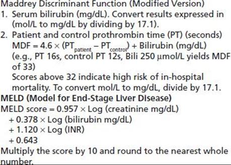
2. Alcohol-related hepatitis is classically defined by symptoms and signs of hepatitis in association with heavy alcohol use and often presents on a background of chronic liver disease. The occasional patient presents for the first time during a bout of acute alcohol-related hepatitis. Alcohol-related hepatitis presents as a spectrum of disease from mild to severe. Milder forms are most common, but severe cases, when present, carry a high short-term mortality of between 30% and 65% (88). Severe cases often present with evidence of advanced liver disease with cirrhosis and superimposed hepatic decompensation. Clinically, these patients complain of anorexia, nausea, and abdominal pain. Fever may be significant in the absence of any infective complications. They have evidence of inflammation and elevated liver enzymes (aspartate amino-transferase [AST] moderately elevated and AST–ALT ratio >2), a raised total bilirubin with jaundice, an elevated INR with bruising, encephalopathy, and often ascites. Neutrophilia is found in severe cases and, when present, should trigger a search for a septic focus, but it may simply reflect the alcohol-related liver inflammation. The severity of alcohol hepatitis can be assessed using an objective rating scale such as the Maddrey Discriminant Function (MDF) or the Model for End-Stage Liver Disease (MELD) score (89). These correlate closely with each other, and both give an indication of prognosis and may direct the use of specific treatments (Table 74-2). Patients may have few signs of chronic liver disease at first presentation, but a majority will have signs of cirrhosis at some stage of their progressive liver disorder. Unlike viral or toxic hepatitis, alcohol-related hepatitis evolves more slowly, and recovery may take many months after abstinence has been achieved.
TABLE 74-2 SCORES THAT ASSESS SHORT-TERM PROGNOSIS OF PATIENTS WITH ALCOHOLIC HEPATITIS
3. Alcohol-related cirrhosis typically presents with complications such as portal hypertension leading to variceal bleeding and/or ascites, liver failure, recurrent infections because of impaired immune function, and hepatocellular carcinoma (HCC). Occasionally, anorexia, nausea, weight loss, and general malaise are present. Alcohol-related cirrhosis is a recognized risk factor for HCC, but that risk may be less than originally thought to be the case (90). Screening for HCC is still recommended in cirrhotic patients, but the yield in pure alcohol-related disease is low (90,91).
Diagnosis and Assessment of Severity
The diagnosis of ALD rests on the history of prolonged heavy alcohol ingestion with a compatible clinical and laboratory picture. Clinical features of chronic liver disease include decreased muscle mass, palmar erythema, presence of spider nevi, hepatic lunules, loss of body hair in males and hirsutism in women, gynecomastia, splenomegaly, and caput medusa, indicating portal hypertension. In decompensated disease, there may be jaundice, encephalopathy with or without asterixis, petechial hemorrhage or ecchymoses, and ascites. Clinicians are increasingly aware of the entity of minimal hepatic encephalopathy, which alters higher center function without being associated with a metabolic flap (92). Liver enzyme tests are a sensitive marker for ALD; however, they are not specific as similar abnormalities may be observed in nonalcoholic steatohepatitis and in patients treated with medications such as anticonvulsants. The γ-glutamyl transpeptidase (γGT) level is almost always raised (93) and often exceeds 1,000 U/L in alcohol-related hepatitis. The transaminases are only moderately elevated. Levels above 500 U/L suggest an additional disorder such as acetaminophen ingestion, viral hepatitis, or liver ischemia.
The AST exceeds the alanine aminotransferase (ALT) level in most cases, and a ratio of 2:1 AST–ALT is typical. Possible explanations for this observation are that AST is a mitochondrial enzyme and alcoholic injury selectively injures mitochondria. In addition, AST is also found in other tissues subject to alcohol injury including skeletal muscle and heart. If the ALT exceeds the AST, chronic hepatitis C, acetaminophen ingestion, or other causes for hepatocellular injury should be considered.
As liver disease severity increases, falling albumin, rising bilirubin, and increasing serum globulin concentrations are seen. In cirrhosis and severe alcohol-related hepatitis, INR levels are prolonged, platelet counts fall, and there may be a macrocytic anemia with spur cells in the most severe cases. In liver failure from these conditions, blood glucose levels may fall due to loss of glycogen stores and failure of gluconeogenesis. Undiagnosed, this can be fatal.
During initial assessment for ALD, other contributing factors to developing chronic liver disease and cirrhosis should be routinely considered. Factors include ingestion of hepatotoxic drugs including herbal preparations and acetaminophen, diabetes mellitus and metabolic syndrome, hepatitis B and C infection, and iron overload. Additional investigations are restricted to atypical cases or those that fail to resolve with abstinence from alcohol. Other explanations for liver disease, including autoimmune hepatitis, Wilson disease, alpha-1-antitrypsin deficiency, and cholestatic liver disease including primary biliary cirrhosis, may need to be considered in specific cases.
Imaging of the abdomen with abdominal ultrasound, computed tomography scan, or magnetic resonance imaging has a role in establishing the presence of chronic liver disease and its complications (fatty change, portal hypertension, HCC); however, imaging is of limited value in establishing the etiology. Abdominal imaging will also allow exclusion of other pathologies that may explain abnormal liver function and enzyme tests. The role of elastography in the evaluation of liver fibrosis is being defined by many studies, and it may well prove to have a role in the assessment of patients with all forms of liver disease (94). The technology is still not approved by the FDA (95).
Once cirrhosis is suspected, referral for a diagnostic endoscopy is regarded as essential to document the presence or absence of varices. If they are present and meet appropriate requirements, prophylactic therapy is indicated (96).
Role of Liver Biopsy
Liver biopsy has been used for decades to provide information regarding the diagnosis and severity of liver disorders. In ALD, biopsy can be of use to find secondary coexisting pathology as well as establishing severity of liver disease (95). Nevertheless, the practice of biopsying the majority of patients with any form of significant liver injury has diminished owing to improvements in noninvasive imaging and diagnostic testing and as patients have become more resistant to the procedure knowing its complications and short falls. Biopsy is also subject to sampling error, and the use of the procedure is restricted now to those patients with complex or incompletely understood disease and in those being considered for liver transplantation (97,98). The morbidity of liver biopsy is 0.5%, and the mortality is 0.01% (99), and these risks generally outweigh the benefit of obtaining tissue pathology. Where clinical uncertainty persists, biopsy should always be considered. In life-threatening cases where urgent liver transplantation is being considered, vitamin K or fresh frozen plasma may be required, to reverse coagulopathy to accomplish biopsies. Transjugular biopsy is validated as a safe procedure in coagulopathic patients (100) but is not widely available and sometimes yields insufficient tissue for diagnosis.
It has been argued that demonstration of the severity of liver damage by biopsy might motivate ambivalent alcohol-dependent patients to adhere more closely to management protocols. However, this has never been validated by clinical studies, and routine use of biopsy could not be justified on this basis.
Treatment
Treatment of Causative Factors
The treatment of ALD rests upon avoidance of further alcohol consumption. Other interventions are reserved for those with particularly severe disease or who are unable to maintain abstinence. There is considerable evidence that survival is increased by maintaining abstinence (101,102). The improvement with abstinence is so consistent that the γGT falls with an apparent half-life of 26 days (103). Failure to do so suggests continuing alcohol consumption or occasionally another coexisting liver disease. Advanced cirrhosis does not resolve with abstinence, indicating that it is an irreversible lesion, but the activity is reduced. Many very ill patients make striking improvements, often returning to compensated cirrhosis. In practice, it is never too late to stop drinking.
The first issue is to define what level of alcohol consumption to recommend for the patient with liver disease. Those with true moderate to severe alcohol use disorder or severe liver disease should be given clear advice to remain abstinent long term. However, many patients have only minor abnormalities in liver enzyme tests without clinical or investigational evidence of cirrhosis. A typical recommendation is a 6-week period of abstinence followed by repeat liver tests. If these normalize and the patient wishes to resume drinking, consumption within recommended lower risk levels may be guided by the results of further liver tests in those who can control their alcohol use. Continuing follow-up in a primary care setting is important, as the major causes of death in mild ALD are extrahepatic problems related to alcohol abuse such as suicide. Another problem for the generalist physician is that many patients with alcohol-induced disorders decline referral to a specialist treatment service. This places a responsibility on internists and other primary care physicians to develop brief counseling skills including motivational interviewing. At a minimum, primary carers can provide feedback concerning medical progress, and this can influence drinking behavior.
Pharmacotherapies to address alcohol use disorders must be used with particular caution in the presence of serious liver disease. A recent study of the use of baclofen in the treatment of alcohol dependence in patients with cirrhosis has provided evidence of very positive benefit (104). Subsequent studies have not replicated this findings, which is unfortunate because of the potential benefit of the lack of liver toxicity of baclofen. Disulfiram is contraindicated in advanced liver disease owing to recognized hepatotoxicity. Another issue is the safety of acamprosate and naltrexone in patients with significant liver disease. Acamprosate does not accumulate even in severe liver disease, as the drug is excreted unchanged in the urine and is not metabolized. According to the manufacturers, acamprosate is contraindicated in severe decompensated (Childs C) liver disease and may exacerbate encephalopathy, but even in that setting, the risks of treatment should be balanced against the risks of continuing alcohol consumption, and there are no published reports of an adverse effect on liver function. Naltrexone is associated with dose-dependent hepatotoxicity (typically at supratherapeutic doses of 300 mg/d), but reactions are most unusual at the standard dose of 50 mg/d. In two studies, liver enzyme tests improved in alcoholics, with no cases of clinically evident hepatotoxicity, indicating the therapeutic effect to reduce alcohol consumption exceeded the potential hepatotoxic effect (105,106). Extended-release injectable naltrexone is a long-acting depot naltrexone product that has been shown to be effective for treatment of alcohol dependence, and a recent report did not detect any hepatotoxicity (107). Nalmefene is a second-generation orally active opiate receptor antagonist that has been reported to have similar efficacy to naltrexone in the treatment of alcohol dependence (108) without reported hepatotoxicity. It remains unapproved for use in the United States or Australia at this time. Clinicians working with alcohol-dependent patients are increasingly using these agents to reduce risks of return to drinking, and with careful monitoring of liver tests, these drugs can be safely used in many cirrhotic patients.
Managing Acute Alcoholic Hepatitis
Treatment of patients who present with alcohol-related hepatitis includes management of decompensated liver disease and the accompanying complications. Nutritional support with parenteral and enteral feeding improves nutritional status, but does not improve survival (109).
Selected cases of alcoholic hepatitis may respond to corticosteroids, but this remains controversial as individual studies and meta-analyses have shown different outcomes (110–112). Currently, the American Association for the Study of Liver Disease practice guidelines recommend prednisolone (40 mg/d for 28 days) for patients presenting with severe alcohol-related hepatitis (MDF >32; see Table 74-2) with or without hepatic encephalopathy and lacking contraindications to steroid use (88,95). Widespread use of corticosteroids has been limited by the knowledge that they may exacerbate sepsis, a common complication of severe liver disease. It is important to acknowledge that unusual infections may complicate alcohol-related disease, so specific tests may need to be requested in those in whom routine cultures fail to identify an infective agent. Infections such as tuberculosis and fungal infections including cryptococcosis need to be considered where patients fail to respond to standard therapy (113). Pentoxifylline (400 mg thrice daily), a phosphodiesterase inhibitor with many effects including decreasing serum levels of TNF-α, has also been reported to reduce the mortality of alcohol-related hepatitis, especially in patients with hepatorenal syndrome (114). Other agents have been trialed; however, none have shown any consistent survival benefit when compared with either placebo or corticosteroids. TNF-α is involved in alcoholic hepatitis, but treatment of this disease with inhibitors has not yielded any survival benefit despite promising early results (115). The trial using etanercept showed a worse 6-month survival compared with placebo (116).
Anabolic–androgenic steroids, which increase muscle mass in healthy subjects, did not show any survival benefit (117), nor have studies using antioxidants (vitamin E, silymarin) (118,119).
Treatment of Alcohol-Induced Cirrhosis
In cases where the disease is particularly severe or does not resolve with abstinence, several therapies have been trialed but have not shown mortality benefit. Propylthiouracil (120–122), colchicine (123,124), and oxandrolone (117,125) have been evaluated for the treatment of ALD, but subsequent studies failed to reproduce beneficial effects of these agents. Some agents that can be viewed as “super-nutrients” have been found to be effective in nonhuman primates. These include S-adenosylmethionine for the treatment of early aspects of alcohol-induced liver injury (126) and polyunsaturated lecithin for the prevention of fibrosis (127,128). There are some data showing that both agents are effective in humans. S-adenosylmethionine treatment led to a significantly lower mortality in patients with Childs class B cirrhosis (129); however, a more recent Cochrane Review did not show any benefit in all-cause mortality, liver-related mortality, or liver transplant (130). Polyenylphosphatidylcholine is derived from soybeans and comprises a mixture of polyunsaturated phosphatidylcholines, about half of which is dilinoleoylphosphatidylcholine. A large Veterans’ Affairs Cooperative Study evaluated this treatment but did not identify any delay in the progression to cirrhosis over the 2 years of the study, though in parallel, the participants substantially reduced their alcohol intake (131). New therapeutic targets derived from the advances in our understanding of the pathophysiology of ALD (132) include:
CXC chemokines (133,134) involved in neutrophil attraction to the liver in AH
IL-22-STAT3 pathway that controls bacterial infection homeostasis and tissue repair (135)
Complement activation that is involved in ethanol-induced liver disease in mice (136,137)
Probiotics and TLR4 antagonists that act to decrease gut microbial and LPS pathways (138,139)
Osteopontin, an extracellular matrix protein that was found to be up-regulated in patients with alcoholic hepatitis and correlates with alcohol-related liver disease severity (140,141)
Endocannabinoids (142)
Nostrin (nitric oxide synthase traffic inducer) that regulates nitric oxide synthesis (143)
At this stage of clinical practice, no new agents are available with a proven record in modifying alcohol-related liver disease (79,132,144).
Managing Complications of Cirrhosis
The management of the complications of cirrhosis, such as ascites and bleeding, lies outside the scope of this chapter. Patients who present with signs of hepatocellular insufficiency or portal hypertension should be evaluated by a gastroenterologist or hepatologist. Salt restriction helps reduce fluid retention, which presents as edema and ascites, whereas prophylactic management of portal hypertension with agents, which lower portal flow such as beta-blockers (propranolol) and long-acting nitrates or esophageal variceal banding, alters prognosis significantly. All patients presenting with ascites for the first time or representing with evidence of deteriorating clinical status should have a diagnostic ascites tap to exclude or diagnose spontaneous bacterial peritonitis (145). Once established, ascites is treated by salt restriction, spironolactone in doses from 50mg daily up to 400mg daily, and, if necessary, furosemide and bed rest. Higher doses of spironolactone may cause painful gynecomastia in men and limits the doses able to be given. In these cases, amiloride may provide a similar diuresis. Paracentesis is effective for diuretic-resistant cases and may be offered in an outpatient setting, but some data suggest that transjugular intrahepatic portal systemic shunting may be as, or more, effective (146). Severe recurrent ascites may respond to transjugular intrahepatic portasystemic shunting or surgical shunts, thus avoiding the risks of repeated paracenteses. Variceal hemorrhage is treated by transfusion, correction of coagulopathy, nitrate or octreotide infusions, and urgent endoscopic banding. After controlling bleeding, secondary prophylaxis with variceal banding and/or beta-blockers reduces the risk of rebleeding. Spontaneous peritonitis is treated by intravenous antibiotics and prophylactic norfloxacin until ascites has resolved. Hepatic encephalopathy may be alleviated by correction of precipitating factors and by decreasing colonic production and absorption of NH3 with lactulose, a nonabsorbable disaccharide that acidifies the colon content through fermentation. Dietary protein should be adjusted to avoid both deficiency and excess as nutritional deficiencies are very common in patients with advanced liver disease. Restriction of protein can impair hepatic recovery and may not necessarily improve encephalopathy (147).
Liver Transplant
Liver transplantation is now an accepted treatment option for individuals with advanced liver disease who have stopped drinking (148), but only 5% of patients with end-stage ALD receive transplants. The procedure remains controversial despite years of experience because of ethical concerns about allocation of precious donor livers to individuals with a (mis)perceived self-induced disease. It is unreasonably simplistic to regard ALD as a “self-induced disorder.” External factors such as family, peers, and society as a whole encourage the availability and use of alcohol. Genetic factors also contribute to the risk of alcohol use disorders, and understanding of this often relapsing condition remains very poorly developed (149). The 5-year survival after transplantation for ALD is comparable to that of nonalcoholics in series from the United States, Europe, and Australia (150,151). Whereas alcoholics may be at higher risk of some posttransplant problems including malignancies possibly related to smoking, there is evidence that the rate of rejection may be lower than for non-ALD (152). Resumption of alcohol consumption remains the major concern and occurs in between 30% and 50% of survivors (153,154). Of those who return to drinking, one-third develop life-threatening alcohol-related morbidity such as pancreatitis, recurrent ALD, and noncompliance with immunosuppression resulting in graft rejection (155). These outcomes are comparable to the posttransplantation recurrence rate of other liver diseases. The rate of recurrent, problematic alcohol consumption is lower than that observed after other treatments for alcohol dependence. This may be due to careful case selection for transplantation and/or the intensity of treatment by the transplant team. Vaillant (156) showed that the prognostic factors that generally predict a favorable outcome for alcoholism treatment are provided by the liver transplant process.
The ideal candidate for transplantation accepts the etiologic role of alcohol in his or her liver disease, has ceased drinking, and has strong family supports with a stable home, employment, and enthusiasm to resume interests. Psychosocial evaluation seeks to stratify patients by their risk of relapse for alcoholism. An objective and valid assessment method is highly desirable but has not been devised. The Michigan Alcoholism Prognosis Scale (157) did not predict postoperative drinking (158), possibly as only those with high scores are transplanted. Other prognostic scales have insufficient predictive value to justify rejection of an otherwise suitable candidate (159,160). Ideal candidates for transplant will have abstained on learning of the nature of their advanced liver disease, will have supportive family members available to assist their preparation for the transplant, and will have had no significant mental health issues other than their dependence on alcohol (159,161). Individual assessment remains critical, and while duration of abstinence pretransplant remains a poor predictor of outcome, most centers still aim for a minimum of 6 months abstinence before surgery.
Alcohol-related hepatitis is generally considered a contraindication to liver transplant due to the absence of 6 months of abstinence. In acute severe presentations in younger patients, transplant has been undertaken with outcomes matching those undertaken for non–alcohol-related disease (162). Some centers are reevaluating transplantation for alcohol-related hepatitis.
VIRAL HEPATITIS
Hepatitis A
Hepatitis A is an RNA virus that is transmitted by fecal– oral contamination. In underdeveloped countries, almost all children develop IgG antibodies by the age of 10, the acute infection resulting in only clinically inapparent or mild hepatitis. With improving hygiene, the seroprevalence has fallen so that adults in the developed world are now generally susceptible to hepatitis A. The clinical consequences of hepatitis A virus (HAV) infection become increasingly severe with advancing age so that hepatitis A is now less common but often more severe than in the past. The illness does not persist as a chronic infection. Parenteral transmission of hepatitis A is rare owing to the short period of viremia but has been described (163).
The prevalence of hepatitis A IgG antibodies is high among IDUs and prison inmates in California (164) and Australia (165). Hepatitis A correlates more closely with institutionalization than sharing of injecting equipment, and vaccination of seronegative prison entrants is undertaken in some jurisdictions.
Prevention of HAV infection is achieved by hygienic precautions to prevent fecal–oral contamination, administration of immunoglobulin to household contacts of cases, and active immunization to those at risk. Hepatitis vaccine is given as two injections by intramuscular injection and is safe and effective. Accepted indications for vaccination include occupational risk, travelers (especially those to high prevalence areas), men who have sex with men, and those with chronic liver disease. Vaccination has also been recommended for IDUs (166), but is not a routine practice in most clinics. HBV vaccination is a higher priority for this group (167).
Hepatitis B
Epidemiology
HBV is the most prevalent chronic viral infection of humans, and >350,000,000 people worldwide are infected. It is readily transmitted among IDUs. Serologic evidence of past hepatitis B infection increases in prevalence with the duration of injecting drug use. Sexual exposure and injecting drug use are now the commonest associations of hepatitis B infection acquired in adults in Western societies (168). In many Western countries, more new diagnoses of HBV occur in people who have migrated from countries of medium to high HBV prevalence than in those born in the country.
Transmission
HBV is highly infectious, and blood and bodily fluids are capable of transmitting the disease. Some fluids are more infectious than others with semen, vaginal fluid, and blood being most infectious. Reports of transmission from exposure to tears and saliva have been published, but the risk is low, and breast-feeding is not discouraged. In countries of high prevalence the majority of infections are transmitted during pregnancy or at delivery (termed vertical transmission) or in the first few years of life (horizontal transmission). Programs that have targeted babies born to HBV-positive mothers have reduced transmission rates, and now, universal vaccination against HBV in many countries is slowly reducing the burden of chronic HBV (CHBV) infection.
Virology
The HBV is a DNA virus with eight genotypes and multiple subtypes. Multiple mutant forms of the virus exist, and the contribution of these to infections worldwide is still being defined. A precore mutant form, which does not produce e antigen and thus leads to no e antibody production, frequently appears as viral clearance is occurring. This form of the virus can cause infection in its own right, emphasizing the need for a full serologic evaluation of new patients. Mutations involving the surface antigen of the virus lead to forms of the virus, which can be more difficult to detect by standard assays (169). Increasingly, it is evident that many infections with the HBV virus are not detected by standard assays and these infections are known as occult HBV infection. Hepatitis C virus (HCV), for example, suppresses HBV replication and may lead to a failure to recognize dual virus infection (170). Infection with the virus is associated with the production of a nuclear covalently closed circular DNA, a template of the virus that allows infection to persist even in those who apparently clear the infection and develop anti-HBs, anti-HBc, and anti-HBe antibodies.
Outcome of Infection
Vertical transmission of HBV or horizontal transmission in early childhood results in a high percentage of CHBV infection. Over 90% of those infected early in life remain infected as the immune system fails to clear the virus. On the other hand, infection in adult life is associated with an effective immune response, and clearance of serologic markers occurs in 90% to 95% of cases.
Clinical Manifestations of Acute Hepatitis B
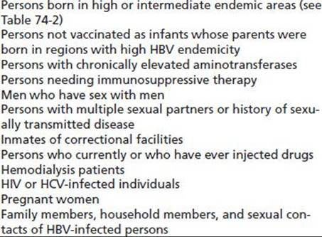
Testing for HBsAg, anti-HBs, and anti-HBc should be performed, and seronegative persons who remain at risk of infection should be vaccinated (Table 74-3) (171). Acute and chronic hepatitis B are diagnosed by serologic tests (HBsAg, anti-HBs, anti-HBc, HBeAg, and anti-HBe) and HBV DNA testing. In combination, these assays allow one to define the phase of the infection, risk of HCC, and need for treatment.
TABLE 74-3 RECOMMENDATIONS FOR WHO SHOULD BE TESTED FOR HBV INFECTION
The incubation period of HBV infection is 6 weeks to 6 months, the period not obviously influenced by the means of infection. Acute hepatitis B may be preceded by a transient serum sickness prodrome, with polyarthralgia, fever, malaise, urticaria, and proteinuria (172). The acute illness is characterized by anorexia, nausea, and sometimes vomiting with malaise, jaundice, pale stools, and dark urine. The infection is frequently subclinical.
Chronic Hepatitis B
Chronic hepatitis B infection is associated with progressive inflammation, which leads to the histologic changes of hepatic fibrosis, cirrhosis, and HCC and the clinical presentation with hepatic failure and portal hypertension in a significant minority. Disease severity is increased by a variety of factors including duration of infection, male sex, ethnic origin, coinfection with other hepatitis viruses or HIV, alcohol ingestion or illicit drug use, and obesity and diabetes. CHBV infection is a more aggressive liver disease than is chronic HCV infection, and 25% or more with CHBV will progress to liver failure or HCC in a 10-year period.
Liver injury is not always apparent in the chronically infected individual, and the disease is now recognized to pass through a series of phases (Fig. 74-2). Inflammation, when present, is the result of a cell-mediated response to infected hepatocytes. In chronic disease, a series of hepatitis flares in the immune clearance or immune escape phases may precede viral clearance and recovery. These flares vary in severity from subclinical through to life threatening.
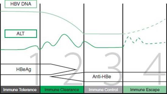
FIGURE 74-2 The four stages of chronic hepatitis B infection. (Adapted from B positive—all you want to know about hepatitis B: a guide for primary care providers. Australian Society for HIV Medicine, 2010.)
Patients with CHBV should be assessed and the phase of the disease and the HBV DNA level documented. Patients in the immune clearance or immune escape phases of the disease (with abnormal ALT levels) or those with clinical evidence of liver disease should be referred for specialist assessment and consideration of antiviral therapy. Patients with CHBV should be offered regular screening for HCC, as the risk is increased in all infected patients but especially in those with cirrhosis. Screening protocols have varied, but current evidence supports six monthly alpha-fetoprotein levels and abdominal ultrasound.
When to Treat
Any decision to treat HBV must involve patient and health care workers as treatment is usually a life time commitment with current medications. Generally, patients in the immune clearance and immune escape phases of infection are candidates for therapy. Treatment is also now considered for patients with high HBV DNA as a strategy to reduce risk of HCC. Treatment decisions are often quite complex, and patients are best managed by referral to a specialist unit for initial assessment and treatment planning. Ongoing prescribing of antiviral agents is now undertaken by primary care physicians in a number of jurisdictions.
A liver biopsy is no longer regarded as a compulsory requirement preceding treatment initiation. Ultrasound elastography, known as FibroScan, is increasingly available in some centers to provide noninvasive estimation of fibrosis and to reduce the need for biopsy.
Current Recommended First-Line Therapies
The current first-line therapies for treatment-naïve patients are the following:
■ Entecavir (ETV)—a nucleoside analogue with a high barrier to resistance (<2% at 6 years in treatment-naïve patients). Lamivudine-resistant strains develop resistance to entecavir more readily, and the drug should not be the first choice in this group of patients.
■ Tenofovir (TDF)—a nucleotide analogue with a high barrier to resistance (<1% at 5 years). This drug should be the first choice in those with lamivudine-resistant strains. This is the drug of choice in pregnancy.
Pegylated interferon (PEG-IFN) does have significant side effects but has a defined 1-year duration of therapy and the possibility of HBsAg clearance. PEG-IFN can be useful in selected younger patients, particularly women who are intending to get pregnant in the future. A response is more likely in HBeAg-positive patients with high ALT and low HBV DNA viral load.
Treatment of active chronic hepatitis B is of value in those patients with active liver inflammation and in those with very high viral load. Treatment with pegylated interferon-α (IFN-α) leads to seroconversion from eAg to eAb status in 30% to 40% compared to spontaneous seroconversion of 15%. The eAg to eAb seroconversion is associated with suppression of viral replication and decreasing hepatic inflammation (173). There is a small but continuing success rate in clearing the virus with loss of HBsAg, which rises from 5% in the first year to >10% over a 5-year follow-up. Treatment can be demonstrated to be cost-effective (174). Loss of HBeAg has been associated with improvements in liver histology and clinical outcome, but patients remain infected and infectious. Pegylated IFN-α should not be used in patients with HBV-related cirrhosis, as it may induce a flare of hepatitis and lead to hepatic decompensation. Such patients should be assessed for liver transplantation.
Oral antiviral agents are now more frequently used than interferon products because of their greater patient acceptability and high viral resistance barriers. Once commenced, these agents need to be continued for prolonged periods as dictated by the viral response to the drugs. Both entecavir and tenofovir produce a rapid and marked fall in viral load in the majority of patients. Viral clearance from serum is the goal of therapy with subsequent seroconversion from eAg positivity to anti-HBe status. In a small percentage, sAg loss occurs. Attempts to stop treatment are appropriate when the patient has had 12 months of normal liver enzymes after seroconversion to anti-HBe status. In those fortunate to have HBsAg clearance, antivirals may be ceased. Patients with HBV requiring treatment are best managed by referral to units with a special interest in HBV management.
Immunosuppressive drugs such as cancer chemotherapies are associated with increased HBV viral load and flares of hepatitis such that prophylactic treatment is recommended.
Hepatitis C Virus
Epidemiology
HCV infection is a major public health problem with over 180 million people infected worldwide (175) and approximately 3.9 million people in the United States (176). HCV remains the leading indication for liver transplantation, and it is projected that the number of people with advanced liver disease and associated HCC will continue to increase for the next two decades.
Transmission
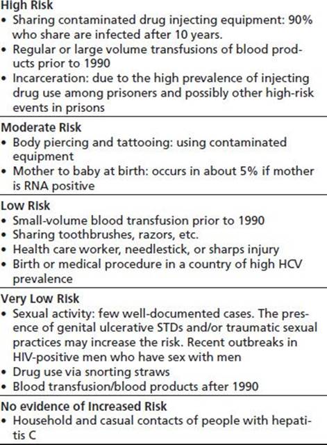
HCV is a blood-borne virus, and in the United States, Europe, and Australia, the most common risk factor for transmission of hepatitis C infection is IDU (Table 74-4), which accounts for the bulk of incident cases (91% in Australia) (177). HCV prevalence is strongly associated with duration of injecting with an incidence of approximately 20% for each year of IDU (178). Most regular IDUs are infected with HCV, and the incidence is 100% in some populations. Measures to limit the spread of this infection appear to be making only a modest impact, and one study demonstrated an incidence of 45 per 100 life years in new injecting users in NSW Australia (179,180). The continuing high incidence appears to be related to the continuing high prevalence of sharing some component of injecting equipment including mixing spoons, filters, swabs, or tourniquets. The wider implementation of infection control procedures such as needle–syringe programs has been associated with a variable reduction of HCV transmission in new recruits to injecting (179,181,182).
TABLE 74-4 INTERPRETATION OF SEROLOGIC MARKERS FOR VIRAL HEPATITIS

HAV, hepatitis A virus; HBV, hepatitis B virus; HCV, hepatitis C virus; PCR, polymerase chain reaction (sensitive molecular diagnostic procedure that can detect minute amounts of specific DNA or RNA).
Sexual Transmission
These rates are generally thought to be very low except among HIV-positive men who have sex with men. Outbreaks of HCV transmission have been reported in Europe, the United Kingdom, the United States, and Australia among highly promiscuous HIV-positive men who have sex with men (183). Heterosexual transmission is rare. An Italian study of the male partners of women infected with contaminated anti-D immunoglobulin showed no evidence of transmission over a combined follow-up period of 862 years (184,185). Similar findings were found in two other studies (186,187). Alter et al. (188) had previously reported that high-risk sexual behavior was a significant risk factor for hepatitis C in the United States based on analysis of the third National Health and Nutrition Examination Survey (NHANES III). This conclusion was challenged (189) because IDU, the major risk factor for hepatitis C infection, was not included in NHANES III so that the effect of other factors such as sexual behavior could have been confounded by IDU (190). Murphy et al. (191) subsequently confirmed that the increased risk of hepatitis C associated with high-risk sexual practices disappeared after adjusting for IDU.
Vertical transmission occurs, and the rate is approximately 5% overall. Untreated HIV increases the risk of transmission. HCV RNA positivity determines transmission risk, and if HCV RNA is undetectable at birth, transmission does not occur (192–194). Hepatitis C RNA has been found in breast milk, but there is no evidence for transmission from breast-feeding, and breast-feeding can be recommended provided the nipple is not cracked. HCV antibody testing of infants should be deferred for 18 months as maternal HCV Ab is detectable until this time in healthy babies. HCV RNA testing at 3 months provides the earliest indication of transmission, but occasionally, babies show a transient HCV viremia, which is cleared without a persisting HCV infection. The majority of infants at risk are not tested in accordance with these guidelines.
Blood Products
The introduction of screening blood donors for HCV infection has markedly reduced the risk of transmission from the use of blood products. Where this is not routinely undertaken, transmission risk remains significant (195).
Occupational and Nosocomial Transmission
Health care and laboratory staff handling blood and blood products are at risk of contracting hepatitis C. Estimates for the risk of transmission from an HCV RNA positive needle-stick injury range from 0% to 10% (185,193). Nosocomial transmission of hepatitis C has been reported rarely in a variety of hospital settings, for example, in plasmapheresis units (196), in a hematology ward (197), in hemodialysis units (198,199), after colonoscopy (200), and after cardio-thoracic surgery (201).
Tattooing
Several studies have demonstrated an association between tattooing and hepatitis C infection. In an Australian study of blood donors, the independent relative risk associated with a history of tattooing was 27 (177). Infection control guidelines for tattooists have been introduced in recent years, but the possibility of hepatitis C transmission continues where these guidelines are not followed.
Hepatitis C in Special Populations
Prisoners
The prevalence of hepatitis C in prisoners is high, largely due to the high proportion of IDUs. In Australia, almost 40% of prison entrants are HCV antibody–positive rising to 65% among prisoners reporting a history of IDU (202,203). Imprisonment has been reported to be associated with hepatitis C even after adjusting for IDU (178,191). It has been suggested that other modes of transmission may occur in prisons owing to high background prevalence with poor hygiene and frequent physical violence (204).
The incidence of HCV among uninfected prisoners is also high (205). Measures that may limit the spread of HCV in the community are frequently not available to prison inmates, including methadone maintenance and needle– syringe programs. Tattooing using unsterilized equipment may also play a role (206).
People Born in Countries of High Hepatitis C Prevalence
In Mediterranean, Eastern European, Asian, South American, and African countries, the prevalence of hepatitis C is much higher than in the United States. The prevalence in Egypt for adults is 20%, making this a major community epidemic. The Egyptian hepatitis C epidemic has been related to mass inoculations for schistosomiasis (207).
Virology
HCV, identified in 1989, is an RNA virus from the flavivirus family. It has a single open-reading frame that generates a large viral polyprotein, which is ligated by viral polymerases to produce structural and nonstructural viral proteins. HCV can now be studied in in vitro and animal models, and this has increased understanding of the infectivity and pathobiology of the virus. HCV antibodies in the blood reliably indicate exposure to the virus, but HCV RNA testing is required to identify ongoing infection. The protective significance of HCV antibodies, produced as a response to infection, is limited. Studies in active IDUs provide variable results, some suggesting they are at reduced risk of reinfection and others showing an increased risk of reinfection when HCV antibodies are present (208–210).
There are six genotypes of the virus. Genotypes are further divided into subtypes (e.g., a, b, c). The most common genotypes in the United States are types 1 and 3. Reinfection after clearance and coinfection with more than one genotype can occur. Genotypes continue to identify treatment responsiveness, but some new agents currently in Phases 2 and 3 studies are proving able to eradicate all genotypes.
Outcome of Infection
Unlike HBV, infection with HCV at any age is associated with a high rate of chronic infection. At most, only 30% to 40% of patients clear the infection in the first 12 months of the disease. Interestingly, some babies born to HCV-positive mothers do demonstrate a transient viremia, which clears without the production of HCV Ab. The significance of this is the focus of research (211).
Clinical Manifestation
Primary Infection
Primary infection with HCV is typically subclinical, but mild hepatitis may occur. Fulminant hepatitis is rare. Peak viremia occurs in the preacute or early in the acute phase (weeks 2 to 3), and antibodies appear as early as 4 weeks (average 6 to 8 weeks). Clinically evident hepatitis reflects a significant immune response to the virus and may be associated with a higher rate of viral clearance than subclinical infection. A number of exposed seronegative individuals have evidence of T-cell immunity to HCV, indicating that the virus can be eliminated without detectable antibodies (212).
Chronic Infection
Between 60% and 75% of patients infected with hepatitis C will fail to clear the virus and become chronically infected (Fig. 74-3). Liver injury predominantly results from immune damage to infected liver cells. After an average of 20 years, 5% to 10% of people will have developed cirrhosis, rising to 20% after 40 years. Progression to cirrhosis is associated with duration of disease, age greater than 40 at the time of infection, average alcohol consumption of more than 50 g/d, coinfection with HBV and/or HIV, and male gender. The route of transmission or viral factors such as genotype or viral titer do not appear to play a role (213). The vast majority of HCV-infected individuals will not die of their HCV disease.

FIGURE 74-3 The natural history of HCV infection. (Adapted from Di Bisceglie AM. Natural history of hepatitis C: its impact on clinical management. Hepatology 2000;31:1014–1018.)
Symptoms of chronic hepatitis C without cirrhosis do not correlate well with disease activity or severity, but they can have a major impact on quality of life. The mechanisms leading to the widely variable symptomatology of HCV infection are poorly understood. Attempts to understand these symptom complexes have led to “symptom clusters” being defined (214). Some neurologic symptoms such as fatigue and altered mood may reflect the presence of the HCV in the CNS (215).
Diagnosis
The measurement of HCV antibodies is the most appropriate test for screening populations for HCV exposure (see Table 74-4). A positive HCV Ab result should be followed by HCV RNA testing. A positive hepatitis C RNA test indicates the presence of active infection, while a negative test in people with risk factors and positive antibody indicates probable clearance of HCV infection. HCV RNA analysis is particularly useful to assess the status of HCV antibody– positive patients with normal liver enzymes. Approximately 50% of these patients are HCV RNA negative. A negative HCV RNA in this context should be repeated 3 to 6 months later, and, if repeatedly negative, the patient can be reassured the virus has been cleared. A majority of hepatitis C antibody–positive patients with abnormal liver enzymes have detectable levels of hepatitis C RNA in their blood.
Hepatitis C genotyping and viral load determination should be carried out on all patients considering antiviral therapy for HCV infection. Both of these measures continue to provide information about the likely outcome of therapy and thus assist patient and doctor in determining whether to proceed with treatment. Genotyping and subtyping may be useful in some forensic situations.
Management Issues
Access to Health Care and Hepatitis C Information for People Who Inject Drugs
People who inject drugs (PWIDs) often lack access to health care for many complex reasons (216). In dealing with a medical problem such as HCV infection, it is important to provide culturally appropriate written material that matches the educational level of the patient, as many cannot discuss their illness with others. Specialized outreach clinics have been established in needle–syringe services, in opioid treatment programs, and in prisons (217). These provide diagnostic evaluation and build a therapeutic relationship to facilitate referral for full evaluation for antiviral therapy along with other substance abuse treatment. Primary care physicians or other health care workers can also engage HCV-infected patients, and clinical guidelines can assist them to provide appropriate management.
Pre- and Posttest Counseling Issues
A diagnosis of hepatitis C often engenders a high level of anxiety that can be exacerbated by misinformation. Adequate time and privacy should be set aside for discussing HCV testing and for obtaining informed consent for the testing process. The results of hepatitis C testing should be given in person, and in doing this, attention should be given to the natural history of the disease, the symptomatology, and privacy issues. Accurate, nonjudgmental language combined with a sincere concern for the patient’s welfare helps to build the patient’s trust. Clarify the meaning of any colloquial, subcultural terms. Patients are fearful of transmitting hepatitis C to their partners, household contacts, and their children. They can be reassured that the risks are minimal, but contact testing should be offered. Full explanations about the advantages and limitations of antiviral therapy allow the patient to make an informed choice about treatment options. It often takes several consultations to be certain that the patient and their close family and friends fully understand the situation.
Assessing the Severity of the Disease
Symptoms, including lethargy, do not correlate with the severity of liver disease. While the classical signs of advanced liver disease, spider nevi, bruises, jaundice, ascites, hepatic lunules, hepatic palms, and hepatic flap are commonly seen in cirrhosis, they are not always present. Plasma ALT is the best laboratory indicator of active viral hepatitis, but the level commonly fluctuates and does not correlate well with the stage of liver disease. A normal ALT level does not exclude active infection or cirrhosis. Patients with normal ALT levels and those who decline treatment may be monitored in primary care settings by ALT levels two to three times per year and referred if the ALT levels rise. Liver biopsy is no longer considered essential before commencing treatment for HCV infection, but, in certain clinical situations, it remains the best investigation for assessing the extent of fibrosis and the likelihood of serious long-term consequences. Newer approaches to defining the extent of hepatic fibrosis using blood tests or devices such as the FibroScan are reducing the need for biopsy. FibroScan is not approved by the FDA at this stage, but an increasing literature demonstrates the value of this noninvasive assessment of liver elasticity in assessing patients with HCV and HBV infections pre- and during treatment (218–220).
Hepatitis A and B Vaccination
When there is no evidence of immunity, vaccination is indicated in those at risk of infection to reduce the risk of further liver injury. Chronic coinfection with multiple hepatitis viruses is associated with accelerated progression to cirrhosis (221). Hepatitis B vaccination should be offered, and patients with hepatitis C respond well, albeit with lower titers, compared to uninfected controls.
Alcohol
Alcohol interacts adversely with chronic hepatitis C in several ways (222). There is now a consensus that daily consumption of alcohol above 40 g has an additive effect on liver inflammation and accelerates the progression of hepatic fibrosis (223). Heavy alcohol use is also associated with increased viral load (224), reduced adherence to therapy (225), increased risk of progression to HCC, and exacerbation of the skin lesions of porphyria cutanea tarda. However, the response to treatment was not reduced in drinkers provided they completed treatment (225).
There is no clear evidence concerning adverse effects of moderate levels of alcohol consumption on people with chronic hepatitis C. The National Institutes of Health (NIH) consensus conference in 1997 recommended a maximum of 10 g/d for all people with chronic hepatitis C and abstinence for those with significant disease and those contemplating antiviral therapy (175,226).
Dietary Guidelines
There is no published evidence to support any specific diet in unselected people with hepatitis C. Hepatic steatosis is a feature of hepatitis C, and obesity and type 2 diabetes mellitus are associated with hepatitis C and accelerated progression of fibrosis (227,228). The potential for dietary interventions in selected subjects to control the activity of hepatitis is under investigation. Vitamin D deficiency is associated with increased fibrotic change in the liver in HCV-infected individuals and is also linked to poorer response to antiviral therapy (229). Caffeine consumption has no adverse effects on liver disease in this population, and it may in fact be associated with milder liver injury (230).
Management of Risk Factors
The presence of HCV infection may increase motivation to participate in alcohol and/or drug dependence treatment, particularly if the patient is seeking antiviral therapy. Avoidance of injecting drug use is the preferred option, but it may not be the choice of the patient. Evidence-based harm reduction and abstinence-based treatments should be offered, as described elsewhere in this volume.
Antiviral Treatment
Treatment of HCV infection is indicated for those with progressive liver disease and also for those who wish to clear the infection even though liver disease is not severe (175,231). The main goal of antiviral therapy is sustained virologic response (SVR), defined as HCV RNA clearance persisting 6 months after treatment completion. Some new guidelines suggest this can be defined at 3 months after treatment completion. Individuals with SVR generally remain PCR negative for life. Several studies have found significant improvements in general health and specific hepatitis C–related symptoms in patients who achieve a sustained response to antiviral therapy (232,233). Those who achieve a sustained response and have no cirrhosis should expect to remain free of cirrhosis and its complications. As in other liver diseases, some patients with cirrhosis who clear the virus may experience reversal of cirrhosis. The risk of HCC in HCV-infected patients appears to be confined to those with cirrhosis or advanced fibrosis.
Recent Developments
The combination of pegylated IFN-α 2a or IFN-α 2b and ribavirin has been the standard therapy for HCV for a decade. This treatment has offered a 45% to 50% SVR for genotype 1 and 4 infections and a 75% to 85% SVR for genotypes 2 and 3 infections. Treatment does have a significant side effect profile, but most patients (90%) do complete the 24-or 48-week course required (234,235). Treatment options for HCV are changing rapidly. Two new drugs (boceprevir, telaprevir) have recently been approved in the United States, United Kingdom, a number of European countries, and Australia for use with interferon and ribavirin in the treatment of patients with HCV genotype 1 infection (236). These drugs are protease inhibitors, acting directly on viral replication. Used alone, they cause a rapid drop in viral load, but viral resistance rapidly develops. Either drug added to standard therapy in genotype 1 patients will increase SVR rates to 65% to 75% with the added advantage of a shorter (24 weeks) course of therapy for many (237–240). Many new oral agents are at various stages of clinical development and in late 2013 the FDA approved both simeprevir and sofosbuvir for use in combination with interferon and ribavirin for treating genotype 1 patients. Many additional agents including daclatasvir and asuneprevir are undergoing clinical development. Several small trials of interferon-free regimens report SVR rates over 90% with well tolerated 12-week treatment. It is expected that within ten years HCV treatment with oral agents will be widely available with high efficacy and favourable side effect profiles. At this stage of HCV treatment evolution, it is most appropriate to refer patients with HCV and abnormal liver tests or signs of chronic liver disease to specialist centers for assessment and advice on appropriate treatment options.
IL-28B and HCV Treatment Responses
A genetic polymorphism, found three kilobases (3 kb) upstream of IL-28B on chromosome 19, encoding interferon- λ-3 (IFN-λ-3), has been shown to identify those who will have improved viral clearance rates (SVR) with pegylated interferon and ribavirin treatment in HCV genotype 1 patients. Within the IL-28B polymorphism, the C/C genotype is associated with a twofold greater rate of SVR than the T/T genotype. As the C/C genotype is present in greater numbers in those with European compared to African American ancestry, this offers an explanation for the differing response rates between these two populations (302,303).
In addition to improved SVR rates, the IL-28B C/C genotype has been found in greater frequency in those who spontaneously cleared HCV infection compared to those with persistent infection (229,242,243). The effectiveness of newer direct-acting antiviral (DAA) combinations without interferon may be less influenced by IL-28B genotypes, suggesting this test will less clinically significant in the future.
Treatment of Special Groups
Human Immunodeficiency Virus Coinfection
A growing number of countries are reporting significant numbers of patients with HIV/HCV and HIV/HBV coinfection reflecting the different risk factors for HCV acquisition in these countries (Table 74-5). It also reflects the fact that PWIDs in countries where there are no needle exchange programs run a high risk of acquiring HIV, which is more prevalent in the injecting communities there. The progression of chronic hepatitis C and B is accelerated in HIV coinfected patients. In patients with very low CD4 counts and clinical AIDS, liver disease is often inactive, and as antiretroviral therapy leads to immune reconstitution, liver disease can become more active. In rare instances, the flare of hepatitis may lead to fulminant liver failure (244–246). Treatment of hepatitis C and hepatitis B may be indicated in patients with early HIV infection and those stable on HAART, but the response rates reported to date have been lower than in monoinfected groups. Consideration must be given to possible drug interactions in those who are on HAART as the increasing complexity of drug schedules now leads to altered drug metabolism and a greater chance of adverse drug reactions. It is of importance to recognize that as HIV treatments have become more effective, death rates from HIV are falling, and death from liver disease is increasing in HIV-affected communities (247).
TABLE 74-5 RISK FACTORS FOR HEPATITIS C
Patients with Compensated Cirrhosis
Patients with compensated cirrhosis may be treated (248). These patients are more likely to require periods of dose modification as the bone marrow suppression caused by interferon and the anemia of ribavirin use are less able to be compensated for in this group. Decompensated cirrhosis remains a contraindication to treatment, and deaths during treatment with current triple treatment regimens have been reported: This group should be managed within a liver transplant center. There is some evidence that treatment reduces the risk of HCC and decompensation, but these are subject to ongoing trials.
Persistently Normal Aminotransferases
Patients who are HCV RNA positive and have persistently normal aminotransferase levels generally have mild disease (249). A patient requesting treatment to clear the virus and reduce risk of transmission should not be denied access to therapy, and outcomes are the same as for people with abnormal liver tests (250). Most patients can be encouraged not to undergo treatment at this stage, but they should be followed up every 4 to 6 months and treated if the ALT becomes abnormal or if signs of progressive liver disease appear.
Patients with Ongoing Substance Abuse
In 1997, the NIH consensus meeting recommended against treating patients with ongoing substance abuse until substance abuse stabilized (175). A growing number of reports demonstrate that those who continue to use can be treated if they commit to the treatment schedule of visits with outcomes equal to those in non–drug-using populations. It has to be stressed that these reports do select patients carefully, and it remains appropriate to encourage those who are actively using illicit drugs on a daily basis to stabilize their use prior to seeking HCV treatment (251,252). There is no clinical evidence that methadone maintenance therapy impairs treatment response, and methadone or buprenorphine is encouraged, when indicated, if hepatitis C treatment is contemplated (253). Most PWIDs provided all the information about HCV treatment choose to defer treatment until they have stabilized their drug use and social situations.
Side Effects of Therapy
Treatment for HCV is associated with a variety of symptoms that may lead to a need to reduce drug dosages. Vast experience now exists in all treatment centers, and over 90% of patients commencing therapy with pegylated interferon and ribavirin will complete the course. Newer agents add to the side effect profile and drug interaction complexity, and while experience is being gained with these drugs (See New Anti-Hepatitis C Virus Agents), treatments should be undertaken by those with skills in the area (254).
Contraindications for Treatment
Decompensated cirrhosis, pregnancy, lactation, active psychiatric illness, and those who drink more than seven standard drinks a week are at higher risk of side effects and lower chance of completing treatment and are generally not treated. Depression may worsen during therapy, and suicide has been reported. Careful psychiatric assessment and ongoing care may be required (255). Contraindications to ribavirin include end-stage renal failure due to drug accumulation, chronic anemias, a history of cardiovascular dysfunction, and inadequate contraception.
Other Treatments
Alternative medications have been used by a high percentage of HCV-infected patients. Studies in the past 5 years have shown that silymarin/silibinin in particular may have specific antiviral activity at several levels (256–262). The role of any of the preparations containing silymarin/silibinin is still debated, but increasing evidence is accumulating to support the use of high-dose oral or IV preparations of silibinin in selected patients. Combination products can lower ALT levels while the product is used, and this may benefit nonresponders (256–264).
Waiting for New Anti-Hepatitis C Virus Agents
There has been an explosion of studies of new DAAs in the treatment of HCV infection (see 'recent developments' above). The hope for shorter course, more effective, pan genotype therapies is likely to be achieved in the coming decade. Results are exciting, but these new regimens are not yet widely available. The patient and clinician face a dilemma whether to treat with currently available but unappealing therapy or wait for newer options. Many patients will be best advised to:
Monitor liver tests.
Monitor liver fibrosis using using a non-invasive technology such as fibroscan or fibrotest with liver imaging if advanced disease is suspected.
Assess and manage relevant comorbidity (such as substance use and metabolic syndrome)
Seek advice from a specialist HCV unit.
Commence treatment with current agents when the severity of liver disease or rate of fibrosis progression dictates the need (241,265–267).
Treatment of Advanced Hepatitis C
HCV cirrhosis progresses to HCC at an annual rate of 3% to 5%, and hepatitis C is among the commonest underlying associations of HCC. Once hepatic decompensation occurs, the 5-year survival falls to 50%, and transplantation should be considered rather than antiviral treatment.
Hepatocellular Carcinoma
Small primary liver cancers can be resected or treated by local therapies. Cirrhotic patients with HCC are considered for transplantation if there are fewer than three tumor nodules smaller than 3 cm or a single nodule less than 5 cm with no extrahepatic spread or vascular invasion. It is currently recommended that such patients undergo six monthly screening with upper abdominal ultrasound and serum alpha-fetoprotein. If abnormalities are found, more extensive evaluation should be undertaken in a specialist liver center.
Liver Transplantation
Hepatitis C is now the leading indication for liver transplantation, and the numbers are expected to rise further during the next decade. Patients with cirrhosis should be considered for transplantation if they develop major complications of their cirrhosis indicating a life expectancy of 1 to 2 years without transplantation. The 3-year post-transplantation survival is 84%, equivalent to survival in patients transplanted with other forms of liver disease (268). Before transplantation, patients should be informed of the high risk of hepatitis C recurrence and its potential consequences, including a 10% risk of recurrent cirrhosis at 5 years. Methadone maintenance is no longer considered an absolute contraindication for transplantation (269–271), but careful individualized assessment is required.
Hepatitis D
The delta agent is a viral RNA particle that cannot replicate without coinfection with hepatitis B. It requires the sAg of HBV to allow entry to the hepatocyte. Outbreaks of delta virus coinfection with hepatitis B have occurred among IDUs and were associated with high mortality. Control of hepatitis B by vaccination will limit the spread of HDV. The diagnosis is by rising titers of IgG antibody or IgM antibody (see Table 74-4). HDV RNA can be assayed, and it may be useful in some circumstances to identify current infection. Delta infection should be considered in any HBV-positive patient with relapse. HBV and delta hepatitis coinfection responds poorly to interferon treatment. Addressing the primary HBV infection with entecavir or tenofovir is appropriate and will improve outcome in most of patients.
Bacterial and Fungal Infections Associated with Injecting Drug Use
A wide array of infections may occur in the IDU, and these may involve the liver (see Chapter 79).
Hepatic Toxicity Associated with Injecting Drug Use
Cocaine Hepatitis
Cocaine hepatic injury appears to be uncommon in humans (272), but about 1% of fulminant hepatic failure is due to cocaine in several series. Most cases occur in association with systemic heat shock–like features of cocaine toxicity such as hyperthermia, rhabdomyolysis, hypoxia, and hypotension (273–275). In experimental animals, cocaine hepatotoxicity is readily demonstrated and is both time and dose dependent (276). It is important to remember that HCV and HBV infections are more likely to explain moderate abnormalities of liver-related blood tests in regular cocaine users.
The clinical presentation of cocaine hepatitis is characterized by a marked increase in serum aminotransferase activities beginning within a few hours of drug ingestion associated with the systemic features of cocaine toxicity listed earlier. Rhabdomyolysis may account for some of the increase in transaminases, as AST and ALT are both present in muscle. The liver biopsy shows coagulative hepatic necrosis typically in a centrilobular distribution, extending to panlobular necrosis in extreme cases. Micro- and macrovesicular steatosis may be present, consistent with involvement of mitochondria in hepatic injury.
The mechanism of hepatic injury is thought to involve hepatic ischemia and/or toxic oxidative metabolites. Hepatic ischemia is a likely mechanism as cocaine is a powerful vasoconstrictor resulting in organ damage from impaired systemic perfusion. The pericentral location of hepatic necrosis supports this concept. Evidence supporting a role for toxic oxidative metabolites comes from experimental animal models in which hepatotoxicity has been prevented by inhibition of enzymes metabolizing cocaine or in animals genetically unable to metabolize cocaine (277). Usually, more than 90% of cocaine is hydrolyzed by plasma pseudocholinesterase (278). The remainder is metabolized by cytochrome P-450 isoenzymes, including CYP 3A1. The hepatotoxic metabolite of cocaine has not been identified with certainty, but the oxidative metabolites of N-hydroxynorcocaine may generate reactive alkylating species. The severity of liver toxicity is correlated to the extent of cocaine oxidation by hepatic cytochrome P-450, which is increased by inducers of the cytochrome P-450 system such as phenobarbital and ethanol and reduced by P-450 inhibitors such as cimetidine. In the mouse, chronic cocaine administration induces P-450 3A and may increase the risk of hepatotoxicity (279). Inhibition or deficiency of pseudocholinesterase increases hepatotoxicity by diverting drug toward the P-450 pathway, and induction of pseudo-cholinesterase may lessen toxicity.
Cocaine is commonly taken with alcohol, and hepatic carboxylesterase generates ethylcocaine (cocaethylene) (280). The same hepatic enzyme also contributes to the nonoxidative metabolism of ethanol to fatty acid ethyl esters (281). Ethylcocaine has similar pharmacologic effects to cocaine but a longer half-life. It may accumulate to higher levels in tissues than cocaine and may be more toxic than cocaine as evidenced by a lower LD50 (282). Nonetheless, a large clinical series found no increase in liver disease among alcoholics using cocaine compared to those not using cocaine (283).
Pretreatment of experimental animals with cimetidine or cysteine protects against cocaine toxicity and provides additional evidence in support of the metabolic theory of toxicity, but pretreatment is not a clinically feasible approach to therapy in humans. No specific therapy has been shown to be effective, but N-acetylcysteine may be considered.
3,4-Methylenedioxymethamphetamine (MDMA; “Ecstasy”)
An increasing number of cases of severe liver failure are being reported (272) leading to fatalities and liver transplantation (284). Two clinical syndromes are emerging (276). One syndrome is similar to cocaine hepatitis and presents shortly after ingestion with systemic toxicity accompanied by severe liver injury. The other presents days to weeks after ingestion with jaundice and pruritus and may proceed to fulminant liver failure. The diagnosis of delayed presentations may be difficult unless specific enquiry is made about MDMA use. Biochemically, marked hyperbilirubinemia is noted with a disproportionate increase in AST as compared to ALT. The severity of hepatic dysfunction does not appear to be dose related (285).
Severe liver injury is a rare event, whereas MDMA use is extremely common, suggesting that other factors may contribute to liver injury. The drug is often taken at “rave” parties where participants dance for hours, predisposing to hyperthermia and volume depletion. Those who suffer from hepatic dysfunction with rhabdomyolysis and hyperpyrexia may have an abnormality of muscle metabolism similar to that seen in malignant hyperthermia syndrome. Other individuals may be susceptible on the basis of delayed drug elimination (286). The cytochrome P-450 isoenzyme CYP2 D6 metabolizes MDMA, and approximately 5% of the population have low activity mutations of this isoenzyme with reduced hydroxylation of MDMA in vitro (286). Increased susceptibility to MDMA toxicity in vivo has been demonstrated in CYP 2D6–deficient mice (287). An immunologic mode of liver injury has been proposed on the basis that rechallenge with ecstasy has produced greater liver damage in the absence of hyperthermia, and liver biopsy features on one patient suggested an autoimmune hepatitis-like injury that resolved spontaneously on withdrawal of the drug (288). There is currently insufficient clinical evidence to evaluate the relative importance of these proposed mechanisms in susceptibility to human disease.
The differential diagnosis of a patient with grossly elevated transaminases includes acute viral hepatitis, toxin ingestion, and ischemia. Unexplained liver test abnormalities particularly in young adults with hepatomegaly should prompt inquiry into illicit drug use and a urinary drug screen. A negative drug screen suggests nondrug causes for liver disease but may also result from delayed presentation. Meticulous supportive care should be employed, with rigorous rehydration and active cooling measures (288). The benefit/risk ratio of orthotopic liver transplantation for fulminant hepatic failure remains in question, but there have been survivors of transplantation, and early discussion of cases in liver failure with a liver transplant unit is advised.
Phencyclidine
A few cases of liver failure associated with malignant hyperthermia have been reported after phencyclidine use (289). In experimental animals, liver toxicity has been demonstrated without hyperthermia.
Cannabis
There is growing concern that regular cannabis use may lead to liver injury either directly or by priming the liver to other insults. The cannabinoid receptor CB1 is linked to profibrogenic activity and is up-regulated in regular users who may therefore be more at risk of fibrotic complications of diseases such as HCV and ALD (290,291). Cannabis use is associated with more severe steatosis in HCV-infected individuals (292). Cannabinoid receptor antagonists are undergoing investigation as treatments for alcoholic and nonalcoholic steatohepatitis (142). Currently, it is appropriate to counsel those with liver disease to reduce cannabis use.
Opioids
Currently, there are no studies that demonstrate hepatotoxicity from pure preparations of opioid agonists. Antagonists such as naltrexone can cause a minor elevation of liver enzymes in a small percentage of patients, but severe liver disease is not described. Patients commencing naltrexone should have liver tests monitored and the dose suspended if enzymes continue to rise after 2 to 3 weeks. Opioid receptor blockade can protect against certain forms of experimental liver injury by modulating Fasmediated apoptosis pathways.
Benzodiazepines
Regular prescribed benzodiazepine use is not associated with liver damage.
Androgenic/Anabolic Steroids
Androgenic steroids are commonly used by athletes and others seeking to build muscle mass. Products may be taken in dietary supplements or by injection. Steroids can produce cholestasis, toxic hepatitis, and hepatic adenomas and carcinomas. Most reports are of small numbers of cases, but the growing number of reports makes it imperative that users are warned of potentially fatal consequences from using these drugs (183,231,293–295). Products purchased via the Internet may contain a range of contaminants that complicate the picture of organ and specifically liver damage. A recent case (not reported) of fulminant liver failure occurring in a young male injecting anabolic steroids was shown to be the result of arsenic toxicity contaminating “black market” pharmaceuticals.
Toxicity from Coinjected Materials
It is often suspected that other materials may contribute substantially to toxicity after injecting of illicit drugs, but this problem appears to be most uncommon.
Injection of drugs intended for oral ingestion may lead to accumulation of talc in a dose-dependent fashion at several sites, particularly the lung and liver (272). There is a striking difference between the toxicity of talc in the lung compared to other tissues that may be simply a dose effect (296). Talc is strongly fibrogenic in the lung leading to pulmonary granulomatous disease with a potentially progressive or fatal outcome (297). Talc liver is inconsequential clinically (298). A series of 70 liver biopsies from IDUs with chronic hepatitis was examined under polarizing microscopy, revealing talc particles in two-thirds with no granulomas (299). Another series reviewed the liver biopsy appearances in chronic hepatitis C with and without known IDU. Talc was found in 9 of 109 biopsies, of which only 2 had reported IDU before biopsy. Of the five patients in whom follow-up interview was possible, three admitted to prior IDU after being shown the liver biopsy findings. Thus, the presence of intrahepatic talc was a useful marker of previous IDU in a setting where liver biopsy was indicated.
Lead poisoning has been reported in several patients after amphetamine injecting (300). Lead acetate used in the synthesis of methamphetamine may contaminate the final product. The effects of acute lead poisoning include hepatitis, encephalopathy, and renal impairment. A recent survey of blood lead levels in 92 amphetamine users presenting to the emergency department found no cases of lead toxicity, indicating that this problem is sporadic (301).
REFERENCES
1.Zakhari S, Li TK. Determinants of alcohol use and abuse: impact of quantity and frequency patterns on liver disease. Hepatology 2007;46(6):2032–2039.
2.Murphy SLJ, Xu J, et al. Deaths: preliminary data for 2010. Natl Vital Stat Rep 2012;2012:60.
3.Rosman AS, Paronetto F, Galvin K, et al. Hepatitis C virus antibody in alcoholic patients. Association with the presence of portal and/or lobular hepatitis. Arch Intern Med 1993;153(8):965–969.
4.Lagging LM, Westin J, Svensson E, et al. Progression of fibrosis in untreated patients with hepatitis C virus infection. Liver 2002;22(2):136–144.
5.Lieber CS, Leo MA. Alcohol and the liver. In: Lieber CS, ed. Medical and nutritional complications of alcoholism. New York: Plenum, 1992:185–239.
6.Holmes J, Meier PS, Booth A, et al. The temporal relationship between per capita alcohol consumption and harm: a systematic review of time lag specifications in aggregate time series analyses. Drug Alcohol Depend 2012;123(1–3):7–14.
7.Thomson SJ, Westlake S, Rahman TM, et al. Chronic liver disease– an increasing problem: a study of hospital admission and mortality rates in England, 1979–2005, with particular reference to alcoholic liver disease. Alcohol Alcohol 2008;43(4):416–422.
8.Liang W, Chikritzhs T, Pascal R, et al. Mortality rate of alcoholic liver disease and risk of hospitalization for alcoholic liver cirrhosis, alcoholic hepatitis and alcoholic liver failure in Australia between 1993 and 2005. Intern Med J 2011;41(1a):34–41.
9.Chikritzhs TN, Allsop SJ, Moodie AR, et al. Per capita alcohol consumption in Australia: will the real trend please step forward? Med J Aust 2010;193(10):594–597.
10.Pequignot G, Tuyns AJ, Berta JL. Ascitic cirrhosis in relation to alcohol consumption. Int J Epidemiol. 1978;7(2):113–120.
11.Norton R, Batey R, Dwyer T, et al. Alcohol consumption and the risk of alcohol related cirrhosis in women. Br Med J (Clin Res Ed) 1987;295(6590):80–82.
12.Batey RG, Burns T, Benson RJ, et al. Alcohol consumption and the risk of cirrhosis. Med J Aust 1992;156(6):413–416.
13.Saunders JB, Davis M, Williams R. Do women develop alcoholic liver disease more readily than men? Br Med J (Clin Res Ed) 1981;282(6270):1140–1143.
14.Morgan MY, Sherlock S. Sex-related differences among 100 patients with alcoholic liver disease. Br Med J 1977;1(6066):939–941.
15.Parrish KM, Dufour MC, Stinson FS, et al. Average daily alcohol consumption during adult life among decedents with and without cirrhosis: the 1986 National Mortality Followback Survey. J Stud Alcohol1993;54(4):450–456.
16.Frezza M, di Padova C, Pozzato G, et al. High blood alcohol levels in women. The role of decreased gastric alcohol dehydrogenase activity and first-pass metabolism [published errata appear in N Engl J Med 1990 May 24;322(21):1540 and 1990 Aug 23;323(8):553] [see comments]. N Engl J Med 1990;322(2):95–99.
17.Crabb DW. First pass metabolism of ethanol: gastric or hepatic, mountain or molehill? Hepatology 1997;25(5):1292–1294.
18.Teschke R, Wiese B. Sex-dependency of hepatic alcohol metabolizing enzymes. J Endocrinol Invest 1982;5(4):243–250.
19.Kwo PY, Ramchandani VA, O’Connor S, et al. Gender differences in alcohol metabolism: relationship to liver volume and effect of adjusting for body mass. Gastroenterology1998;115(6):1552–1557.
20.Kono H, Wheeler MD, Rusyn I, et al. Gender differences in early alcohol-induced liver injury: role of CD14, NF-kappaB, and TNF-alpha. Am J Physiol Gastrointest Liver Physiol2000;278(4):G652–661.
21.Iimuro Y, Frankenberg MV, Arteel GE, et al. Female rats exhibit greater susceptibility to early alcohol-induced liver injury than males. Am J Physiol 1997;272(5 Pt 1):G1186–1194.
22.Anstee QM, Daly AK, Day CP. Genetics of alcoholic and nonalcoholic fatty liver disease. Semin Liver Dis 2011;31(2):128–146.
23.Hrubec Z, Omenn GS. Evidence of genetic predisposition to alcoholic cirrhosis and psychosis: twin concordances for alcoholism and its biological end points by zygosity among male veterans. Alcohol Clin Exp Res 1981;5(2):207–215.
24.Stickel F, Hampe J. Genetic determinants of alcoholic liver disease. Gut 2012;61(1):150–159.
25.Zintzaras E, Stefanidis I, Santos M, et al. Do alcohol-metabolizing enzyme gene polymorphisms increase the risk of alcoholism and alcoholic liver disease? Hepatology 2006;43(2):352–361.
26.Seth D, Haber PS, Syn WK, et al. Pathogenesis of alcohol-induced liver disease: classical concepts and recent advances. J Gastroenterol Hepatol 2011;26(7):1089–1105.
27.Tian C, Stokowski RP, Kershenobich D, et al. Variant in PNPLA3 is associated with alcoholic liver disease. Nat Genet 2010;42(1):21–23.
28.Stickel F, Buch S, Lau K, et al. Genetic variation in the PNPLA3 gene is associated with alcoholic liver injury in caucasians. Hepatology 2011;53(1):86–95.
29.Mokdad AH, Serdula MK, Dietz WH, et al. The continuing epidemic of obesity in the United States. JAMA 2000;284(13):1650–1651.
30.Negro F. HCV infection and metabolic syndrome: which is the chicken and which is the egg? Gastroenterology 2012;142(6):1288–1292.
31.Tilg H, Diehl AM. Mechanisms of disease: cytokines in alcoholic and nonalcoholic steatohepatitis. N Engl J Med 2000;343(20):1467–1476. [Record as supplied by publisher].
32.Tsukamoto H, Towner SJ, Ciofalo LM, et al. Ethanol-induced liver fibrosis in rats fed high fat diet. Hepatology 1986;6(5):814–822.
33.Naveau S, Giraud V, Borotto E, et al. Excess weight risk factor for alcoholic liver disease. Hepatology 1997;25:108–111.
34.Raynard B, Balian A, Fallik D, et al. Risk factors of fibrosis in alcohol-induced liver disease. Hepatology 2002;35(3):635–638.
35.Clouston AD, Jonsson JR, Powell EE. Steatosis as a cofactor in other liver diseases: hepatitis C virus, alcohol, hemochromatosis, and others. Clin Liver Dis 2007;11(1):173–189, x.
36.Alter HJ, Seeff LB. Recovery, persistence, and sequelae in hepatitis C virus infection: a perspective on long-term outcome [In Process Citation]. Semin Liver Dis 2000;20(1):17–35.
37.Sata M, Fukuizumi K, Uchimura Y, et al. Hepatitis C virus infection in patients with clinically diagnosed alcoholic liver diseases. J Viral Hepat 1996;3(3):143–148.
38.Sherlock S, James D. Diseases of the liver and biliary system. 10th ed. Chapter 12. Hepatitis B. 1997.
39.Fong TL, Govindarajan S, Valinluck B, et al. Status of hepatitis B virus DNA in alcoholic liver disease: a study of a large urban population in the United States. Hepatology1988;8(6):1602–1604.
40.Younossi ZM. Epidemiology of alcohol-induced liver disease. Clin Liver Dis 1998;2(4):661–671.
41.Black M. Acetaminophen hepatotoxicity. Annu Rev Med 1984;35: 577–593.
42.Seeff LB, Cuccherini BA, Zimmerman HJ, et al. Acetaminophen hepatotoxicity in alcoholics. A therapeutic misadventure. Ann Intern Med 1986;104(3):399–404.
43.Sato C, Matsuda Y, Lieber CS. Increased hepatotoxicity of acetaminophen after chronic ethanol consumption in the rat. Gastroenterology 1981;80(1):140–148.
44.Israel Y, Speisky H, Lanca AJ. Metabolism of hepatic glutathione and its relevance in alcohol-induced liver damage. Cell Mol Aspects Cirrhosis 1992;216:25–37.
45.Whitcomb DC, Block GD. Association of acetaminophen hepatotoxicity with fasting and ethanol use [see comments]. JAMA 1994;272(23):1845–1850.
46.Lieber CS, DeCarli LM. An experimental model of alcohol feeding and liver injury in the baboon. J Med Primatol 1974;3(3):153–163.
47.Lieber CS, DeCarli LM, Rubin E. Sequential production of fatty liver, hepatitis, and cirrhosis in sub-human primates fed ethanol with adequate diets. Proc Natl Acad Sci U S A1975;72:437–441.
48.Nanji AA, Griniuviene B, Sadrzadeh SM, et al. Effect of type of dietary fat and ethanol on antioxidant enzyme mRNA induction in rat liver. J Lipid Res 1995;36(4):736–744.
49.Nanji AA, Sadrzadeh SM, Yang EK, et al. Dietary saturated fatty acids: a novel treatment for alcoholic liver disease. Gastroenterology 1995;109(2):547–554.
50.Mohammad MK, Zhou Z, Cave M, et al. Zinc and liver disease. Nutr Clin Pract 2012;27(1):8–20.
51.Hill DB, Deaciuc IV, Nanji AA, et al. Mechanisms of hepatic injury in alcoholic liver disease. Clin Liver Dis 1998;3:703–721.
52.Song Z, Zhou Z, Deaciuc I, et al. Inhibition of adiponectin production by homocysteine: a potential mechanism for alcoholic liver disease. Hepatology 2008;47(3):867–879.
53.Schuppan D, Afdhal NH. Liver cirrhosis. Lancet 2008;371(9615): 838–851.
54.Albano E. New concepts in the pathogenesis of alcoholic liver disease. Expert Rev Gastroenterol Hepatol 2008;2(6):749–759.
55.Seth D, D’Souza El-Guindy NB, Apte M, et al. Alcohol, signaling, and ECM turnover. Alcohol Clin Exp Res 2010;34(1):4–18.
56.Fernandez-Checa JC, Kaplowitz N, Garcia-Ruiz C, et al. Mitochondrial glutathione: importance and transport. Semin Liver Dis 1998;18(4):389–401.
57.Ambade A, Mandrekar P. Oxidative stress and inflammation: essential partners in alcoholic liver disease. Int J Hepatol 2012;2012:853175.
58.Thurman RG. II. Alcoholic liver injury involves activation of Kupffer cells by endotoxin. Am J Physiol 1998;275(4 Pt 1):G605–611.
59.Cao Q BR. The role of T lymphocytes in the pathogenesis of ALD. London, UK: Elsevier, 2005.
60.Hoek JB, Kholodenko BN. The intracellular signaling network as a target for ethanol. Alcohol Clin Exp Res 1998;22(5 Suppl):224S–230S.
61.Hoek JB, Thomas AP, Rooney TA, et al. Ethanol and signal transduction in the liver. Faseb J 1992;6(7):2386–2396.
62.Mello T, Ceni E, Surrenti C, et al. Alcohol induced hepatic fibrosis: role of acetaldehyde. Mol Aspects Med 2008;29(1–2):17–21.
63.Svegliati-Baroni G, Ridolfi F, Di Sario A, et al. Intracellular signaling pathways involved in acetaldehyde-induced collagen and fibronectin gene expression in human hepatic stellate cells. Hepatology2001;33(5):1130–1140.
64.Korsten MA, Matsuzaki S, Feinman L, et al. High blood acetaldehyde levels after ethanol administration. Difference between alcoholic and nonalcoholic subjects. N Engl J Med1975;292(8):386–389.
65.Xu D, Thiele GM, Beckenhauer JL, et al. Detection of circulating antibodies to malondialdehyde-acetaldehyde adducts in ethanol-fed rats. Gastroenterology 1998;115(3):686–692.
66.Lieber CS, Leo MA. Metabolism of ethanol and some associated adverse effects on the liver and the stomach. Recent Dev Alcohol 1998;14:7–40.
67.Lieber CS. Cytochrome P-4502E1: its physiological and pathological role. Physiol Rev 1997;77(2):517–544.
68.Colell A, Garcia-Ruiz C, Miranda M, et al. Selective glutathione depletion of mitochondria by ethanol sensitizes hepatocytes to tumor necrosis factor. Gastroenterology 1998;115(6):1541–1551.
69.Fernandez-Checa JC, Kaplowitz N, Garcia-Ruiz C, et al. GSH transport in mitochondria: defense against TNF-induced oxidative stress and alcohol-induced defect. Am J Physiol1997;273(1 Pt 1):G7–G17.
70.Schletter J, Heine H, Ulmer AJ, et al. Molecular mechanisms of endotoxin activity. Arch Microbiol 1995;164(6):383–389.
71.Kopp EB, Medzhitov R. The Toll-receptor family and control of innate immunity. Curr Opin Immunol 1999;11:13–18.
72.Bode C, Kugler V, Bode JC. Endotoxemia in patients with alcoholic and non-alcoholic cirrhosis and in subjects with no evidence of chronic liver disease following acute alcohol excess. J Hepatol1987;4(1):8–14.
73.Enomoto N, Ikejima K, Bradford B, et al. Alcohol causes both tolerance and sensitization of rat Kupffer cells via mechanisms dependent on endotoxin. Gastroenterology1998;115(2):443–451.
74.Hanck C, Rossol S, Bocker U, et al. Presence of plasma endotoxin is correlated with tumour necrosis factor receptor levels and disease activity in alcoholic cirrhosis. Alcohol Alcohol1998;33(6):606–608.
75.Bhagwandeen BS, Apte M, Manwarring L, et al. Endotoxin induced hepatic necrosis in rats on an alcohol diet. J Pathol 1987;152(1):47–53.
76.Adachi Y, Moore LE, Bradford BU, et al. Antibiotics prevent liver injury in rats following long-term exposure to ethanol. Gastroenterology 1995;108(1):218–224.
77.Jarvelainen HA, Fang C, Ingelman-Sundberg M, et al. Effect of chronic coadministration of endotoxin and ethanol on rat liver pathology and proinflammatory and anti-inflammatory cytokines [see comments]. Hepatology 1999;29(5):1503–1510.
78.McClain C, Hill D, Schmidt J, et al. Cytokines and alcoholic liver disease. Semin Liver Dis 1993;13(2):170–182.
79.Gao B. Hepatoprotective and anti-inflammatory cytokines in alcoholic liver disease. J Gastroenterol Hepatol 2012;27 (Suppl 2):89–93.
80.Streetz KL, Luedde T, Manns MP, et al. Interleukin 6 and liver regeneration. Gut 2000;47(2):309–312.
81.Mohr L, Tanaka S, Wands JR. Ethanol inhibits hepatocyte proliferation in insulin receptor substrate 1 transgenic mice. Gastroenterology 1998;115(6):1558–1565.
82.Friedman SL. Seminars in medicine of the Beth Israel Hospital, Boston. The cellular basis of hepatic fibrosis. Mechanisms and treatment strategies. N Engl J Med 1993;328(25):1828–1835.
83.Wang JH, Batey RG, George J. Role of ethanol in the regulation of hepatic stellate cell function. World J Gastroenterol 2006;12(43):6926–6932.
84.Parola M, Robino G. Oxidative stress-related molecules and liver fibrosis. J Hepatol 2001;35(2):297–306.
85.Apte MV, Haber PS, Applegate TL, et al. Periacinar stellate shaped cells in rat pancreas: identification, isolation and culture. Gut 1998;43:128–133.
86.Campbell GR, Campbell JH. Smooth muscle diversity: Implications for the question; What is a smooth muscle cell? Biomed Res 1997;8(2):81–125.
87.Bonis PA, Friedman SL, Kaplan MM. Is liver fibrosis reversible? N Engl J Med 2001;344(6):452–454.
88.Carithers RL, Jr, Herlong HF, Diehl AM, et al. Methylprednisolone therapy in patients with severe alcoholic hepatitis. A randomized multicenter trial [see comments]. Ann Intern Med1989;110(9):685–690.
89.Day CP. Treatment of alcoholic liver disease. Liver Transpl 2007;13(11 Suppl 2):S69–75.
90.Jepsen P, Ott P, Andersen PK, et al. Risk for hepatocellular carcinoma in patients with alcoholic cirrhosis: a Danish nationwide cohort study. Ann Intern Med 2012;156(12):841–847, W295.
91.Bassendine MF. Alcohol—a major risk factor for hepatocellular carcinoma? J Hepatol 1986;2(3):513–519.
92.Bajaj JS, Pinkerton SD, Sanyal AJ, et al. Diagnosis and treatment of minimal hepatic encephalopathy to prevent motor vehicle accidents: a cost-effectiveness analysis. Hepatology2012;55(4):1164–1171.
93.Moussavian SN, Becker RC, Piepmeyer JL, et al. Serum gamma-glutamyl transpeptidase and chronic alcoholism. Influence of alcohol ingestion and liver disease. Dig Dis Sci1985;30(3):211–214.
94.Berzigotti A. Transient elastography and prognosis of cirrhosis. Hepatology 2012;55(5):1629–1631.
95.O’Shea RS, Dasarathy S, McCullough AJ. Alcoholic liver disease. Hepatology 2010;51(1):307–328.
96.Albillos A, Penas B, Zamora J. Role of endoscopy in primary prophylaxis for esophageal variceal bleeding. Clin Liver Dis 2010;14(2):231–250.
97.Poynard T, Ratziu V, Bedossa P. Appropriateness of liver biopsy. Can J Gastroenterol 2000;14(6):543–548.
98.Ratziu V, Charlotte F, Heurtier A, et al. Sampling variability of liver biopsy in nonalcoholic fatty liver disease. Gastroenterology 2005;128(7):1898–1906.
99.Spinzi G, Terruzzi V, Minoli G. Liver biopsy. N Engl J Med 2001;344(26):2030.
100.McAfee JH, Keeffe EB, Lee RG, et al. Transjugular liver biopsy. Hepatology 1992;15(4):726–732.
101.Alexander JF, Lischner MW, Galambos JT. Natural history of alcoholic hepatitis. II. The long-term prognosis. Am J Gastroenterol 1971;56(6):515–525.
102.Lucey MR. Management of alcoholic liver disease. Clin Liver Dis 2009;13(2):267–275.
103.Orrego H, Blake JE, Israel Y. Relationship between gamma-glutamyl transpeptidase and mean urinary alcohol levels in alcoholics while drinking and after alcohol withdrawal. Alcohol Clin Exp Res1985;9(1):10–13.
104.Addolorato G, Leggio L, Ferrulli A, et al. Effectiveness and safety of baclofen for maintenance of alcohol abstinence in alcohol-dependent patients with liver cirrhosis: randomised, double-blind controlled study. Lancet 2007;370(9603):1915–1922.
105.Brahen LS, Capone TJ, Capone DM. Naltrexone: lack of effect on hepatic enzymes. J Clin Pharmacol 1988;28(1):64–70.
106.Croop RS, Faulkner EB, Labriola DF. The safety profile of naltrexone in the treatment of alcoholism. Results from a multicenter usage study. The Naltrexone Usage Study Group. Arch Gen Psychiatry1997;54(12):1130–1135.
107.Mitchell MC, Memisoglu A, Silverman BL. Hepatic safety of injectable extended-release naltrexone in patients with chronic hepatitis C and HIV infection. J Stud Alcohol Drugs2012;73(6):991–997.
108.Mason BJ, Ritvo EC, Morgan RO, et al. A double-blind, placebo-controlled pilot study to evaluate the efficacy and safety of oral nalmefene HCl for alcohol dependence. Alcohol Clin Exp Res1994;18(5):1162–1167.
109.Stickel F, Hoehn B, Schuppan D, et al. Review article: nutritional therapy in alcoholic liver disease. Aliment Pharmacol Ther 2003;18(4):357–373.
110.Christensen E, Gluud C. Glucocorticoids are ineffective in alcoholic hepatitis: a meta-analysis adjusting for confounding variables. Gut 1995;37(1):113–118.
111.Rambaldi A, Saconato HH, Christensen E, et al. Systematic review: glucocorticosteroids for alcoholic hepatitis–a Cochrane Hepato-Biliary Group systematic review with meta-analyses and trial sequential analyses of randomized clinical trials. Aliment Pharmacol Ther 2008;27(12):1167–1178.
112.Imperiale TF, McCullough AJ. Do corticosteroids reduce mortality from alcoholic hepatitis? A meta-analysis of the randomized trials. Ann Intern Med 1990;113(4):299–307.
113.The tuberculosis epidemic: legal and ethical issues for alcohol and other drug treatment providers. Substance Abuse and Mental Health Services Administration. Center for substance abuse treatment. Report SMA 95-3047. Substance abuse and mental health services administration. In: treatment Cfsa, ed. USA 1995.
114.Akriviadis E, Botla R, Briggs W, et al. Pentoxifylline improves short-term survival in severe acute alcoholic hepatitis: a double-blind, placebo-controlled trial. Gastroenterology2000;119(6):1637–1648.
115.Naveau S, Chollet-Martin S, Dharancy S, et al. A double-blind randomized controlled trial of infliximab associated with prednisolone in acute alcoholic hepatitis. Hepatology2004;39(5):1390–1397.
116.Boetticher NC, Peine CJ, Kwo P, et al. A randomized, double-blinded, placebo-controlled multicenter trial of etanercept in the treatment of alcoholic hepatitis. Gastroenterology2008;135(6):1953–1960.
117.Mendenhall CL, Anderson S, Garcia-Pont P, et al. Short-term and long-term survival in patients with alcoholic hepatitis treated with oxandrolone and prednisolone. N Engl J Med1984;311(23):1464–1470.
118.Mezey E, Potter JJ, Rennie-Tankersley L, et al. A randomized placebo controlled trial of vitamin E for alcoholic hepatitis. J Hepatol 2004;40(1):40–46.
119.Pares A, Planas R, Torres M, et al. Effects of silymarin in alcoholic patients with cirrhosis of the liver: results of a controlled, double-blind, randomized and multicenter trial. J Hepatol1998;28(4):615–621.
120.Orrego H, Kalant H, Israel Y, et al. Effect of short-term therapy with propylthiouracil in patients with alcoholic liver disease. Gastroenterology 1979;76:105–115.
121.Orrego H, Blake JE, Blendis LM, et al. Long-term treatment of alcoholic liver disease with propylthiouracil. N Engl J Med 1987;317(23):1421–1427.
122.Fede G, Germani G, Gluud C, et al. Propylthiouracil for alcoholic liver disease. Cochrane Database Syst Rev 2011(6):CD002800.
123.Morgan TR, Weiss DG, Nemchausky B, et al. Colchicine treatment of alcoholic cirrhosis: a randomized, placebo-controlled clinical trial of patient survival. Gastroenterology2005;128(4):882–890.
124.Rambaldi A, Gluud C. Colchicine for alcoholic and nonalcoholic liver fibrosis and cirrhosis. Cochrane Database Syst Rev 2005(2):CD002148.
125.Rambaldi A, Gluud C. Anabolic-androgenic steroids for alcoholic liver disease. Cochrane Database Syst Rev 2006(4):CD003045.
126.Lieber CS, Casini A, DeCarli LM, et al. S-adenosyl-L-methionine attenuates alcohol-induced liver injury in the baboon. Hepatology 1990;11(2):165–172.
127.Lieber CS, DeCarli LM, Mak KM, et al. Attenuation of alcohol-induced hepatic fibrosis by polyunsaturated lecithin. Hepatology 1990;12(6):1390–1398.
128.Lieber CS, Robins SJ, Li J, et al. Phosphatidylcholine protects against fibrosis and cirrhosis in the baboon [see comments]. Gastroenterology 1994;106(1):152–159.
129.Mato JM, Camara J, Fernandez de Paz J, et al. S-adenosylmethionine in alcoholic liver cirrhosis: a randomized, placebo-controlled, double-blind, multicenter clinical trial. J Hepatol1999;30(6):1081–1089.
130.Rambaldi A, Gluud C. S-adenosyl-L-methionine for alcoholic liver diseases. Cochrane Database Syst Rev 2006(2):CD002235.
131.Lieber CS, Weiss DG, Groszmann R, et al. Veterans Affairs Cooperative Study of polyenylphosphatidylcholine in alcoholic liver disease. Alcohol Clin Exp Res 2003;27(11):1765–1772.
132.Gao B, Bataller R. Alcoholic liver disease: pathogenesis and new therapeutic targets. Gastroenterology 2011;141(5):1572–1585.
133.Dominguez M, Miquel R, Colmenero J, et al. Hepatic expression of CXC chemokines predicts portal hypertension and survival in patients with alcoholic hepatitis. Gastroenterology2009;136(5):1639–1650.
134.Colmenero J, Bataller R, Sancho-Bru P, et al. Hepatic expression of candidate genes in patients with alcoholic hepatitis: correlation with disease severity. Gastroenterology2007;132(2):687–697.
135.Ki SH, Park O, Zheng M, et al. Interleukin-22 treatment ameliorates alcoholic liver injury in a murine model of chronic-binge ethanol feeding: role of signal transducer and activator of transcription 3. Hepatology 2010;52(4):1291–1300.
136.Pritchard MT, McMullen MR, Stavitsky AB, et al. Differential contributions of C3, C5, and decay-accelerating factor to ethanol-induced fatty liver in mice. Gastroenterology2007;132(3):1117–1126.
137.Cohen JI, Roychowdhury S, McMullen MR, et al. Complement and alcoholic liver disease: role of C1q in the pathogenesis of ethanol-induced liver injury in mice. Gastroenterology2010;139(2):664–674, 674.e1.
138.Mencin A, Kluwe J, Schwabe RF. Toll-like receptors as targets in chronic liver diseases. Gut. 2009;58(5):704–720.
139.Bass NM, Mullen KD, Sanyal A, et al. Rifaximin treatment in hepatic encephalopathy. N Engl J Med 2010;362(12):1071–1081.
140.Altamirano J, Bataller R. Alcoholic liver disease: pathogenesis and new targets for therapy. Nat Rev Gastroenterol Hepatol 2011;8(9):491–501.
141.Morales O. Osteopontin is a novel therapeutic target in patients with alcoholic hepatitis. J Hepatol. 2010;52:S24–S41.
142.Tam J, Liu J, Mukhopadhyay B, et al. Endocannabinoids in liver disease. Hepatology 2011;53(1):346–355.
143.Mookerjee RP, Wiesenthal A, Icking A, et al. Increased gene and protein expression of the novel eNOS regulatory protein NOSTRIN and a variant in alcoholic hepatitis. Gastroenterology 2007;132(7):2533–2541.
144.Frazier TH, Stocker AM, Kershner NA, et al. Treatment of alcoholic liver disease. Therap Adv Gastroenterol. 2011;4(1):63–81.
145.Runyon BA. Management of adult patients with ascites due to cirrhosis: an update. Hepatology 2009;49(6):2087–2107.
146.Saab S, Nieto JM, Lewis SK, et al. TIPS versus paracentesis for cirrhotic patients with refractory ascites. Cochrane Database Syst Rev 2006(4):CD004889.
147.Bemeur C, Desjardins P, Butterworth RF. Role of nutrition in the management of hepatic encephalopathy in end-stage liver failure. J Nutr Metab 2010;2010:489823.
148.Kumar S, Stauber RE, Gavaler JS, et al. Orthotopic liver transplantation for alcoholic liver disease. Hepatology 1990;11(2):159–164.
149.Leshner AI. Addiction is a brain disease, and it matters. Science 1997;278(5335):45–47.
150.Wiesner RH, Lombardero M, Lake JR, et al. Liver transplantation for end-stage alcoholic liver disease: an assessment of outcomes. Liver Transpl Surg 1997;3(3):231–239.
151.Haber PS, McCaughan GW. “I’ll never touch it again, doctor!”— harmful drinking after liver transplantation. Hepatology 2007;46(4):1302–1304.
152.Van Thiel DH, Bonet H, Gavaler J, et al. Effect of alcohol use on allograft rejection rates after liver transplantation for alcoholic liver disease. Alcohol Clin Exp Res 1995;19(5):1151–1155.
153.DiMartini A, Day N, Dew MA, et al. Alcohol consumption patterns and predictors of use following liver transplantation for alcoholic liver disease. Liver Transpl 2006;12(5):813–820.
154.Faure S, Herrero A, Jung B, et al. Excessive alcohol consumption after liver transplantation impacts on long-term survival, whatever the primary indication. J Hepatol 2012;57(2):306–312.
155.Neuberger J, Tang H. Relapse after transplantation: European studies. Liver Transpl Surg 1997;3(3):275–279.
156.Vaillant GE. The natural history of alcoholism and its relationship to liver transplantation. Liver Transpl Surg 1997;3(3):304–310.
157.Lucey MR, Merion RM, Henley KS, et al. Selection for and outcome of liver transplantation in alcoholic liver disease. Gastroenterology 1992;102(5):1736–1741.
158.Lucey MR, Carr K, Beresford TP, et al. Alcohol use after liver transplantation in alcoholics: a clinical cohort follow-up study. Hepatology 1997;25(5):1223–1227.
159.Kelly M, Chick J, Gribble R, et al. Predictors of relapse to harmful alcohol after orthotopic liver transplantation. Alcohol Alcohol 2006;41(3):278–283.
160.De Gottardi A, Spahr L, Gelez P, et al. A simple score for predicting alcohol relapse after liver transplantation: results from 387 patients over 15 years. Arch Intern Med2007;167(11):1183–1188.
161.Lucey MR. Liver transplantation in patients with alcoholic liver disease. Liver Transpl 2011;17(7):751–759.
162.Singal AK, Bashar H, Anand BS, et al. Outcomes after liver transplantation for alcoholic hepatitis are similar to alcoholic cirrhosis: exploratory analysis from the UNOS database. Hepatology2012;55(5):1398–1405.
163.Hollinger FB, Khan NC, Oefinger PE, et al. Posttransfusion hepatitis type A. JAMA 1983;250(17):2313–2317.
164.Tennant F, Moll D. Seroprevalence of hepatitis A, B, C, and D markers and liver function abnormalities in intravenous heroin addicts. J Addict Dis 1995;14(3):35–49.
165.Crofts N, Cooper G, Stewart T, et al. Exposure to hepatitis A virus among blood donors, injecting drug users and prison entrants in Victoria. J Viral Hepat 1997;4(5):333–338.
166.Iwarson S. New target groups for vaccination against hepatitis A: homosexual men, injecting drug users and patients with chronic hepatitis. Scand J Infect Dis 1998;30(3):316–318.
167.Shapiro CN, Coleman PJ, McQuillan GM, et al. Epidemiology of hepatitis A: seroepidemiology and risk groups in the USA. Vaccine 1992;10 (Suppl 1):S59–S62.
168.Lamagni TL, Davison KL, Hope VD, et al. Poor hepatitis B vaccine coverage in injecting drug users: England, 1995 and 1996. Commun Dis Public Health 1999;2(3):174–177.
169.Huang CH, Yuan Q, Chen PJ, et al. Influence of mutations in hepatitis B virus surface protein on viral antigenicity and phenotype in occult HBV strains from blood donors. J Hepatol2012;57(4):720–729.
170.Raimondo G, Pollicino T, Romano L, et al. A 2010 update on occult hepatitis B infection. Pathol Biol (Paris) 2010;58(4):254–257.
171.Lok AS, McMahon BJ. Chronic hepatitis B: update 2009. Hepatology 2009;50(3):661–662.
172.Sherlock S, Dooley J. Virus hepatitis. Diseases of the liver and biliary system. 10th ed. Oxford: Blackwell, 1997.
173.Korenman J, Baker B, Waggoner J, et al. Long-term remission of chronic hepatitis B after alpha-interferon therapy. Ann Intern Med 1991;114(8):629–634.
174.Dusheiko GM, Roberts JA. Treatment of chronic type B and C hepatitis with interferon alfa: an economic appraisal [see comments]. Hepatology 1995;22(6):1863–1873.
175.NIH. National Institutes of Health Consensus Development Conference Panel statement: management of hepatitis C. Hepatology 1997;26(3 Suppl 1):2S–10S.
176.Alter MJ. Epidemiology of hepatitis C. Hepatology 1997;26(3 Suppl 1):62S–65S.
177.Caldor JM, Archer GT, Burring ML, et al. Risk factors for hepatitis C virus infection in blood donors: a case–control study. Med J Aust 1992;157(4):227–230.
178.Van Beek I, Dwyer R, Dore GJ, et al. Infection with HIV and hepatitis C virus among injecting drug users in a prevention setting: retrospective cohort study [see comments]. BMJ1998;317(7156):433–437.
179.MacDonald MA, Wodak AD, Dolan KA, et al. Hepatitis C virus antibody prevalence among injecting drug users at selected needle and syringe programs in Australia, 1995–1997. Collaboration of Australian NSPs [see comments]. Med J Aust 2000;172(2):57–61.
180.Maher L, Li J, Jalaludin B, et al. High hepatitis C incidence in new injecting drug users: a policy failure? Aust N Z J Public Health 2007;31(1):30–35.
181.Goldberg D, Burns S, Taylor A, et al. Trends in HCV prevalence among injecting drug users in Glasgow and Edinburgh during the era of needle/syringe exchange. Scand J Infect Dis2001;33(6):457–461.
182.Hagan H, McGough JP, Thiede H, et al. Syringe exchange and risk of infection with hepatitis B and C viruses. Am J Epidemiol 1999;149(3):203–213.
183.Danta M, Brown D, Bhagani S, et al. Recent epidemic of acute hepatitis C virus in HIV-positive men who have sex with men linked to high-risk sexual behaviours. AIDS 2007;21(8):983–991.
184.Sachithanandan S, Fielding JF. Low rate of HCV transmission from women infected with contaminated anti-D immunoglobulin to their family contacts. Ital J Gastroenterol Hepatol1997;29(1):47–50.
185.National Health and Medical Research Council NHMRC. A strategy for the detection and management of hepatitis C in Australia. Commonwealth of Australia. 1997.
186.Meisel H, Reip A, Faltus B, et al. Transmission of hepatitis C virus to children and husbands by women infected with contaminated anti-D immunoglobulin. Lancet1995;345(8959):1209–1211.
187.Bresters D, Mauser-Bunschoten EP, Reesink HW, et al. Sexual transmission of hepatitis C virus. Lancet 1993;342(8865):210–211.
188.Alter MJ, Kruszon-Moran D, Nainan OV, et al. The prevalence of hepatitis C virus infection in the United States, 1988 through 1994. N Engl J Med 1999;341(8):556–562.
189.Murphy EL, Bryzman S, Williams AE. Prevalence of hepatitis C virus infection in the United States. N Engl J Med 1999;341(27):2093; discussion 2094–2095.
190.Dore GJ, Law MG, Kaldor JM. Prevalence of hepatitis C virus infection in the United States. N Engl J Med 1999;341(27):2093– 2094; discussion 2094–2095.
191.Murphy EL, Bryzman SM, Glynn SA, et al. Risk factors for hepatitis C virus infection in United States blood donors. NHLBI Retrovirus Epidemiology Donor Study (REDS). Hepatology2000;31(3):756–762.
192.Conte D, Fraquelli M, Prati D, et al. Prevalence and clinical course of chronic hepatitis C virus (HCV) infection and rate of HCV vertical transmission in a cohort of 15,250 pregnant women. Hepatology2000;31(3):751–755.
193.Dore GJ, Kaldor JM, McCaughan GW. Systematic review of role of polymerase chain reaction in defining infectiousness among people infected with hepatitis C virus. BMJ1997;315(7104):333–337.
194.Spencer JD, Latt N, Beeby PJ, et al. Transmission of hepatitis C virus to infants of human immunodeficiency virus-negative intravenous drug-using mothers: rate of infection and assessment of risk factors for transmission. J Viral Hepat 1997;4(6):395–409.
195.Whyte GS, Savoia HF. The risk of transmitting HCV, HBV or HIV by blood transfusion in Victoria. Med J Aust 1997;166(11):584–586.
196.Padron GJ, Rodriguez Z, Rivera L, et al. [Hepatitis C virus in plasmapheresis donors]. Sangre (Barc) 1995;40(3):187–190.
197.Allander T, Gruber A, Naghavi M, et al. Frequent patient-to-patient transmission of hepatitis C virus in a haematology ward. Lancet 1995;345(8950):603–607.
198.Dussol B, Berthezene P, Brunet P, et al. Hepatitis C virus infection among chronic dialysis patients in the south of France: a collaborative study. Am J Kidney Dis 1995;25(3):399–404.
199.Okuda K, Hayashi H, Kobayashi S, et al. Mode of hepatitis C infection not associated with blood transfusion among chronic hemodialysis patients. J Hepatol 1995;23(1):28–31.
200.Bronowicki JP, Venard V, Botte C, et al. Patient-to-patient transmission of hepatitis C virus during colonoscopy. N Engl J Med 1997;337(4):237–240.
201.Review C. Hepatitis C transmission from health care worker to patient. PHLS Communicable Diseases Surveillance Centre, 1995:3.
202.Crofts N, Stewart T, Hearne P, et al. Spread of bloodborne viruses among Australian prison entrants. BMJ 1995;310(6975):285–288.
203.Butler TG, Dolan KA, Ferson MJ, et al. Hepatitis B and C in New South Wales prisons: prevalence and risk factors. Med J Aust 1997;166(3):127–130.
204.Haber PS, Parsons SJ, Harper SE, et al. Transmission of hepatitis C within Australian prisons [see comments]. Med J Aust 1999;171(1):31–33.
205.Champion JK, Taylor A, Hutchinson S, et al. Incidence of hepatitis C virus infection and associated risk factors among Scottish prison inmates: a cohort study. Am J Epidemiol2004;159(5):514–519.
206.Post JJ, Dolan KA, Whybin LR, et al. Acute hepatitis C virus infection in an Australian prison inmate: tattooing as a possible transmission route. Med J Aust 2001;174(4):183–184.
207.Frank C, Mohamed MK, Strickland GT, et al. The role of parenteral antischistosomal therapy in the spread of hepatitis C virus in Egypt. Lancet 2000;355(9207):887–891.
208.Grebely J, Conway B, Raffa JD, et al. Hepatitis C virus reinfection in injection drug users. Hepatology 2006;44(5):1139–1145.
209.Grebely J, Knight E, Ngai T, et al. Reinfection with hepatitis C virus following sustained virological response in injection drug users.J Gastroenterol Hepatol 2010;25(7):1281–1284.
210.Grebely J, Pham ST, Matthews GV, et al. Hepatitis C virus reinfection and superinfection among treated and untreated participants with recent infection. Hepatology 2012;55(4):1058–1069.
211.Ruiz-Extremera A, Munoz-Gamez JA, Salmeron-Ruiz MA, et al. Genetic variation in interleukin 28B with respect to vertical transmission of hepatitis C virus and spontaneous clearance in HCV-infected children. Hepatology 2011;53(6):1830–1838.
212.Koziel MJ, Wong DK, Dudley D, et al. Hepatitis C virus-specific cytolytic T lymphocyte and T helper cell responses in seronegative persons. J Infect Dis 1997;176(4):859–866.
213.Poynard T, Ratziu V, Charlotte F, et al. Rates and risk factors of liver fibrosis progression in patients with chronic hepatitis c. J Hepatol 2001;34(5):730–739.
214.Lang CA, Conrad S, Garrett L, et al. Symptom prevalence and clustering of symptoms in people living with chronic hepatitis C infection. J Pain Symptom Manage 2006;31(4):335–344.
215.Fletcher NF, McKeating JA. Hepatitis C virus and the brain. J Viral Hepat 2012;19(5):301–306.
216.Stephenson J. Former addicts face barriers to treatment for HCV. JAMA 2001;285(8):1003–1005.
217.Ferguson L, Batey R. Prisons, prisoners, and hepatitis C. J Gastroenterol Hepatol 2010;25(7):1184–1186.
218.Cross TJ, Calvaruso V, Maimone S, et al. Prospective comparison of Fibroscan, King’s score and liver biopsy for the assessment of cirrhosis in chronic hepatitis C infection. J Viral Hepat 2010;17(8):546–554.
219.Vergniol J, Foucher J, Castera L, et al. Changes of non-invasive markers and FibroScan values during HCV treatment. J Viral Hepat 2009;16(2):132–140.
220.Boursier J, Bertrais S, Oberti F, et al. Comparison of accuracy of fibrosis degree classifications by liver biopsy and non-invasive tests in chronic hepatitis C. BMC Gastroenterol2011;11:132.
221.Weltman MD, Brotodihardjo A, Crewe EB, et al. Coinfection with hepatitis B and C or B, C and delta viruses results in severe chronic liver disease and responds poorly to interferon-alpha treatment. J Viral Hepat 1995;2(1):39–45.
222.Degos F. Hepatitis C and alcohol. J Hepatol 1999;31(Suppl 1): 113–118.
223.Ostapowicz G, Watson KJ, Locarnini SA, et al. Role of alcohol in the progression of liver disease caused by hepatitis C virus infection. Hepatology 1998;27(6):1730–1735.
224.Cromie SL, Jenkins PJ, Bowden DS, et al. Chronic hepatitis C: effect of alcohol on hepatic activity and viral titre. J Hepatol 1996;25(6):821–826.
225.Anand BS, Currie S, Dieperink E, et al. Alcohol use and treatment of hepatitis C virus: results of a national multicenter study. Gastroenterology 2006;130(6):1607–1616.
226.Schiff ER. Hepatitis C and alcohol. Hepatology 1997;26(Suppl 1): 39S-42S.
227.Adinolfi LE, Gambardella M, Andreana A, et al. Steatosis accelerates the progression of liver damage of chronic hepatitis C patients and correlates with specific HCV genotype and visceral obesity. Hepatology 2001;33(6):1358–1364.
228.Clouston AD, Jonsson JR, Purdie DM, et al. Steatosis and chronic hepatitis C: analysis of fibrosis and stellate cell activation. J Hepatol 2001;34(2):314–320.
229.Petta S, Ferraro D, Camma C, et al. Vitamin D levels and IL28B polymorphisms are related to rapid virological response to standard of care in genotype 1 chronic hepatitis C. Antivir Ther 2012;17(5):823–831.
230.Modi AA, Feld JJ, Park Y, et al. Increased caffeine consumption is associated with reduced hepatic fibrosis. Hepatology 2010;51(1):201–209.
231.International Consensus Conference on Hepatitis C. Paper presented at: International Consensus Conference on Hepatitis C, 1999; Paris.
232.Neary MP, Cort S, Bayliss MS, et al Sustained virologic response is associated with improved health-related quality of life in relapsed chronic hepatitis C patients. Semin Liver Dis1999;19(Suppl 1):77–85.
233.Ware JE, Jr., Bayliss MS, Mannocchia M, et al. Health-related quality of life in chronic hepatitis C: impact of disease and treatment response. The Interventional Therapy Group. Hepatology1999;30(2):550–555.
234.Ramachandran P, Fraser A, Agarwal K, et al. UK consensus guidelines for the use of the protease inhibitors boceprevir and telaprevir in genotype 1 chronic hepatitis C infected patients. Aliment Pharmacol Ther 2012;35(6):647–662.
235.Rosa I. Management of side-effects. Clin Res Hepatol Gastroenterol 2011;35 (Suppl 2):S69–S74.
236.Gidding HF, Law MG, Amin J, et al. Hepatitis C treatment outcomes in Australian clinics. Med J Aust 2012;196(10):633–637.
237.Farnik H, Zeuzem S. New antiviral therapies in the management of HCV infection. Antivir Ther 2012;17(5):771–783.
238.Pawlotsky JM. Therapy of hepatitis C: from empiricism to eradication. Hepatology Feb 2006;43(2 Suppl 1):S207–S220.
239.McHutchison JG, Gordon SC, Schiff ER, et al. Interferon alfa-2b alone or in combination with ribavirin as initial treatment for chronic hepatitis C. Hepatitis Interventional Therapy Group. N Engl J Med1998;339(21):1485–1492.
240.Poynard T, Marcellin P, Lee SS, et al. Randomised trial of interferon alpha2b plus ribavirin for 48 weeks or for 24 weeks versus interferon alpha2b plus placebo for 48 weeks for treatment of chronic infection with hepatitis C virus. International Hepatitis Interventional Therapy Group (IHIT). Lancet 1998;352(9138):1426–1432.
241.Dore GJ. The changing therapeutic landscape for hepatitis C. Med J Aust 2012;196(10):629–632.
242.Thomas DL, Thio CL, Martin MP, et al. Genetic variation in IL28B and spontaneous clearance of hepatitis C virus. Nature 2009;461(7265):798–801.
243.Lange CM, Zeuzem S. IL28B single nucleotide polymorphisms in the treatment of hepatitis C. J Hepatol 2011;55(3):692–701.
244.French MA. Immune reconstitution inflammatory syndrome: immune restoration disease 20 years on. Med J Aust 2012;196(5):318–321.
245.Cameron BA, Emerson CR, Workman C, et al. Alterations in immune function are associated with liver enzyme elevation in HIV and HCV co-infection after commencement of combination antiretroviral therapy. J Clin Immunol 2011;31(6):1079–1083.
246.Anderson AM, Mosunjac MB, Palmore MP, et al. Development of fatal acute liver failure in HIV-HBV coinfected patients. World J Gastroenterol 2010;16(32):4107–4111.
247.Ioannou GN, Bryson CL, Weiss NS, et al. The prevalence of cirrhosis and hepatocellular carcinoma in patients with HIV infection. Hepatology 2013;57:249–257.
248.Heathcote EJ, Shiffman ML, Cooksley WG, et al. Peginterferon alfa-2a in patients with chronic hepatitis C and cirrhosis. N Engl J Med 2000;343(23):1673–1680.
249.Persico M, Persico E, Suozzo R, et al. Natural history of hepatitis C virus carriers with persistently normal aminotransferase levels. Gastroenterology 2000;118(4):760–764.
250.Sangiovanni A, Morales R, Spinzi G, et al. Interferon alfa treatment of HCV RNA carriers with persistently normal transaminase levels: a pilot randomized controlled study. Hepatology1998;27(3):853–856.
251.Davis GL, Rodrigue JR. Treatment of chronic hepatitis C in active drug users. N Engl J Med 2001;345(3):215–217.
252.Edlin BR, Seal KH, Lorvick J, et al. Is it justifiable to withhold treatment for hepatitis C from illicit-drug users? N Engl J Med 2001;345(3):211–215.
253.Novick DM. The impact of hepatitis C virus infection on methadone maintenance treatment. Mt Sinai J Med 2000;67(5–6):437–443.
254.Kiser JJ, Burton JR, Anderson PL, et al. Review and management of drug interactions with boceprevir and telaprevir. Hepatology 2012;55(5):1620–1628.
255.Dieperink E, Willenbring M, Ho SB. Neuropsychiatric symptoms associated with hepatitis C and interferon alpha: a review. Am J Psychiatry 2000;157(6):867–876.
256.Rutter K, Scherzer TM, Beinhardt S, et al. Intravenous silibinin as ’rescue treatment’ for on-treatment non-responders to pegylated interferon/ribavirin combination therapy. Antivir Ther2011;16(8):1327–1333.
257.Payer BA, Reiberger T, Rutter K, et al. Successful HCV eradication and inhibition of HIV replication by intravenous silibinin in an HIV-HCV coinfected patient. J Clin Virol2010;49(2):131–133.
258.Ferenci P, Scherzer TM, Kerschner H, et al. Silibinin is a potent antiviral agent in patients with chronic hepatitis C not responding to pegylated interferon/ribavirin therapy. Gastroenterology2008;135(5):1561–1567.
259.Fried MW, Navarro VJ, Afdhal N, et al. Effect of silymarin (milk thistle) on liver disease in patients with chronic hepatitis C unsuccessfully treated with interferon therapy: a randomized controlled trial. JAMA2012;308(3):274–282.
260.Ahmed-Belkacem A, Ahnou N, Barbotte L, et al. Silibinin and related compounds are direct inhibitors of hepatitis C virus RNA-dependent RNA polymerase. Gastroenterology2010;138(3):1112–1122.
261.Wagoner J, Morishima C, Graf TN, et al. Differential in vitro effects of intravenous versus oral formulations of silibinin on the HCV life cycle and inflammation. PLoS One2011;6(1):e16464.
262.Wagoner J, Negash A, Kane OJ, et al. Multiple effects of silymarin on the hepatitis C virus lifecycle. Hepatology 2010;51(6):1912–1921.
263.Batey RG, Salmond SJ, Bensoussan A. Complementary and alternative medicine in the treatment of chronic liver disease. Curr Gastroenterol Rep 2005;7(1):63–70.
264.Batey RG, Bensoussan A, Fan YY, et al. Preliminary report of a randomized, double-blind placebo-controlled trial of a Chinese herbal medicine preparation CH-100 in the treatment of chronic hepatitis C. J Gastroenterol Hepatol 1998;13(3):244–247.
265.Wartelle-Bladou C, Le Folgoc G, Bourliere M, et al. Hepatitis C therapy in non-genotype 1 patients: the near future. J Viral Hepat 2012;19(8):525–536.
266.Sarrazin C, Hezode C, Zeuzem S, et al. Antiviral strategies in hepatitis C virus infection. J Hepatol 2012;56(Suppl 1):S88–S100.
267.Bourliere M, Khaloun A, Wartelle-Bladou C, et al. Chronic hepatitis C: treatments of the future. Clin Res Hepatol Gastroenterol 2011;35(Suppl 2):S84–S95.
268.Levy MT, Chen JJ, McGuinness PH, et al. Liver transplantation for hepatitis C-associated cirrhosis in a single Australian centre: referral patterns and transplant outcomes. J Gastroenterol Hepatol1997;12(6):453–459.
269.Liu LU, Schiano TD, Lau N, et al. Survival and risk of recidivism in methadone-dependent patients undergoing liver transplantation. Am J Transplant 2003;3(10):1273–1277.
270.Rothstein KD, Kanchana TP, Kaul V, et al. Is liver transplantation appropriate in patients on methadone maintenance? Hepatology 2000;32(4, part 2):245A.
271.Koch M, Banys P. Liver transplantation and opioid dependence.JAMA 2001;285(8):1056–1058.
272.Riordan SM, Skouteris GG, Williams R. Metabolic activity and clinical efficacy of animal and human hepatocytes in bioartificial support systems for acute liver failure [editorial]. Int J Artif Organs1998;21(6):312–318.
273.Dettmeyer R, Schlamann M, Madea B. Cocaine-associated abscesses with lethal sepsis after splenic infarction in an 17-year-old woman. Forensic Sci Int 2004;140(1):21–23.
274.Elnenaei MO, Heneghen MA, Moniz C. Life-threatening hyperkalaemia and multisystem toxicity following first-time exposure to cocaine. Ann Clin Biochem 2012;49(Pt 2):197–200.
275.Silva MO, Roth D, Reddy KR, et al. Hepatic dysfunction accompanying acute cocaine intoxication. J Hepatol 1991;12(3):312–315.
276.Selim K, Kaplowitz N. Hepatotoxicity of psychotropic drugs. Hepatology 1999;29(5):1347–1351.
277.Duysen EG, Li B, Carlson M, et al. Increased hepatotoxicity and cardiac fibrosis in cocaine-treated butyrylcholinesterase knockout mice. Basic Clin Pharmacol Toxicol 2008;103(6):514–521.
278.Mallat A, Dhumeaux D. Cocaine and the liver. J Hepatol 1991;12(3):275–278.
279.Henry JA, Jeffreys KJ, Dawling S. Toxicity and deaths from 3,4-methylenedioxymethamphetamine “ecstasy” [see comments]. Lancet 1992;340(8816):384–387.
280.Hearn WL, Flynn DD, Hime GW, et al. Cocaethylene: a unique cocaine metabolite displays high affinity for the dopamine transporter. J Neurochem 1991;56(2):698–701.
281.Heith AM, Morse CR, Tsujita T, et al. Fatty acid ethyl ester synthase catalyzes the esterification of ethanol to cocaine. Biochem Biophys Res Commun 1995;208(2):549–554.
282.Andrews P. Cocaethylene toxicity. J Addict Dis 1997;16(3):75–84.
283.Worner TM. Hepatotoxicity is not increased in alcoholics with positive urinary cocaine metabolites. Drug Alcohol Depend 1994;35(3):191–195.
284.Brauer RB, Heidecke CD, Nathrath W, et al. Liver transplantation for the treatment of fulminant hepatic failure induced by the ingestion of ecstasy. Transpl Int 1997;10(3):229–233.
285.Ellis AJ, Wendon JA, Portmann B, et al. Acute liver damage and ecstasy ingestion. Gut 1996;38(3):454–458.
286.Tucker GT, Lennard MS, Ellis SW, et al. The demethylenation of methylenedioxymethamphetamine (“ecstasy”) by debrisoquine hydroxylase (CYP2D6). Biochem Pharmacol 1994;47(7): 1151–1156.
287.Colado MI, Williams JL, Green AR. The hyperthermic and neurotoxic effects of ‘Ecstasy’ (MDMA) and 3,4 methylenedioxyamphetamine (MDA) in the Dark Agouti (DA) rat, a model of the CYP2D6 poor metabolizer phenotype. Br J Pharmacol 1995;115(7):1281–1289.
288.Jones AL, Simpson KJ. Review article: mechanisms and management of hepatotoxicity in ecstasy (MDMA) and amphetamine intoxications. Aliment Pharmacol Ther 1999;13(2):129–133.
289.Armen R, Kanel G, Reynolds T. Phencyclidine-induced malignant hyperthermia causing submassive liver necrosis. Am J Med 1984;77(1):167–172.
290.Teixeira-Clerc F, Julien B, Grenard P, et al. The endocannabinoid system as a novel target for the treatment of liver fibrosis]. Pathol Biol (Paris) 2008;56(1):36–38.
291.Ishida JH, Peters MG, Jin C, et al. Influence of cannabis use on severity of hepatitis C disease. Clin Gastroenterol Hepatol 2008;6(1):69–75.
292.Hezode C, Zafrani ES, Roudot-Thoraval F, et al. Daily cannabis use: a novel risk factor of steatosis severity in patients with chronic hepatitis C. Gastroenterology 2008;134(2):432–439.
293.Kafrouni MI, Anders RA, Verma S. Hepatotoxicity associated with dietary supplements containing anabolic steroids. Clin Gastroenterol Hepatol 2007;5(7):809–812.
294.Sanchez-Osorio M, Duarte-Rojo A, Martinez-Benitez B, et al. Anabolic-androgenic steroids and liver injury. Liver Int 2008;28(2): 278–282.
295.Auguet T, Vidal F, Lopez-Dupla M, et al. A study on the TNF-alpha system in Caucasian Spanish patients with alcoholic liver disease. Drug Alcohol Depend 2008;92(1–3):91–99.
296.Kringsholm B, Christoffersen P. The nature and the occurrence of birefringent material in different organs in fatal drug addiction. Forensic Sci Int 1987;34(1–2):53–62.
297.Pare JP, Cote G, Fraser RS. Long-term follow-up of drug abusers with intravenous talcosis. Am Rev Respir Dis 1989;139(1):233–241.
298.Molos MA, Litton N, Schubert TT. Talc liver. J Clin Gastroenterol 1987;9(2):198–203.
299.Allaire GS, Goodman ZD, Ishak KG, et al. Talc in liver tissue of intravenous drug abusers with chronic hepatitis. A comparative study. Am J Clin Pathol 1989;92(5):583–588.
300.Dusheiko G. Side effects of alpha interferon in chronic hepatitis C. Hepatology 1997;26(3 Suppl 1):112S-121S.
301.Norton RL, Burton BT, McGirr J. Blood lead of intravenous drug users. J Toxicol Clin Toxicol 1996;34(4):425–430.
302.Ge D, Fellay J, Thompson AJ, et al. Genetic variation in IL28B predicts hepatitis C treatment-induced viral clearance. Nature 2009;461(7262):399–401.
303.Suppiah V, Moldovan M, Ahlenstiel G, et al. IL28B is associated with response to chronic hepatitis C interferon-alpha and ribavirin therapy. Nat Genet 2009;41(10):1100–1104.