
Definition
• An infection caused by fungi of the Zygomycota phylum
Pathogenesis
• The infection is acquired through inhalation or ingestion of spores
• The organism has a particular predilection for invading blood vessels, which causes ischemic infarction and necrosis
• The organism produces ketone reductase, which allows it to survive high-glucose and acidic conditions (e.g., diabetic ketoacidosis)
• Iron availability is crucial for the growth of the organism; iron supplements increase the susceptibility to zygomycosis
Clinical features
Epidemiology
• Human zygomycosis is caused by organisms from two orders (Mucorales and Entomophthorales) in Zygomycetes class; the order Mucorales is further subclassified as Absidia, Apophysomyces, Rhizopus, Rhizomucor, and Mucor genera
• Commonly used term mucormycosis is a misnomer because most infections are caused by Rhizopus and Absidia spp.
• Zygomycosis is an uncommon opportunistic fungal infection that is very similar to invasive aspergillosis clinically and histopathologically
• Almost all cases occur in patients that are immunocompromised or with underlying diseases
• Male:female ratio is 2:1
• Five major clinical forms:
• Rhinocerebral zygomycosis:
– Most common form
– Most frequent in patients with diabetic ketoacidosis
• Pulmonary zygomycosis:
– Second most common form
– Patients with hematological malignancy, profound neutropenia, stem cell transplantation, or high-dose steroid therapy
• Gastrointestinal (abdominopelvic and gastric) zygomycosis:
– Least common form (<10%)
– Patients with malnutrition, patients with renal failure receiving peritoneal dialysis, or low birth weight infants
– Infection caused by ingestion of the spores
• Cutaneous zygomycosis:
– Approximately 20% of all cases
– Patients with local trauma, burns (primary cutaneous), or hematologic malignancy with skin involvement (secondary cutaneous)
• Disseminated zygomycosis:
– Generally starts in the lung and hematogenously spreads to the brain, liver, spleen, kidneys, heart, and skin
– Deferoxamine therapy seems to be a significant risk factor
Presentation
• Patients with pulmonary zygomycosis usually present with a fever, cough, chest pain, dyspnea, and hemoptysis (sometime massive, even life-threatening)
• Usually multiple, bilateral, nodular lesions with infarction, cavitary or fistula formation, and frequent direct extension to the mediastinum, pericardium, thoracic soft tissue, chest wall, diaphragm, and even heart
Prognosis and treatment
• Treatment: prompt and intensive antifungal therapy and surgical débridement
• High mortality rate (overall, 50%; rhinocerebral, 85%) despite aggressive treatment
Pathology
Histology
• Necrotizing bronchopneumonia with infarction
• Angioinvasion is common
• Characteristic fungal hyphae: irregular and broad (6-25 μm), pauciseptate, right angle branching
• Rare chlamydoconidia can be seen when exposed to air
Immunopathology/special stains
• GMS and PAS stains highlight the fungal organisms
Main differential diagnoses
• Aspergillosis: septated, acute angle branching
• Candida: pseudohyphae and budding yeasts
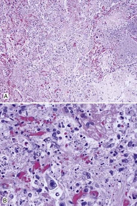
Fig 1 Zygomycosis. This post–renal transplant patient was coinfected with Zygomycetes and cytomegalovirus (CMV). A, Low power showing hemorrhagic infarction; and B, high power showing CMV viral inclusion and irregular fungal hyphae.
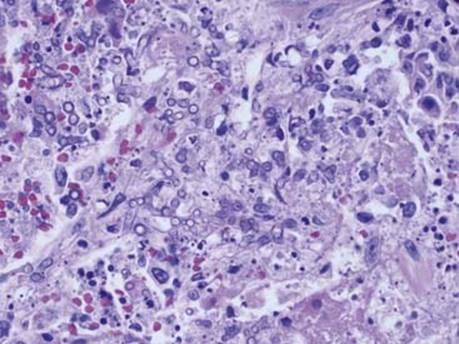
Fig 2 Zygomycosis. Cross-sectioned hyphae are round to oval with clear centers.
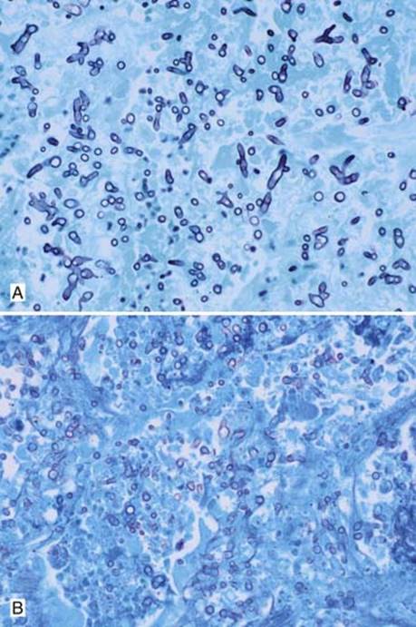
Fig 3 Zygomycosis. Irregular hyphae with right angle branching and many cross-sections are seen on GMS (A) and PAS light-green (B) stains.
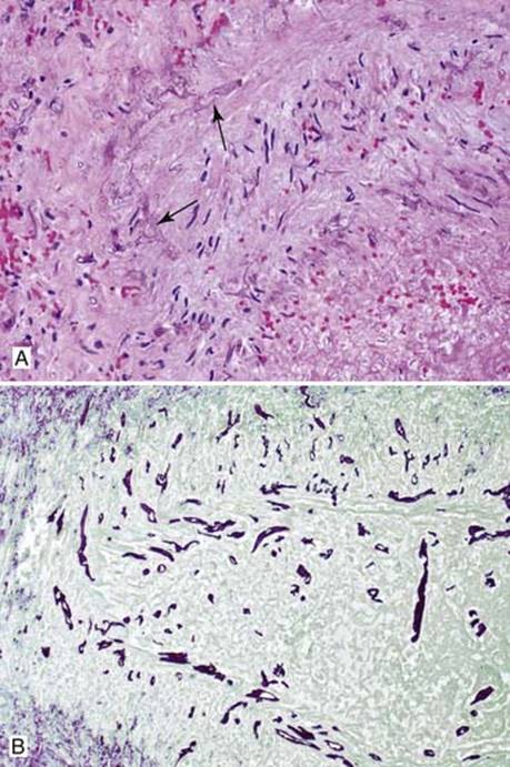
Fig 4 Zygomycosis. High powers showing the characteristic broad, wavy, ribbonlike hyphae infiltrating arterial wall with H&E (arrows) (A) and GMS (B) stains.