
Definition
• PCP in humans is an opportunistic infection caused by Pneumocystis jiroveci (formerly P. carinii), an ascomycetous fungus, in patients with impaired immunity
Pathogenesis
• The transmission of Pneumocystis organisms between individuals is not fully understood. Current studies favor an airborne transmission model for PCP with person-to-person spread being the most likely
• Once Pneumocystis is inhaled, it escapes the defenses of the upper respiratory tract and is deposited in alveoli. There are at least two unique morphological life cycle forms of Pneumocystis, the trophic form and the cyst form. The trophic form measures 1 to 4 μm in diameter, and the mature cyst is 8 to 10 μm in diameter. The trophic form preferentially attaches to the alveolar type I pneumocyte, which in turn promotes the proliferation of the organism. Pneumocystis maintains an extracellular existence within alveoli and probably obtains essential nutrients from the alveolar fluid or living cells
• Both cellular and humoral immune systems are important in defense against Pneumocystis infection. The CD4+ T lymphocyte is critical as most patients with PCP have a very low CD4+ T-cell count. Impaired alveolar macrophage function and altered B-cell immune response have also been implicated in the pathogenesis
Clinical features
Epidemiology
• Pneumocystis was first identified as a form of trypanosome in guinea pigs by Chagas and in rats by Antonio Carinii. Subsequently, it was considered to be a protozoan and is now reclassified as a fungus based on nucleotide sequencing of the 16 S rRNA sequence and the cell wall composition
• During the early stage of the AIDS epidemic, there were approximately 20,000 cases of PCP per year in the United States. The incidence of PCP declined with prophylaxis and institution of highly active antiretroviral therapy
• PCP continues to be an AIDS-defining illness. Other non-HIV immunosuppressed patients, particularly those receiving immunosuppressive agents in the settings of malignancy, organ transplantation, and various rheumatologic and hematologic conditions are also susceptible to PCP
Presentation
• Patients typically present with an insidious cough (often nonproductive), dyspnea, unexplained fever for longer than 2 weeks, chest pain, substernal tightness, and fatigue
• The chest radiograph typically shows bilateral alveolar disease, first appearing in the perihilar area and then progressing peripherally, often with sparing of apical areas
• Chest radiographs may be normal in appearance or reveal asymmetry, nodules, cavitation, or bullae
Prognosis and treatment
• The first choice of drug for both treatment and prophylaxis of PCP is trimethoprim-sulfamethoxazole. For those who are allergic to sulfa, the second choice is a combination of primaquine and clindamycin. The drugs mainly act by inhibiting the folate synthesis machinery of the organism
• Although there is emerging drug resistance due to mutations in the dihydrofolate reductase enzyme (the target of trimethoprim), the response to treatment is excellent in most patients
Pathology
Histology
• Laboratory diagnosis of PCP is usually achieved by demonstrating the presence of Pneumocystis organisms in induced sputum or bronchoalveolar lavage fluid or lung biopsy specimens
• The classic findings of PCP in sections of lung stained with H&E are widening of the alveolar septa with an infiltrate of mononuclear cells and foamy honeycombed acellular exudates within the alveolar spaces. This exudate consists of aggregated cysts and trophozoites that are not visualized on H&E
• Occasionally granulomas, infarcts, or plasmacytic inflammation can be seen
Immunopathology/special stains
• The cyst form of P. jiroveci can be illustrated in tissue with the use of GMS stain. The cysts stain brown to black and have characteristic cup-shaped, boat-shaped, or crescent-shaped morphological features. The trophic form has a central nucleus. Pneumocystis cysts do not bud, and this feature can be used to distinguish it from other tissue-based fungi
• Calcofluor white, a fluorescent stain, which nonspecifically binds to beta-linked polysaccharides, such as chitin and cellulose, can also be used for detection of Pneumocystis in fluid specimens
• Although immunofluorescent stains are sensitive for detection of the organisms, Calcofluor and GMS stains are considered to be more specific in fluids
Main differential diagnoses
• Clinical and radiological differential diagnoses include viral, bacterial, mycobacterial, fungal, and protozoal pneumonias, as well as malignancies
• Pulmonary edema: pale pink uniform intraalveolar fluid
• Histoplasmosis: oval yeasts pointed at one end and rounded at the other; occasional budding
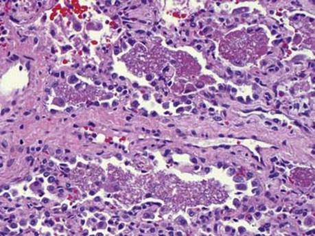
Fig 1 Pneumocystis pneumonia. H&E stained section of lung with fluffy, intraalveolar exudates and sparse inflammatory infiltrate pathognomonic of PCP (also referred to as PJP). Note blue dotlike structures in the acellular exudates.
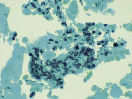
Fig 2 Pneumocystis pneumonia. GMS stained section with numerous, intraalveolar, boat-shaped and crescentic cyst forms of Pneumocystis.
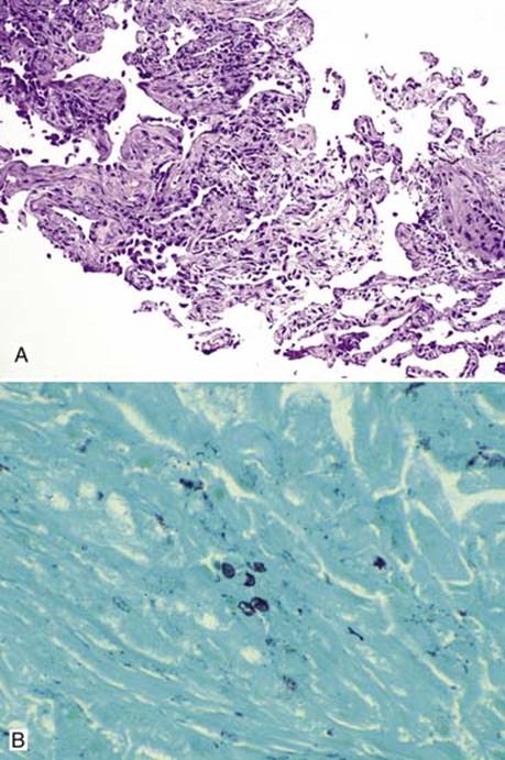
Fig 3 Pneumocystis pneumonia. A, Apparently normal looking lung biopsy specimen in a posttransplantation patient with mild type II pneumocytic hyperplasia. B, GMS stained section from the same case shows sparse, intraalveolar cystic forms of Pneumocystis, arguing the case for performing GMS stains on all lung biopsy specimens in a posttransplant setting.
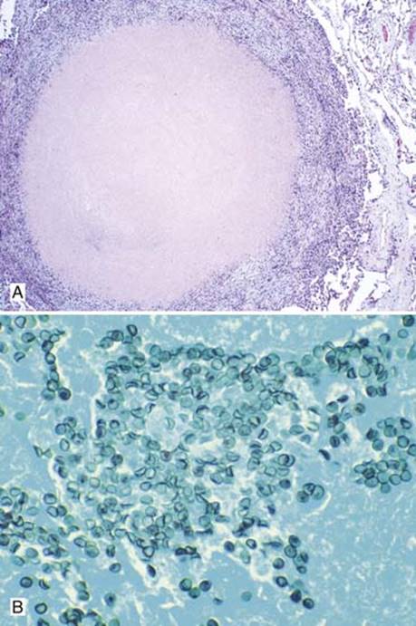
Fig 4 Pneumocystis pneumonia. This patient with PCP had multiple granulomas, one of which is seen here on H&E (A). GMS stain shows many characteristic organisms in the center of the granuloma (B).
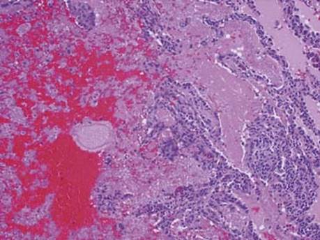
Fig 5 Pneumocystis pneumonia. There is an area of hemorrhagic infarct (far left) adjoining the fluffy exudates of PCP (center right). Compare and contrast the exudates of PCP to the pink, uniform acellular pulmonary edema fluid seen in the upper right corner.