
Definition
• GVHD is an immunologically mediated disorder where donated cells (usually hematopoietic stem cells or bone marrow allografts) attack the normal tissues of the recipient
Pathogenesis
• Donor T cells identify recipient histocompatibility antigens as “foreign” and mount an immune response against these recipient cells
• Other pathogenetic factors may play a role in causing lung injury in this clinical setting. Myeloablative pretransplant conditioning (by total body irradiation or chemotherapy) can cause diffuse alveolar damage (DAD). Diffuse alveolar hemorrhage (DAH) occurs presumably secondary to endothelial damage (after endotheliitis, radiation, or chemotherapy) or venoocclusive disease. Infections such as aspergillosis, cytomegaloviral pneumonia, or Pneumocystis jiroveci pneumonia can cause clinical illness prompting lung biopsy. These patients often receive a wide variety of immunosuppressive and chemotherapeutic agents, which can cause an equally wide range of drug-induced lung injury
Clinical features
Epidemiology
• Pulmonary complications occur in up to 60% of stem cell transplant (SCT) recipients and are an important cause of morbidity and mortality. The differential diagnosis in these patients includes infections, drug toxicity, and immunologically mediated lung damage. Lung pathology may be due to a combination of these etiologic factors in a given patient.
• Most patients with pulmonary GVHD have involvement of other sites, especially skin (rash), gastrointestinal tract (diarrhea), and liver (jaundice, elevated liver enzymes). However, up to 30% of patients may not show extrapulmonary evidence of GVHD
• GVHD has also been anecdotally described after solid organ transplantation. Although this is most common after liver transplantation, features of GVHD including skin rash, cytopenia, diarrhea, and fever have also been reported after lung and other solid organ transplantation
Presentation
• Presentation may be acute, subacute, or chronic, depending on the underlying pathology
• Acute presentation is seen in a setting of infectious pneumonia, DAD, and DAH. These patients have severe dyspnea and hypoxemia, with bilateral diffuse interstitial and alveolar infiltrates on imaging studies. Risk factors for early DAD include advanced age of the recipient, preexisting renal failure, and alveolar hemorrhage
• Late phase complications (100 days post-SCT) are more likely to be due to GVHD. Patients with lymphocytic bronchitis/bronchiolitis with interstitial pneumonitis present with a relatively subacute illness. These patients have variable cough and dyspnea. Imaging studies vary from normal to mild interstitial changes and air-trapping with hyperinflation
• Obliterative (constrictive or cicatricial) bronchiolitis presents with increasing coughing and dyspnea. The cardinal imaging finding is hyperinflation with relatively minimal interstitial change
Prognosis and treatment
• Early events such as DAD and DAH are life-threatening and require supportive care
• Although immunosuppressive therapy is essential for reducing the likelihood and severity of GVHD, infectious complications are a management challenge
• Obliterative bronchiolitis is irreversible and may necessitate lung transplantation
Pathology
Histology
• Because immunopathogenesis of GVHD is similar to that of transplantation pathology (which is due to a host-versus-graft response), histological features also overlap between these two conditions
• It is essential to first exclude opportunistic infections (especially fungal and viral) by meticulous examination of well-prepared sections and the use of microbiology techniques (including culture and polymerase chain reaction)
• Histological features of DAD and DAH are similar to those seen in other settings
• Acute GVHD: lymphocytic bronchitis or bronchiolitis is characterized by a peribronchial or peribronchiolar infiltration by lymphocytes and plasma cells. These cells infiltrate the airway walls (intraepithelial lymphocytosis). Epithelial apoptosis may be present. Commonly, the infiltrate extends into the surrounding interstitium. The interstitial infiltrate may be as dense as in lymphoid interstitial pneumonia
• Perivascular mononuclear infiltrates are also often present, with or without endotheliitis
• Organizing pneumonia (previously called bronchiolitis obliterans organizing pneumonia) is characterized by the presence of loose plugs of fibroblastic tissue filling airspaces and/or bronchioles. In the setting of GVHD, there is often accompanying lymphocytic bronchiolitis
• Chronic GVHD: obliterative bronchiolitis ranges from small airway disease with submucosal fibrosis and smooth muscle hyperplasia to complete cicatricial replacement of the airway lumen. The airway epithelium may show squamous metaplasia. Scar tissue may extend centrifugally into the surrounding parenchyma resulting in a stellate appearance
• Other histological manifestations of pulmonary GVHD include nonspecific interstitial pneumonia (cellular and fibrotic), thromboemboli, and venoocclusive disease
• Similar histological appearances may be seen in lung allograft rejection and after T-cell–based immunotherapies (such as for malignant melanoma)
Immunopathology/special stains
• Special stains for organisms (GMS, AFB, CMV, EBER), depending on histological findings
Main differential diagnoses
• Infections (as already mentioned)
• Drug-induced lung injury
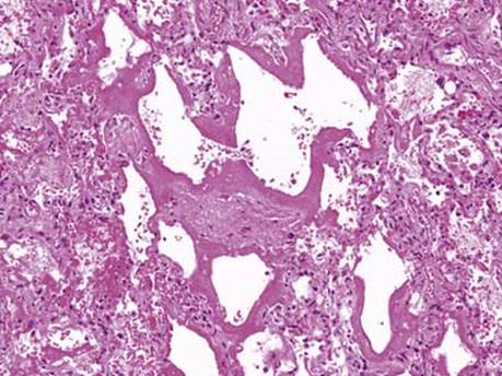
Fig 1 Graft-versus-host disease. DAD with well-formed hyaline membranes may be seen in the early days after SCT and has multifactorial etiopathogenesis.

Fig 2 Graft-versus-host disease. Organizing pneumonia, seen here as a fibroblast plug within airspace, may be a manifestation of GVHD itself or may be the result of an infection in an immunocompromised patient.

Fig 3 Graft-versus-host disease. Lymphocytic bronchiolitis is more characteristic of GVHD and follows engraftment of the donor marrow. This photograph shows an intense infiltration of the bronchial wall by lymphocytes with epithelial damage and hyperplasia. The infiltrating T cells have a cytotoxic phenotype (CD3+, CD8+).
(Case courtesy of Dr. Dani S. Zander, Hershey, Pa.)

Fig 4 Graft-versus-host disease. This airway shows epithelial apoptosis, similar to what is seen in the gastrointestinal tract and skin as a manifestation of GVHD. Lymphocytic infiltration of the airway is also present, although much less than in Fig 3.
(Case courtesy of Dr. Dani S. Zander, Hershey, Pa.)

Fig 5 Graft-versus-host disease. The bronchiolitis may be severe enough to overrun the airway. In this example, the only clues to an airway are the accompanying artery and the residual anthracotic dust.
(Case courtesy of Dr. Dani S. Zander, Hershey, Pa.)

Fig 6 Graft-versus-host disease. A cellular nonspecific interstitial pneumonia pattern is seen in this patient with GVHD.
(Case courtesy of Dr. Dani S. Zander, Hershey, Pa.)

Fig 7 Graft-versus-host disease. This airway (A) shows an early manifestation of constrictive bronchiolitis. The airway wall is damaged and is being replaced by fibroblastic tissue (top left). With progression, the airway lumen gets obliterated and is replaced by scar tissue (B), so-called obliterative or cicatricial bronchiolitis.
(A, Case courtesy of Dr. Dani S. Zander, Hershey, Pa.)
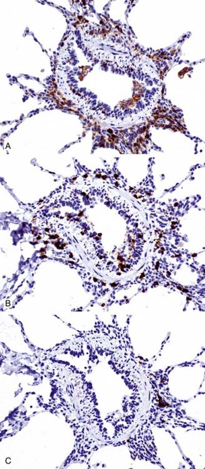
Fig 8 Graft-versus-host disease. IHC staining shows intraepithelial lymphocytes to be a mixture of CD4 (A) and CD8 (B) positive cells. CD20 positive cells (C) are seen only in the peribronchial region and not in the airway epithelium.