
Definition
• Lobar hyperinflation or hyperplasia with mass effect presenting in an otherwise normal infant
Pathogenesis
• Partial or complete obstruction of the bronchus supplying the involved lobe
• Intrinsic factors such as congenital bronchial atresia, stenosis, or mucous plugging
• Extrinsic factors such as vascular malformation or neoplasms
• Most are idiopathic
Clinical features
Epidemiology
• Most patients present in the first 6 months of life
• Boys are more frequently affected than girls
Presentation
• Respiratory distress
• Left upper lobe is most commonly involved, followed by the right middle and right upper lobes in congenital cases
• Lower lobe involvement is seen in the rare acquired forms
Prognosis and treatment
• Cure is achieved by surgical excision
• Some patients are managed conservatively by decompressing the affected lobe through the use of selective intubation
Pathology
Histology
• I: Classic pattern (congenital lobar overinflation)
• Uniformly overinflated alveoli
• Has normal number of alveoli, but alveoli can reach 3 to 10 times the normal size
• II: Polyalveolar pattern
• Variable areas of overinflated alveoli
• Increase in the absolute count of acini/alveoli
• Radial count is used to count alveoli by making a line from the last respiratory bronchiole to the pleura or closest interlobular septum
• Normally the count varies between 5 to 12; in infantile lobar emphysema the counts can reach 20 to 30 alveoli
Immunopathology/special stains
• Not contributory
Main differential diagnoses
• Congenital pulmonary adenomatoid malformation (congenital cystic adenomatoid malformation): five types with variable abnormalities in airways and alveoli
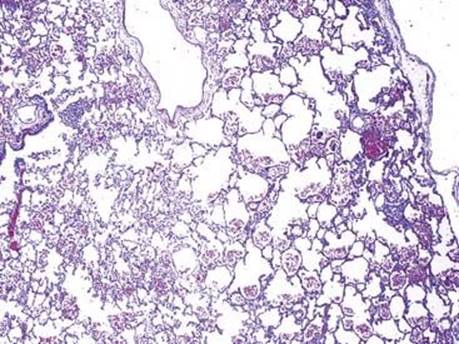
Fig 1 Infantile (congenital) lobar emphysema. Scanning view shows overinflated lung tissue and an increase in the number of alveoli characteristic of the polyalveolar pattern.

Fig 2 Infantile (congenital) lobar emphysema. Medium-power view shows overinflation of some of the alveolar spaces that can be seen in the polyalveolar pattern.

Fig 3 Infantile (congenital) lobar emphysema. Low-power view shows distended alveoli that are normal in number characteristically seen in the classic pattern.

Fig 4 Infantile (congenital) lobar emphysema. All alveoli are distended in this classic pattern.

Fig 5 Infantile (congenital) lobar emphysema. High-power view shows markedly distended alveolar space that can be seen in both classic and polyalveolar patterns. Note absence of inflammation and fibrosis.