
Definition
• Dissection of air into septal connective tissue, peribronchial tissue, and the perivascular sheath
Pathogenesis
• Rupture of alveoli due to mechanical ventilation allows dissection of air around bronchovascular bundles
Clinical features
Epidemiology
• Incidence is decreasing but may still occur in 5% to 10% of premature infants undergoing ventilation for respiratory distress syndrome
• Rare in healthy infants without mechanical ventilation
Presentation
• X-ray film demonstrates linear streaks and cystic spaces radiating from the hilum
• Persistent IPE (PIPE) occurs in infants with acute IPE (AIPE) for more than a week. It is usually localized but may be diffuse in association with bronchopulmonary dysplasia
Prognosis and treatment
• AIPE may dissect centrally to produce pneumomediastinum, pneumopericardium, or pneumoperitoneum or may dissect peripherally to produce pneumothorax
• AIPE may be reabsorbed or persist as PIPE
• Treatment with oxygen is usually sufficient, and resection is rarely required
Pathology
Gross
• AIPE demonstrates 0.3 to 1.0 cm air-filled subpleural blebs adjacent to interlobular septa and along bronchovascular bundles
• Diffuse PIPE demonstrates 0.1 to 0.3 cm air-filled cysts along the interlobular septa
• Localized PIPE has larger (3 to 4 cm) interconnecting air-filled cysts along interlobular septa
Histology
• In AIPE, irregular air spaces adjacent to bronchovascular bundles are lined by connective tissue
• May rupture into lymphatics, when the outline is round, but does not extend into pleural lymphatics
• In PIPE, irregular air spaces are lined by fibrous tissue with scattered foreign body giant cells
Immunopathology/special stains
• Immunohistochemistry for endothelial and epithelial markers should be negative in the lining of the cyst unless there is rupture into the lymphatics
Main differential diagnoses
• Congenital pulmonary lymphangiectasis: cystic spaces are lined by lymphatic endothelium and extend laterally along the pleura away from interlobular septa
• Peripheral cysts: represent dilated alveoli and are lined by epithelium
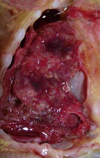
Fig 1 Interstitial pulmonary emphysema. Multiple subpleural blebs due to air tracking are seen in this patient with IPE.
(Courtesy of Dr. John Hicks, Texas.)
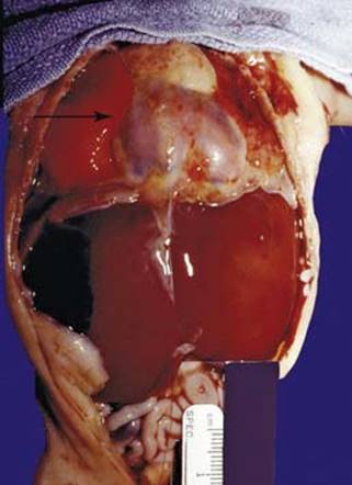
Fig 2 Interstitial pulmonary emphysema. IPE dissected centrally to produce pneumopericardium (arrow) in this infant.
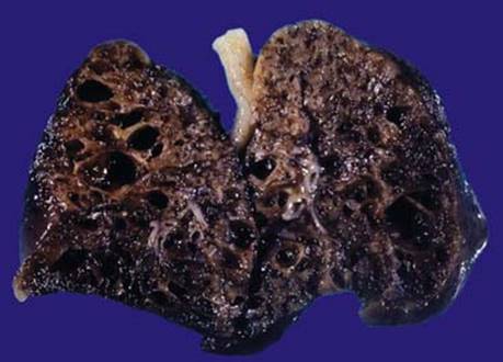
Fig 3 Interstitial pulmonary emphysema. Diffuse IPE can create large cysts along the septa as seen in the lung of this infant.
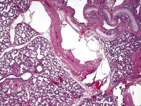
Fig 4 Interstitial pulmonary emphysema. Irregularly shaped cystic spaces along the bronchovascular bundle compress a large artery.
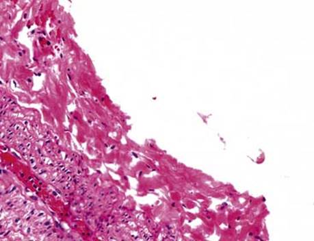
Fig 5 Interstitial pulmonary emphysema. The cystic spaces lack endothelial or epithelial lining.
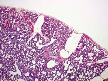
Fig 6 Interstitial pulmonary emphysema. The air spaces extend along the bronchovascular bundles to the pleura, and the round shape suggests that air has dissected into the lymphatics, but pleural lymphatics lateral to the septa are not involved.
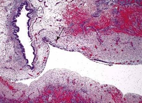
Fig 7 Interstitial pulmonary emphysema. In persistent or chronic IPE, air spaces are lined by foreign body giant cells (arrow) as seen in this low power.
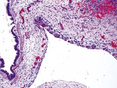
Fig 8 Interstitial pulmonary emphysema. High power of Fig 7.