
Definition
• A malignant epithelial tumor with squamous cell differentiation
Clinical features
Epidemiology
• Accounts for about 20% of all invasive lung cancers
• More than 90% are associated with cigarette smoking
Presentation
• Most patients have central tumors and present with signs of obstruction: recurrent infection, hemoptysis, coughing
• Patients with peripheral SCCs may have idiopathic pulmonary fibrosis and may present with shortness of breath and dry coughing
• Associated with hypercalcemia resulting from parathyroid hormone–related protein secreted by the tumor
Prognosis and treatment
• Surgical resection and radiation therapy
• Prognosis varies by histological variant and grade; in general, it is better than adenocarcinoma
Pathology
Gross
• Two thirds of cases are centrally located, arising from proximal bronchi
• One third of cases are peripherally located
• Tumors are firm gray-white masses with areas of necrosis and cavitation
• Central lesions often have endobronchial growth, which may occlude the lumen of airways and cause obstructive changes
Histology
• Tumor is graded according to degree of squamous differentiation and anaplasia of tumor cells
• Well-differentiated SCC shows prominent squamous cell differentiation with keratinization, pearl formation, and intercellular bridges
• Squamous cell differentiation may be subtle or only focal in poorly differentiated SCC
• Tumor cells are polygonal, hyperchromatic, and have irregular nuclei and prominent nucleoli; the amount of cytoplasm is variable from abundant to scanty
• Histologic variants:
• Papillary variant
• Clear-cell variant
• Small-cell variant
• Basaloid variant
• Alveolar space–filling type of peripheral SCC
• Lymphoepithelioma-like carcinoma
• Spindle cell carcinoma: see Sarcomatoid Carcinoma
Immunopathology/special stains
• Positive for CK5/6, p63, high-molecular-weight keratin 34βE12, and CEA
• Central tumors usually negative for CK7 and TTF-1
• Increased CK7 expression in peripheral tumors
Main differential diagnoses
• Metastatic SCC: immunohistochemical studies do not help in differentiating primary from metastatic carcinoma, which needs clinical correlation
• Thymic SCC: mediastinal mass; CD5 is usually positive in tumor cells in addition to SCC markers (p63 and CK5/6)
• Adenosquamous carcinoma: be careful when there is entrapment of benign bronchial epithelium or alveolar structures in an SCC
• Poorly differentiated adenocarcinoma: positive for TTF-1 and negative for p63 and CK5/6
• Large-cell undifferentiated carcinoma: positive for pankeratins but negative for TTF-1, p63 and CK5/6
• Mucoepidermoid carcinoma:
• Rare central tumor
• No keratinization or squamous pearl formation
• Squamous, intermediate, and mucinous cells are necessary for diagnosis; the latter can be confirmed by mucicarmine or PAS stains
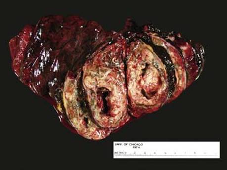
Fig 1 Squamous cell carcinoma. Gross photograph of SCC with central necrosis and cavitation.
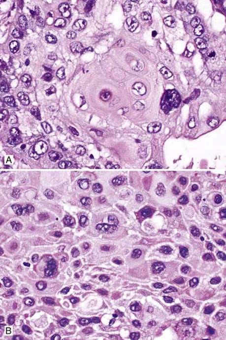
Fig 2 Squamous cell carcinoma. Well-differentiated SCC with keratinization (A) and intercellular bridges (B); note mild cytological atypia.
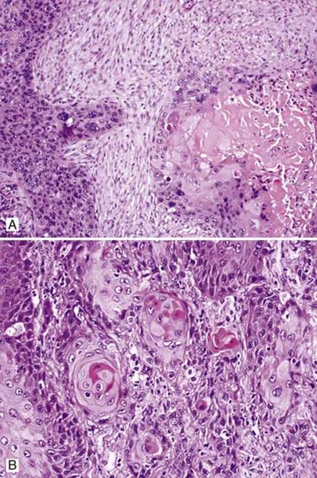
Fig 3 Squamous cell carcinoma. Moderately differentiated SCC showing squamous differentiation with keratin pearl formation and nuclear pleomorphism (A and B).

Fig 4 Squamous cell carcinoma. Poorly differentiated SCC showing no squamous differentiation (A); marked nuclear anaplasia with abnormal mitotic figure (B); cytokeratin 5/6 showing cytoplasmic staining with membranous accentuation (C); and p63 with nuclear staining (D).

Fig 5 Squamous cell carcinoma. SCC with vascular invasion.