
Recent advances in understanding of the molecular biology of adenocarcinoma of the lung have led to a major revision in histological classification. Parameters that need to be reported in biopsy and resection specimens are different. Refer to respective chapters for details. Major changes are summarized as follows:
• Bronchioloalveolar carcinoma (BAC) (≤3 cm) is replaced by adenocarcinoma in situ (AIS), nonmucinous or mucinous
• Adenocarcinoma (≤3 cm) with invasive component ≤0.5 cm is called minimally invasive adenocarcinoma (MIA), nonmucinous or mucinous
Definition
• A malignant epithelial invasive tumor with glandular differentiation or mucin production
• Preinvasive lesions are atypical adenomatous hyperplasia and AIS (formerly BAC) (see respective chapters)
Pathogenesis
• Adenocarcinomas arise from precursor cells normally present in the bronchial or bronchiolar epithelium (central type) or in the alveoli (peripheral type)
• Stepwise accumulation of mutations occurs in tumors associated with smoking (see specific mutations further on)
Clinical features
Epidemiology
• Most common type of lung cancer; accounts for approximately 40% of all invasive lung cancers in the United States
• Most common lung cancer in women and nonsmokers
• Incidence increased in past decade
• Variable (weaker) association with smoking
Presentation
• More commonly (three quarters) peripheral; patients may present with a cough with or without blood-streaked sputum, shortness of breath, chest pain, fatigue, and weight loss
• Some patients may be asymptomatic; tumor discovered incidentally
• Metastases to lymph nodes, brain, bone, or adrenal glands present in about half the patients at presentation
Prognosis and treatment
• Surgical resection with or without neoadjuvant therapy for early stage adenocarcinomas
• Neoadjuvant therapy with posttherapy surgical resection for higher stage local disease
• Neoadjuvant therapy only for metastatic disease
• Neoadjuvant therapy may include chemotherapy, radiation therapy, kinase inhibitors, monoclonal antibodies, and hormonal therapy
• Molecular test for mutations is very important in deciding whether the patient is suitable for kinase inhibitor treatment
• Prognosis varies by tumor grading and staging
Pathology
Gross
• Tan-fleshy mass; larger tumors with necrosis
• Overlying pleura in peripheral tumor often has puckering due to fibrosis or invasion
Histology
• Tumor is characterized by glandular differentiation, usually with gland formation or mucin production; often with central scar
• MIA
• Tumor is ≤3 cm with invasive component ≤0.5 cm (if multiple invasive foci, largest should be ≤0.5 cm; do not add together)
• No invasion into lymphatics, blood vessels, or pleura
• No tumor necrosis
• Noninvasive component is called lepidic pattern
• Any nonlepidic pattern is considered invasive (see patterns further on)
• Almost 100% survival reported
• Tumor is commonly graded according to degree of differentiation and anaplasia of tumor cells; however, architectural patterns, as given further on, have more prognostic significance
• A spectrum of morphological features is seen. The major growth pattern should be given in the diagnosis with the percentage of minor pattern(s):
• Acinar pattern
– Most common pattern seen in invasive adenocarcinoma
– Tumor cells form invasive small glandular structures like acini or tubules in a background of desmoplastic stroma
• Papillary pattern
– Papillae formation with fibrovascular cores; often with secondary and tertiary branching patterns
– Psammoma bodies may present
– Pure papillary growth pattern should be distinguished from metastatic tumor
• Micropapillary pattern
– Small papillary clusters with no fibrovascular cores; tumor cells usually small with minimal atypia; the tumor clusters may appear to float within alveoli
– Important to identify this pattern
– The tumor has a worse prognosis, even if a small percentage of this pattern is present
– Associated with higher frequency of lymphatic invasion and lymph node metastases, which may have more of the micropapillary pattern than the primary tumor
• Lepidic (formerly bronchioloalveolar) pattern
– Tumor cells grow along preexisting alveolar walls
– Usually seen at the periphery of the tumor, which is composed of one or more of the aforementioned patterns
• Solid pattern
– Tumor cells forming sheets with no acinar, papillary, or micropapillary formation
– Signifies poor prognosis, and some mutations are more frequent
• Signet-ring pattern
– Usually focal; associated with other patterns
– Tumor cells containing large intracytoplasmic mucin vacuoles with peripherally displaced nuclei
– Solid pattern with more than 10% signet ring is more likely to have the EML4-ALK mutation
• Clear cell pattern
– Clear cell changes are mostly focal and may occur in any histological pattern
– Rarely, it is the major component of the tumor, and metastatic renal cell carcinoma is the main differential diagnosis
– No clinical or prognostic significance of clear cell features
• Histological variants (subtypes):
• Invasive mucinous adenocarcinoma (formerly mucinous BAC)
– Larger than 3 cm, invasive focus >0.5 cm, or multifocal or uncircumscribed tumor of any size with miliary spreading into adjacent lung
– Tumor cells are characteristically tall and columnar with basal small nuclei and abundant cytoplasmic mucin; sometimes resembling goblet cells; nuclear atypia is minimal
– TTF-1 is mostly negative (0%-30% of cases are positive)
– K-ras mutations are present in most (80%-100%)
• Colloid adenocarcinoma
– Similar to its counterpart in other body sites
– Contains pools of mucin with islands of epithelial cells
– The neoplastic cells may be extremely bland
– May be circumscribed with central cystic space containing mucin (formerly mucinous cystadenocarcinoma)
• Fetal adenocarcinoma
– Synonyms: pulmonary adenocarcinoma of fetal type, pulmonary endodermal tumor resembling fetal lung
– Rare tumor; mean age, 40 years
– Resembles epithelial component of pulmonary blastoma
– May occur in familial adenomatous polyposis and with increased β-catenin signaling
– Better prognosis than pulmonary blastoma
– Histology:
– Composed of tubules with nonciliated columnar cells with clear cytoplasm resembling fetal lung tubules
– Similar to endometrioid adenocarcinoma with supranuclear or subnuclear glycogen vacuoles, and squamous morules
• Adenocarcinoma with enteric differentiation
– Heterogeneous tumor with some components similar to lung adenocarcinoma; often with lepidic growth; at least 50% of the tumor shows enteric pattern
– The enteric pattern is histologically and immunologically similar to colorectal primary; expresses one enteric marker such as CDX-2, CK20, or MUC2; usually CK7 positive; half are TTF-1 positive
Immunopathology/special studies
• Mucin production can be demonstrated with mucicarmine, PAS, or Alcian blue special stains
• Immunoreactive with epithelial markers: keratins CAM5.2 and AE1/AE3, EMA, CEA, MOC31, BG8, CD15 (Leu-M1), Ber-Ep4, and B72.3
• Usually positive for CK7 and negative for CK20
• TTF-1 is positive in 75% to 95% of lung adenocarcinomas; focal p63 staining is present in up to 30% of adenocarcinomas
• In small biopsy and cytological specimens, report should include if diagnosis is made on H&E or immunohistochemical (IHC) staining
• If no glandular differentiation is present, TTF-1-positive and p63- and CK5/6-negative tumors should be reported as “non–small-cell carcinoma, favor adenocarcinoma”
• If TTF-1 and p63 are positive in different areas of the tumor, adenosquamous carcinoma diagnosis should be suggested
• Use of the term non–small-cell carcinoma, not otherwise specified, should be limited to those tumors that show no histological or IHC differentiation
• Molecular studies: EGFR mutation, K-ras mutation, and EML4-ALK translocation are mutually exclusive
• EGFR mutation:
– Seen in <10% of all adenocarcinomas
– Most common in female nonsmokers with Asian ethnicity (up to 50%)
– Present in 40% of nonsmoking whites
– Good response to targeted therapy with kinase inhibitors
• K-ras mutation:
– 30% of adenocarcinomas in smokers
– 5% in nonsmokers
– K-ras mutation confers radioresistance, which can be reversed by kinase suppressor of Ras1
• EML4-ALK:
– 5% of adenocarcinomas; often younger, male, and never or light smoker
– Variable histological patterns
– Resistance may develop during ALK inhibitor therapy
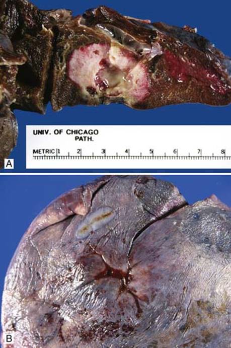
Fig 1 Adenocarcinoma. A, Cut surface of a peripheral adenocarcinoma with relatively well-circumscribed edge. B, Gross picture of puckering of pleural surface.
Main differential diagnoses
• Metastatic adenocarcinoma
• Features favoring metastatic adenocarcinoma:
– Multiple lung nodules or masses
– Well-circumscribed nodules without a central scar
– For specific antibodies useful in differentiating primary from metastatic see chapter on metastatic carcinoma
• Epithelioid malignant mesothelioma:
• Positive calretinin (cytoplasmic staining with nuclear accentuation), WT-1 (nuclear), keratin CK5/6 (cytoplasmic), and D2-40 (membranous) immunostains
• Negative MOC31, BG8, TTF-1, and negative intracytoplasmic mucin
• Bronchiolar metaplasia
• May be so extensive that it mimics well-differentiated adenocarcinoma; especially difficult to differentiate in frozen sections
• Bland columnar cells with cilia or cuboidal epithelium

Fig 2 Adenocarcinoma. Adenocarcinoma of lung showing acinar (A), papillary (B), micropapillary (C), solid (D), signet ring (E), and lepidic (F) patterns.

Fig 3 Adenocarcinoma. Adenocarcinoma of lung with a micropapillary growth pattern: A, low power; B, medium power; C, high power. Note the psammoma bodies in B and C.
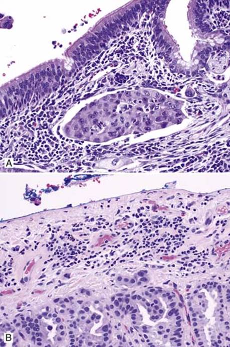
Fig 4 Adenocarcinoma. Adenocarcinoma with lymphovascular (A) and visceral pleural (B) invasion.

Fig 5 Adenocarcinoma. Mucinous adenocarcinoma: A and B, low power; C and D, medium power; E, high power. Note mucin-containing tumor cells with both invasive and lepidic growth and mucin accumulation in alveolar spaces. F, IHC stain for TTF-1 stains residual small pneumocyte nuclei while tumor cells are negative.
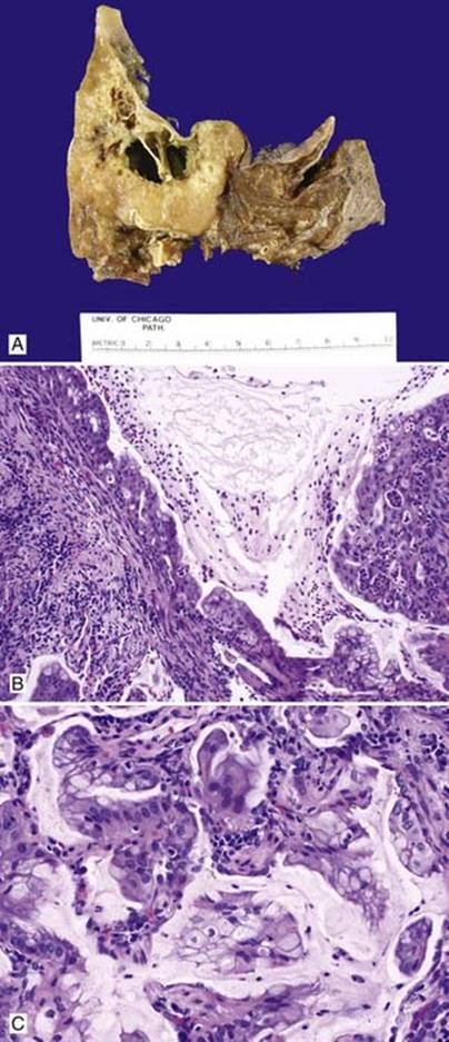
Fig 6 Adenocarcinoma. Colloid adenocarcinoma with cystic change (previously called mucinous cystadenocarcinoma): A, gross picture of cut surface showing solid and cystic components; B,intermediate power; C, high power of the same tumor.

Fig 7 Adenocarcinoma. Adenocarcinoma with clear cell feature: A, low power; B, high power; C, positive TTF-1 immunostain.
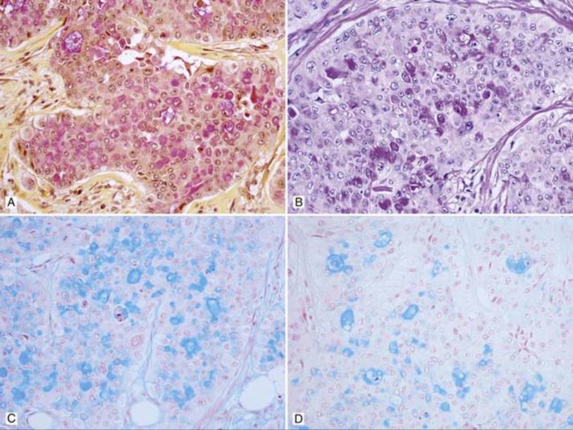
Fig 8 Adenocarcinoma. Intracytoplasmic mucin of adenocarcinoma is positive for A, mucicarmine; B, PAS-D; C, Alcian blue pH 2.5; and D, Alcian blue pH 2.5 after hyaluronidase digestion.
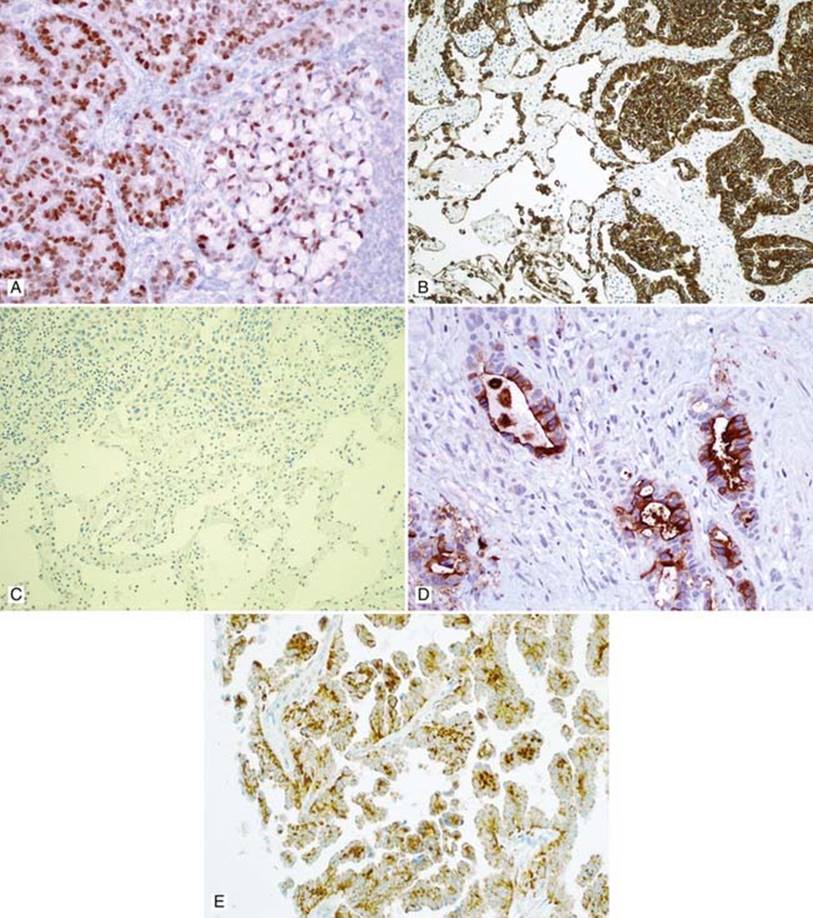
Fig 9 Adenocarcinoma. Adenocarcinoma with A, positive TTF-1; B, positive keratin CK7; C, negative CK20; and D, positive CEA immunostains. E, WT-1 stains the cytoplasm of this papillary adenocarcinoma. Note: WT-1 shows nuclear staining in malignant mesothelioma.
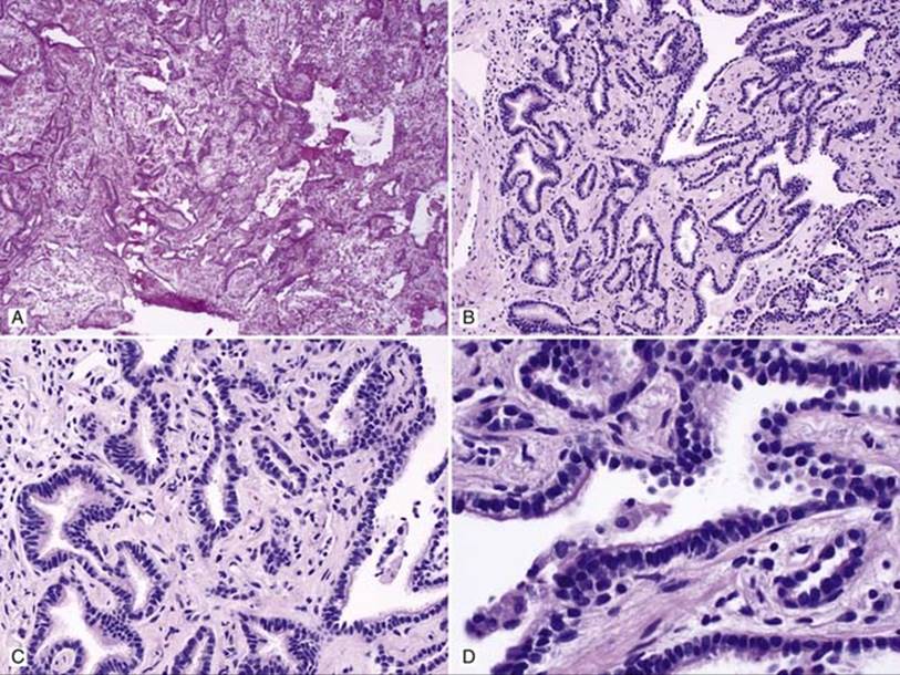
Fig 10 Adenocarcinoma. Extensive bronchiolar metaplasia mimics well-differentiated invasive adenocarcinoma: A, frozen section; B, low power; C, medium power; D, high power showing benign bronchial epithelium with cilia.