
Definition
• A poorly differentiated SCC with small tumor cells
Clinical features
Epidemiology
• Same as other SCCs
Presentation
• Same as other SCCs
Prognosis and treatment
• Surgical resection and radiation therapy
• Unfavorable prognosis: early metastases to lymph nodes and distant sites
Pathology
Histology
• Hyperchromatic and irregular small tumor cells that can mimic small-cell carcinoma
• Lacks nuclear features of small-cell carcinoma: molding or salt and pepper chromatin
• Compared with small-cell carcinoma, tumor cells contain coarse and vesicular chromatin, prominent nucleoli, moderate cytoplasm, and distinct cell borders
• Focal squamous differentiation (keratinization or intercellular bridges) may be present
Immunopathology/special stains
• Very helpful in distinguishing from small-cell carcinoma
• Positive for CK5/6 and p63
• Negative for TTF-1 and neuroendocrine markers
Main differential diagnoses
• Small-cell carcinoma: inconspicuous or absent nucleoli; p63 and CK5/6 negative; usually positive for TTF-1 and neuroendocrine markers
• Combined small-cell carcinoma with SCC: immunohistochemical (IHC) stains should demonstrate both components

Fig 1 Small-cell variant of squamous cell carcinoma. There are small nests of malignant cells that are about two to three times the size of lymphocytes.

Fig 2 Small-cell variant of squamous cell carcinoma. High power shows scant cytoplasm, salt and pepper chromatin, and frequent mitotic figures, which suggest the differential diagnosis of small-cell SCC versus small-cell carcinoma.
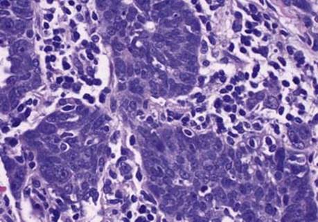
Fig 3 Small-cell variant of squamous cell carcinoma. Another area of the same tumor has some nuclei with prominent nucleoli, which is characteristic of small-cell SCC.

Fig 4 Small-cell variant of squamous cell carcinoma. IHC stains are very helpful in showing A, positive CK5/6; B, positive p63; and C, negative TTF-1 in small-cell SCC.