
Definition
• A histological variant of squamous cell carcinoma with characteristic syncytial growth pattern and heavy infiltration of lymphocytes
Clinical features
Epidemiology
• Rare cases reported
• More common in Asian women than in whites
• May be associated with Epstein-Barr virus (EBV) infection
Presentation
• Same as other non–small-cell carcinomas
Prognosis and treatment
• Surgical resection
• Better prognosis than other non–small-cell carcinomas
Pathology
Gross
• Well-circumscribed lung nodule
Histology
• Tumor is composed of syncytial sheets of large undifferentiated tumor cells with heavy inflammatory infiltration
• Usually with a pushing border
• The large epithelial cells have oval to round nuclei with open or vesicular chromatin and prominent nucleoli
• The inflammatory infiltrates are predominantly mature lymphocytes with a mixture of plasma cells, neutrophils, and, rarely, eosinophils
Immunopathology/special stains
• The tumor cells are positive for p63 and keratin CK5/6 and negative for LCA; in situ hybridization study for EBV (EBER) may be positive
• Lymphocytes are CD8+ T cells
Main differential diagnoses
• Metastatic nasopharyngeal carcinoma: similar morphological features; clinical history is critical
• Lymphoma: keratin negative

Fig 1 Lymphoepithelioma-like carcinoma. Syncytial sheets of poorly differentiated cells with abundant inflammatory cells (low power).

Fig 2 Lymphoepithelioma-like carcinoma. Large, poorly differentiated cells with vesicular chromatin, prominent nucleoli, and indistinct cell borders (A and B, medium power); note mixed inflammatory cells (lymphocytes, plasma cells, and neutrophils) in the background.

Fig 3 Lymphoepithelioma-like carcinoma. Abnormal mitotic figure in the center (high power).

Fig 4 Lymphoepithelioma-like carcinoma. Large tumor cells staining positive for p63 (A) and CK5/6 (B).

Fig 5 Lymphoepithelioma-like carcinoma. Large tumor cells (nest in center) and surrounding inflammatory cells are negative for TTF-1; note that the positive-staining cells are residual alveolar pneumocytes.
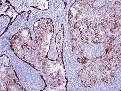
Fig 6 Lymphoepithelioma-like carcinoma. Keratin AE1/AE3 stains both tumor cells and residual benign alveolar lining cells.

Fig 7 Lymphoepithelioma-like carcinoma. In situ hybridization for EBV (EBER) shows strong positivity in all tumor cells; note the negative-staining lymphocytes.