
Definition
• A subtype of sarcomatoid carcinoma with at least 10% of malignant spindle cells and/or giant cells
Clinical features
Epidemiology
• Extremely rare
Presentation
• Similar to other subtypes of sarcomatoid carcinoma
Prognosis and treatment
• Similar to other subtypes of sarcomatoid carcinoma
Pathology
Histology
• The tumor may contain other components of non–small-cell carcinoma, specifically squamous cell carcinoma, adenocarcinoma, or large-cell carcinoma, the presence of which should be documented, or the tumor may be composed only of spindle cells and giant cells
• The spindle cell component is the same as the tumor cells of spindle cell carcinoma
• The giant cell component is the same as the tumor cells of giant cell carcinoma; often with emperipolesis
• Squamous cell carcinoma may rarely present as pseudoangiosarcomatous carcinoma with anaplastic epithelioid cells arranged in pseudopapillae or anastomosing spaces filled with red blood cells
Immunopathology/special stains
• No studies are needed to prove epithelial differentiation in spindle and/or giant cells if there is a component of non–small-cell carcinoma (squamous cell carcinoma, adenocarcinoma, or large-cell carcinoma)
• When the tumor is composed only of spindle and giant cells, multiple keratins and EMA are necessary to demonstrate their epithelial differentiation: tumor may show only focal staining with one marker
• Some of the bizarre giant multinucleated cells may show immunoreactivity for β-human chorionic gonadotropin (hCG) and placental-like alkaline phosphatase (PLAP)
• Electron microscopy may be helpful in demonstrating epithelial differentiation in spindle and/or giant cells in difficult cases
Main differential diagnoses
• Metastatic pleomorphic carcinoma from other sites
• Clinical correlation is very important
• Metastatic melanoma
• Positive for melanoma markers (S100, Melan-A, HMB45, or MITF)
• Extrauterine epithelioid trophoblastic tumor
• Extremely rare
• Women of reproductive age with elevated serum β-hCG levels
• Some patients may only have isolated lung lesions with no intrauterine disease resembling primary lung carcinomas, especially squamous cell carcinoma and pleomorphic carcinoma
• Tumor cells are imunoreactive for cytokeratin, p63, EMA, β-hCG, and PLAP
• Inhibin immunostain is positive in contrast to primary lung carcinomas

Fig 1 Pleomorphic carcinoma. Pleomorphic carcinoma composed of spindle cells and giant pleomorphic cells.
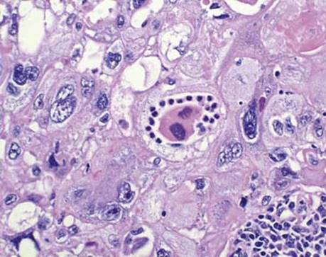
Fig 2 Pleomorphic carcinoma. Pleomorphic carcinoma with emperipolesis (intracytoplasmic neutrophils).
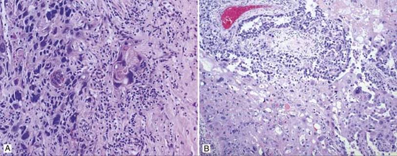
Fig 3 Pleomorphic carcinoma. Pleomorphic carcinoma with non–small-cell carcinoma components: A, squamous cell carcinoma with keratin pearl formation; B, adenocarcinoma.

Fig 4 Pleomorphic carcinoma. Pleomorphic carcinoma positive for CK7 (A) and MOC-31 (B).
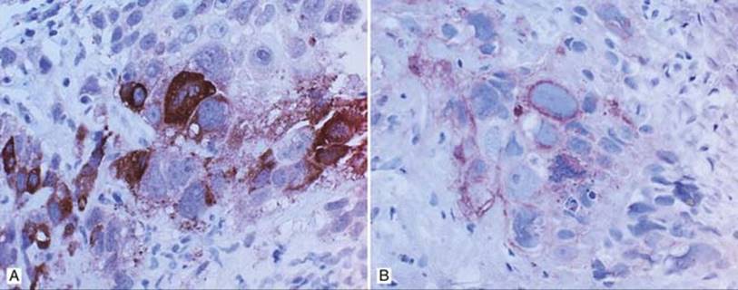
Fig 5 Pleomorphic carcinoma. The pleomorphic multinucleated and/or mononucleated giant cells in this pleomorphic carcinoma are positive for β-hCG (A) and PLAP (B).