
Definition
• A subtype of sarcomatoid carcinoma composed entirely of spindle-shaped carcinoma cells; also considered to be a variant of squamous cell carcinoma
Clinical features
Epidemiology
• Extremely rare
Presentation
• Similar to other subtypes of sarcomatoid carcinoma
Prognosis and treatment
• Similar to other subtypes of sarcomatoid carcinoma
Pathology
Histology
• Tumor is composed of malignant spindle cells arranged in irregular fascicles or a storiform pattern; tumor cells are epithelioid to spindled with hyperchromatic nuclei and conspicuous nucleoli
• No carcinoma patterns (including squamous cell carcinoma, adenocarcinoma, or large-cell carcinoma) or giant cell component present
• May have lymphoplasmacytic infiltration
Immunopathology/special stains
• Multiple epithelial markers including keratins, p63, and EMA are necessary to prove the tumor’s epithelial differentiation: tumor may show only focal staining with one of the markers
• Electron microscopy may be helpful in demonstrating epithelial differentiation with paranuclear filaments and tonofibrils
Main differential diagnoses
• Sarcomatoid malignant mesothelioma
• It can be extremely difficult because sarcomatoid malignant mesothelioma may not express mesothelial markers
• Radiological findings can be useful
• Metastatic sarcomatoid renal cell carcinoma: clinical history of renal cell carcinoma
• Metastatic spindle cell melanoma: usually positive for S100 and negative for Melan-A and HMB45
• Primary or metastatic sarcomas
• Solitary fibrous tumor
• Synovial sarcoma
• Inflammatory myofibroblastic tumor
• Usually younger patients
• Contains spindle cells with fine chromatin and inconspicuous nucleoli with minimal atypia; mixed with abundant inflammatory cells
• ALK immunostain may be positive, which correlates with rearrangement of ALK gene
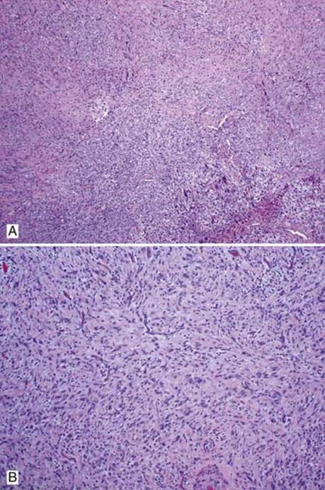
Fig 1 Spindle cell carcinoma. A, Epithelioid to spindled tumor cells. B, Malignant spindle cells arranged in short fascicles with vague storiform pattern; note the lymphocytic infiltration in the background.
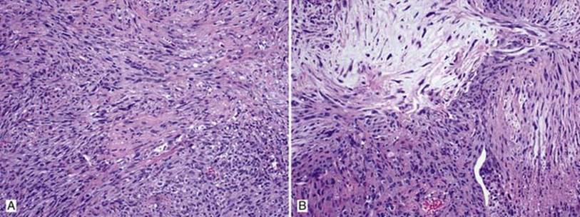
Fig 2 Spindle cell carcinoma. A and B, Medium power shows spindled to epithelioid tumor cells with hyperchromatic nuclei; note focal myxoid change in B.
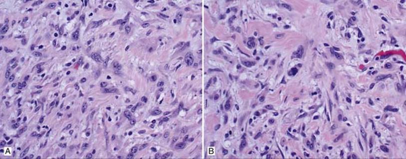
Fig 3 Spindle cell carcinoma. A and B, High power shows tumor cells with enlarged irregular nuclei, vesicular chromatin, and conspicuous nucleoli; note the abnormal mitotic figure in B.
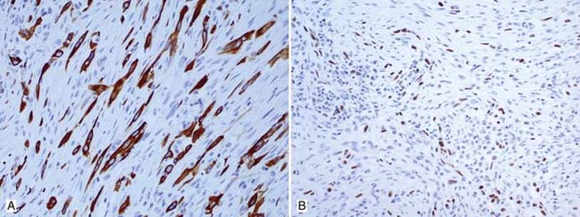
Fig 4 Spindle cell carcinoma. Spindle cell carcinoma is positive for cytokeratin 5/6 (A) and p63 (focal) (B).