
Definition
• Abnormal location of pulmonary veins within bronchovascular bundles and increased alveolar capillaries
Pathogenesis
• Some cases are associated with mutations in STRA6 on chromosome 15
Clinical features
Epidemiology
• Rare congenital disease
• Familial cases have been described
Presentation
• Presents soon after birth with marked respiratory distress and pulmonary hypertension
• In approximately 50% of cases other anomalies are identified, such as duodenal atresia, congenital heart disease, asplenia, phocomelia, and ureteric and urethral obstruction
Prognosis and treatment
• No effective therapy; lung transplantation could be an option
• Patients usually die within the first few days or weeks of life
Pathology
Histology
• Thickened alveolar septa and increased number of alveolar capillaries
• Abnormally located capillaries within central portion of septa instead of near alveolar lumen
• Pulmonary arteries with medial hypertrophy
• Abnormally muscularized arterioles within bronchovascular bundles
• Dilated pulmonary veins abnormally located near pulmonary arteries in the bronchovascular bundles; although this feature can be focal or diffuse, it is pathognomic of alveolar capillary dysplasia
Immunopathology/special stains
• Not contributory

Fig 1 Alveolar capillary dysplasia. Low-power view shows ectatic abnormally located pulmonary vein (arrow) adjacent to pulmonary arteries. Dilated lymphatics are also present (arrowheads).
(Courtesy of Dr. John Hicks, Children’s Hospital, Houston, Tex.)
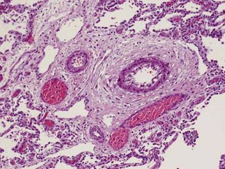
Fig 2 Alveolar capillary dysplasia. Medium-power view shows thickened, muscularized arterioles and adjacent ectatic abnormally located pulmonary veins. Note centrally located capillaries in alveolar septae in lower left and upper right.
(Courtesy of Dr. John Hicks, Children’s Hospital, Houston, Tex.)

Fig 3 Alveolar capillary dysplasia. Medium-power view shows an ectatic vein abnormally located next to the pulmonary artery.
(Courtesy of Dr. John Hicks, Children’s Hospital, Houston, Tex.)

Fig 4 Alveolar capillary dysplasia. High-power view shows a bronchovascular bundle with thickened muscularized arterioles and ectatic abnormally located venules.
(Courtesy of Dr. John Hicks, Children’s Hospital, Houston, Tex.)

Fig 5 Alveolar capillary dysplasia. High power of alveolar walls showing centrally located dilated capillaries.
(Courtesy of Dr. John Hicks, Children’s Hospital, Houston, Tex.)