
Definition
• Rare congenital defect in surfactant proteins or in proteins involved in processing and homeostasis of surfactants
• Formerly categorized as congenital pulmonary alveolar proteinosis
Pathophysiology
• Numerous different genetic defects have been described in surfactant protein (Sp)B, SpC, and ABCA3 (involved in surfactant processing and homeostasis)
• Mutations can be autosomal dominant, autosomal recessive, or sporadic
Clinical features
Epidemiology
• Rare cause of neonatal respiratory distress or pediatric interstitial lung disease
• Most often due to genetic defect of ABCA3 or SpB, and rarely SpC
• More common in patients with family history of lung disease or consanguinity
Presentation
• Term neonates presenting with severe respiratory distress lasting longer than 1 week, chest radiography similar to respiratory distress syndrome of premature neonates, and with no improvement after surfactant administration
• Patients with genetic mutation of SpC have variable clinical presentation and age at onset of symptoms, which include poor growth, decreased activity, dyspnea, and nonproductive cough
• Chest radiographic findings are typically much more severe than suggested by the clinical signs and symptoms
Prognosis and treatment
• Poor survival (days to months) in neonates with severe disease
• No specific treatment available
• Lung transplant has had some success but still carries significant mortality
Pathology
Histology
• Accumulation of PAS positive, mucicarmine negative eosinophilic granular alveolar material
• Type II pneumocyte hyperplasia, interstitial fibrosis, and alveolar simplification, which have been descriptively referred to as being consistent with pulmonary alveolar proteinosis, nonspecific interstitial pneumonia, desquamative interstitial pneumonia, or chronic pneumonitis of infancy
• Electron microscopy: disorganized or incomplete lamellar bodies (SpB or SpC defects), small lamellar bodies with eccentric electron dense inclusions with a “fried-egg” appearance (ABCA3 defects)
Immunopathology (including immunohistochemistry)
• Alveolar eosinophilic material is negative for SpB in cases with SpB deficiency and negative for SpC in cases of SpC deficiency
Main differential diagnoses
• Neonatal respiratory distress syndrome
• Pulmonary alveolar proteinosis, secondary type

Fig 1 Surfactant dysfunction disorders. Simplification of alveolar architecture, interstitial fibrosis, and type II pneumocyte hyperplasia, seen at low power in this case of ABCA3 mutation.

Fig 2 Surfactant dysfunction disorders. At higher power, brightly eosinophilic granular material is seen in some of the alveolar spaces.
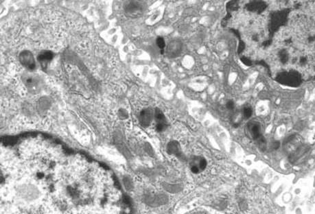
Fig 3 Surfactant dysfunction disorders. Electron microscopy in this case with an ABCA3 defect reveals a paucity of lamellar bodies, which are small and irregular; several have eccentric electron dense inclusions.

Fig 4 Surfactant dysfunction disorders. Wedge biopsy from a 4-week-old with progressive respiratory distress shows typical intraalveolar granular eosinophilic material.

Fig 5 Surfactant dysfunction disorders. Different area from same patient as in Fig 4 shows foamy macrophages in the alveolar spaces and reactive type 2 pneumocytes, illustrating the variability of surfactant accumulation.