
Definition
• A malignant epithelial tumor with characteristic cribriform, tubular, or solid growth patterns and both pseudoglandular and true glandular lumina formation, similar to its counterpart in the salivary gland
Clinical features
Epidemiology
• Rare, <1% of lung tumors
• No sex predominance
• Affects patients 40 to 50 years old
• Not associated with smoking
Presentation
• Symptoms of proximal airway obstruction: wheezing, coughing, shortness of breath, or hemoptysis
Prognosis and treatment
• Surgical resection with supplemental radiation; frozen section is recommended to evaluate the resection margins
• Prone to multiple recurrences and late metastasis
• Poor long-term prognosis in patients with positive margins, solid component, and high stage
Pathology
Gross
• Gray-white endobronchial polypoid lesion or submucosal plaque within main bronchus
Histology
• Tumor is typically arranged in tubular, cribriform, or solid growth patterns
• The presence of pseudoglandular lumina with basement membrane material and true glandular lumina with mucin is characteristic
• The tumor contains a variable amount of luminal cells and myoepithelial cells; luminal cells are small and dark with scant cytoplasm
• Frequent perineural invasion
Immunopathology/special stains
• The ductal (epithelial) cells are positive for cytokeratin, EMA, CEA, and c-kit (CD117); negative for S100 and SMA
• The myoepithelial cells are positive for cytokeratin, S100, SMA, p63, and calponin
• The basement membrane material within the pseudoglandular lumina stains positively for type IV collagen and laminin; mucicarmine and PAS-D stain mucin within the true glandular lumina
Main differential diagnoses
• Metastatic adenoid cystic carcinoma:
• More common than primary counterpart
• History of salivary gland adenoid cystic carcinoma
• Pleomorphic adenoma: benign well-circumscribed lesion with chondroid matrix
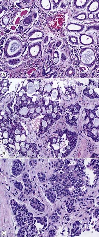
Fig 1 Adenoid cystic carcinoma. Adenoid cystic carcinoma with tubular growth pattern (A), cribriform architecture (B), and nested and solid growth patterns (C).

Fig 2 Adenoid cystic carcinoma. Adenoid cystic carcinoma with areas of pseudoglandular lumina with basement membrane material (left) and true glandular lumina with mucin (right).

Fig 3 Adenoid cystic carcinoma. Adenoid cystic carcinoma with perineural invasion.

Fig 4 Adenoid cystic carcinoma. Adenoid cystic carcinoma with A, H&E showing double layer of cells, inner epithelial, and outer myoepithelial cells; B, CD117 (c-kit) immunostain showing positivity in inner ductal/luminal cells; C, p63 staining (nuclear) in outer myoepithelial cells; and D, calponin staining (cytoplasmic) in outer myoepithelial cells.