
Definition
• Small nests of neuroendocrine cell proliferation near a bronchiole surrounded by fibrous stroma
Clinical features
Epidemiology
• Unknown incidence rate; incidental microscopic finding; often found in lung resection specimens
• More common in adults
Presentation
• Asymptomatic; rarely present with obstructive symptoms
Prognosis and treatment
• Excellent prognosis; no need for treatment
Pathology
Histology
• Size less than 5 mm; usually adjacent to airway
• Small nests of neuroendocrine cells within fibrous stroma
Immunopathology/special stains
• Positive for neuroendocrine markers (chromogranin and synaptophysin) and cytokeratins
Main differential diagnoses
• Carcinoid tumor: >5 mm
• Neuroendocrine cell hyperplasia: no fibrous stroma; limited to epithelial side of basement membrane
• Meningothelial nodule: more eosinophilic cytoplasm; not related to airway; negative for cytokeratin and neuroendocrine markers
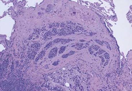
Fig 1 Carcinoid tumorlet. Low magnification shows nests of neuroendocrine cells between bronchial epithelium and cartilage, surrounded by fibrosis.
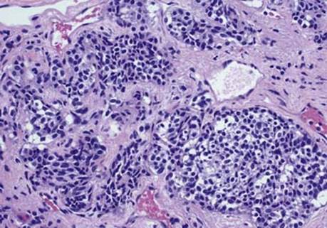
Fig 2 Carcinoid tumorlet. Medium magnification shows nests of neuroendocrine cells with round, oval, or spindled nuclei and abundant amphophilic cytoplasm; note thin-walled blood vessels adjacent to the nests.
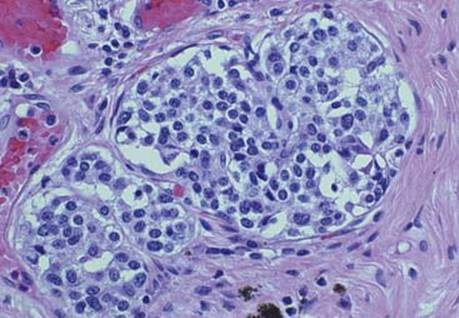
Fig 3 Carcinoid tumorlet. High magnification shows neuroendocrine cells with round nuclei, “salt and pepper” chromatin, and granular cytoplasm.

Fig 4 Carcinoid tumorlet. The cells of carcinoid tumorlets are strongly and diffusely positive for neuroendocrine markers synaptophysin (A), chromogranin (B), and CD56 (C).