
Definition
• Low (typical) to intermediate grade (atypical) neuroendocrine carcinoma of the lung
Clinical features
Epidemiology
• Carcinoids occur at numerous body sites; lung is the second most common site after gastrointestinal tract
• Accounts for 2% to 5% of all primary lung cancers
• No causal association with cigarette smoking
• More common in adults; seen at a younger age than patients with other lung carcinomas; can occur in children
• Paraneoplastic syndrome can occur but is rare in pulmonary carcinoid tumors
• Carcinoid syndrome is rare and only occurs with widespread metastases
• Can occur in patients with multiple endocrine neoplasia syndrome type I
Presentation
• Varies by location (central or peripheral)
• Central tumors usually present with obstructive symptoms: coughing, wheezing, dyspnea, chest pain, hemoptysis, or recurrent pneumonia
• Peripheral tumors are usually asymptomatic
• The seventh edition of the American Joint Committee on Cancer TNM staging is now applicable for carcinoid tumor (effective January 1, 2010)
Prognosis and treatment
• Good prognosis after surgical resection, with or without chemotherapy
• Typical carcinoids have better prognosis than atypical carcinoids
Pathology
Gross
• Central tumors (70%) arise in the main or major bronchi and are frequently seen as endobronchial mass lesions
• Peripheral tumors (30%) arise in segmental bronchi or beyond and are usually subpleural
• Tumors are yellow-tan, well-circumscribed nodules or masses with glistening cut surface
Histology
• Subdivided into typical and atypical carcinoids (see separate sections)
• Monotonous tumor cells with organoid, trabecular, insular, ribbonlike or rosettelike growth patterns that suggest neuroendocrine differentiation
• Polygonal cells with moderate eosinophilic granular cytoplasm, round to oval nuclei, “salt and pepper” chromatin, and inconspicuous nucleoli
• Rarely, the tumor cells can have clear cytoplasm or may contain melanin pigment
• May have marked nuclear atypia and pleomorphism; however, not useful in distinguishing typical from atypical carcinoid
• Spindle cell morphology is more common in peripheral lesions
• The stroma is vascular and hyalinized, occasionally with bone and cartilage formation and rarely with amyloid deposition
• Neuroendocrine cell hyperplasia may coexist in adjacent lung parenchyma
Immunopathology/special stains
• Positive for neuroendocrine markers (chromogranin, synaptophysin, and CD56), cytokeratins (80%), TTF-1 (80%-95%), and CD99
• Sustentacular cells are positive for S100
Main differential diagnoses
• Large-cell neuroendocrine carcinoma
• Small-cell carcinoma
• Metastatic low-grade neuroendocrine cell carcinoma
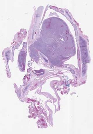
Fig 1 Carcinoid tumor. Carcinoid tumor arising from the bronchus, seen as a polypoid endobronchial mass, with extrabronchial invasion.
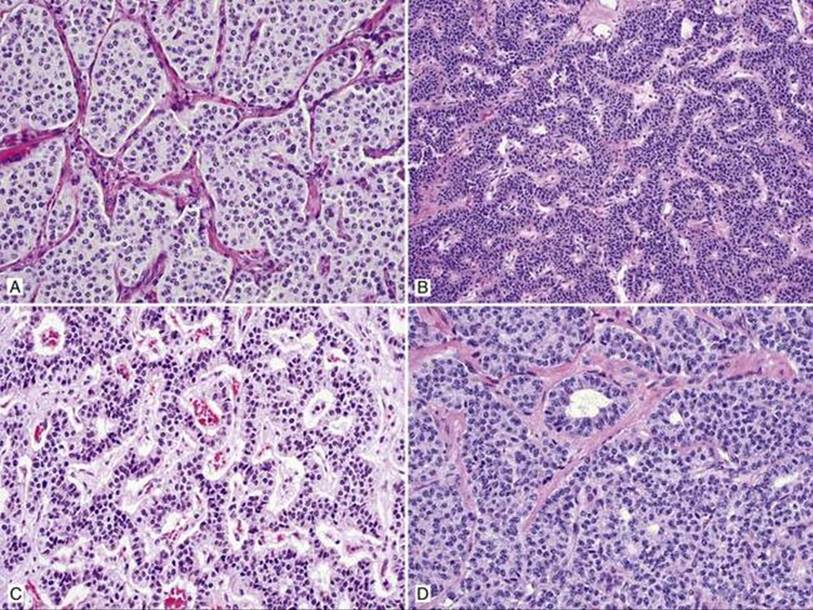
Fig 2 Carcinoid tumor. Carcinoid tumor with polygonal cells arranged in different growth patterns: A, nested; B, trabecular; C, ribbonlike; D, rosettelike.
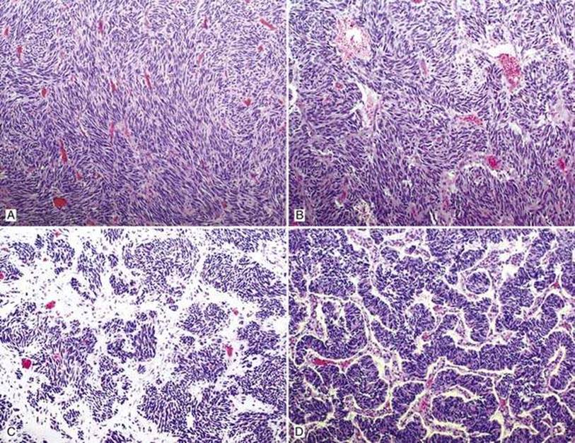
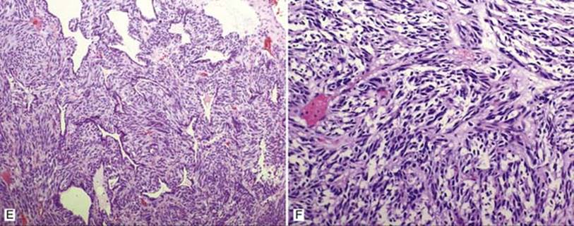
Fig 3 Carcinoid tumor. Carcinoid tumor with spindle cell morphology: A, arranged in long fascicles; B, in short fascicles with vague storiform pattern; C, with loose myxoid stroma; D, ribbonlike (gyriform) pattern; E, with entrapped alveoli lined by pneumocytes; F, clear cell change.
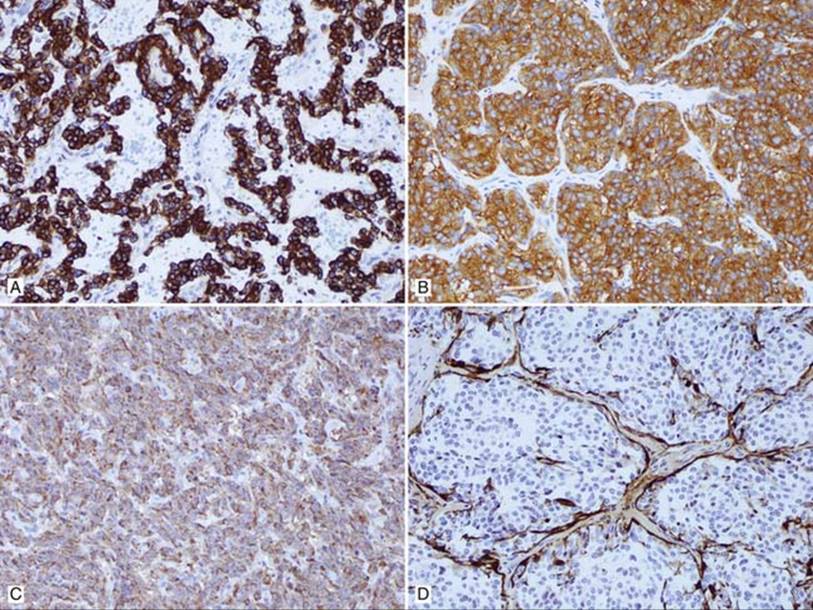
Fig 4 Carcinoid tumor. Carcinoid tumor with immunohistochemical stains showing A, keratin CAM5.2 strongly positive; B, synaptophysin strongly positive; C, chromogranin moderately positive with granular cytoplasmic staining; D, S100 negative in tumor cells but positive in sustentacular cell network.