
Definition
• A high-grade neuroendocrine carcinoma with large cells and prominent nucleoli
Clinical features
Epidemiology
• Rare; accounts for about 3% of all lung cancers
• Mean age at diagnosis, 64 years
• Strong association with smoking
Presentation
• Central tumors present with obstructive symptoms: cough, hemoptysis, recurrent infections
• Peripheral tumors present with chest pain
Prognosis and treatment
• Surgical resection and chemotherapy
• Aggressive tumor with dismal prognosis, similar to small-cell carcinoma
Pathology
Gross
• Usually large, bulky central tumor with extensive necrosis
• Grossly indistinguishable from other lung carcinomas
Histology
• High-grade tumors with organoid, trabecular, palisading growth patterns or rosette formation
• Large cells with abundant cytoplasm; nuclei can be vesicular or have “salt and pepper” chromatin
• Prominent nucleoli help to distinguish from small-cell carcinoma
• High mitotic activity: >10 mitoses per 10 high-power fields (hpf) (average, 60-70 mitoses per 10 hpf)
• Extensive necrosis
• Difficult to diagnose: neuroendocrine growth pattern is first identified by light microscopy, then must be confirmed by clear-cut positive immunoreactivity with at least one specific neuroendocrine marker (chromogranin, synaptophysin, or CD56)
Immunopathology/special stains
• Diffusely positive for cytokeratins and carcinoembryonic antigen (CEA), which highlight the large amount of cytoplasm
• 50% of tumors express TTF-1
• Positive for neuroendocrine markers, but staining is often patchy and relatively weak compared with carcinoid tumors
• Chromogranin is less likely to be positive than synaptophysin, but it is more specific
Main differential diagnoses
• Atypical carcinoid: 2 to 10 mitoses per 10 hpf, and/or punctate necrosis
• Small-cell carcinoma: no nucleoli, scanty cytoplasm
• Poorly differentiated adenocarcinoma with focal neuroendocrine differentiation: <10% of cells show positivity for neuroendocrine markers
• Large-cell carcinoma: majority have no evidence of neuroendocrine differentiation by light microscopy, immunohistochemistry (IHC), or electron microscopy; some tumors may show focal/weak IHC staining for neuroendocrine markers

Fig 1 Large-cell neuroendocrine carcinoma. Nested organoid pattern at low (A) and medium (B) powers. Note the prominent nucleoli.

Fig 2 Large-cell neuroendocrine carcinoma. Tumor has peripheral palisading.

Fig 3 Large-cell neuroendocrine carcinoma. Extensive necrosis at low (A) and high (B) powers.
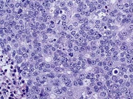
Fig 4 Large-cell neuroendocrine carcinoma. High power shows nuclei with vesicular chromatin and conspicuous nucleoli; note high mitotic activity.

Fig 5 Large-cell neuroendocrine carcinoma. Large-cell neuroendocrine carcinoma is diffusely positive for cytokeratin AE1/AE3. Note the normal bronchial epithelial cells (top left) and pneumocytes (top right) are also immunoreactive.
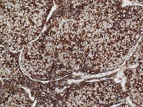
Fig 6 Large-cell neuroendocrine carcinoma. The tumor is diffusely positive for CEA, which highlights the abundant cytoplasm.

Fig 7 Large-cell neuroendocrine carcinoma. Large-cell neuroendocrine carcinoma with positive staining for chromogranin (A), synaptophysin (B), and CD56, membranous pattern (C).
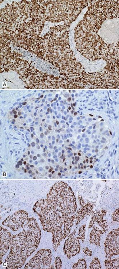
Fig 8 Large-cell neuroendocrine carcinoma. Positive TTF-1 immunostain can be diffuse (A) or focal (B). C, The tumor cells are strongly positive for p53.