
Definition
• A high-grade neuroendocrine carcinoma with small cells and indistinct/absent nucleoli
Clinical features
Epidemiology
• Accounts for about 15% of lung cancers in the United States
• Almost exclusively in smokers
• Peak age, 55 to 65 years
• Slightly more common in men than women
Presentation
• Usually presents with bloody sputum, chest pain, coughing, shortness of breath, weight loss, wheezing
• Hoarseness and vocal cord paralysis are common
• Widespread metastases occur early in the course of the disease, with spreading to mediastinal lymph nodes, liver, bones, adrenal glands, and brain
• Commonly associated with paraneoplastic syndromes: most frequently the syndrome of inappropriate secretion of antidiuretic hormone and the syndrome of ectopic adrenocorticotropic hormone production
• Infrequently associated with Lambert-Eaton myasthenic syndrome: patients with small-cell lung carcinoma (SCLC) produce autoantibodies that bind to cancer cells and nonneoplastic cells of the central nervous system or neuromuscular junction, resulting in cerebellar degenerative syndrome
• The seventh edition of the American Joint Committee on Cancer TNM lung cancer staging is now applicable for SCLC (effective January 1, 2010) (see section on lung carcinoma staging)
• It was staged as limited or extensive disease (prognostic data based on old system)
• Limited disease (30% to 40% of patients): disease confined to one hemithorax
– With regional lymph node metastases, including
– Hilar and ipsilateral lymph nodes
– Contralateral, mediastinal, or supraclavicular nodes (since the prognosis is still better than that of distant metastasis)
– With ipsilateral pleural effusion
• Extensive disease (60% to 70% of patients): any cases that cannot be included in the limited stage
Prognosis and treatment
• A small percentage of limited disease may be successfully resected; 5-year survival rate of limited disease is about 20%
• Extensive disease is usually treated with chemotherapy; initially responds to chemotherapy
• Extensive disease has a dismal prognosis with a 2-year survival rate of about 5%
Pathology
Gross
• About 70% of cases are seen as central peribronchial masses; endobronchial lesions are uncommon
• White-tan, soft, friable perihilar mass with extensive necrosis and frequent nodal metastases
• About 5% are seen as peripheral coin lesions
Histology
• Tumor arises in peribronchial location and infiltrates the bronchial submucosa
• Tumor cell size alone is not a reliable diagnostic criterion: it can be small or medium; tumor cells are relatively larger in well-preserved resection specimens
• Nuclear molding is prominent due to scanty cytoplasm and indistinct cell borders
• Tumor cell nuclei may be round, oval, or spindle-shaped, with finely granular chromatin and a classic “salt and pepper” appearance
• Absent or inconspicuous nucleoli is the most useful diagnostic feature
• Often associated with extensive necrosis and brisk mitotic activity; average, >60 mitoses per 10 high-power fields (hpf)
• Tumor cells can be easily crushed (especially in biopsy specimens) and difficult to distinguish from lymphocytes
• Azzopardi effect: DNA from necrotic tumor cells gets deposited in the walls of vessels and connective tissue; however, this effect can be seen in any high-grade tumor
• Growth patterns may be similar to other neuroendocrine cell tumors: nesting, trabecular, or rosette formation or sheetlike without a neuroendocrine pattern
• Combined small-cell carcinoma:
• Less than 3% of total small-cell lung cancers
• Small-cell carcinoma combined with non–small-cell component
• Non–small-cell component can be any of the non–small-cell carcinomas; more commonly, adenocarcinoma, squamous cell carcinoma, or large-cell carcinoma; less commonly, spindle cell or giant cell carcinoma
• For combined small-cell and large-cell carcinoma, the large-cell component should be at least 10% or more
• Non–small-cell component needs to be specified in the diagnosis; however, SCLC is mentioned first
• Prognosis is determined by SCLC
Immunopathology/special stains
• Cytokeratin (including CK7) and EMA: positive in almost all cases
• TTF-1: positive in about 90% of cases; note: extrapulmonary small-cell carcinomas can be positive for TTF-1 in up to one third of cases
• Neuroendocrine markers:
• About 90% of SCLCs are immunoreactive with one or more of the markers; <10% are negative for all markers
• Synaptophysin is more likely to be positive but is less specific than chromogranin
• Negative neuroendocrine staining does not exclude SCLC; the diagnosis is made by light microscopy
• Keratin, synaptophysin, and chromogranin often have a dotlike staining pattern and highlight that the tumor cells have only scant cytoplasm
Main differential diagnoses
• Lymphocytic infiltrates in a small biopsy: LCA positive, cytokeratin negative
• Small-cell variant of squamous cell carcinoma: p63 and CK5/6 positive; usually negative for TTF-1, CK7, and neuroendocrine markers
• Thymic carcinoma: mediastinal mass; positive for CD5 and cytokeratins; negative for TTF-1 and neuroendocrine markers
• Other neuroendocrine tumors:
• Large-cell neuroendocrine carcinoma: lower nuclear/cytoplasmic ratio, prominent nucleoli, lack of nuclear molding
• Atypical carcinoid: lower mitotic count (2 to 10 mitosis per 10 hpf) or punctate necrosis
• Other small round blue cell tumors:
• Lymphoma: LCA positive, cytokeratin negative
• Primitive neuroectodermal tumors: especially in younger patients; less mitotically active; cytokeratin and TTF-1 negative; MIC-2 (CD99) and FLI-1 positive
• Metastatic Merkel cell carcinoma: CK20 positive; CK7 and TTF-1 negative
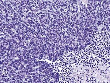
Fig 1 Small-cell carcinoma. SCLC showing spindled to oval nuclei with high mitotic activity, apoptosis, and necrosis.
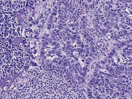
Fig 2 Small-cell carcinoma. High-power view of small-cell carcinoma with finely granular chromatin, inconspicuous nucleoli, and scant cytoplasm; note prominent nuclear molding and larger size of tumor cells as compared with mature lymphocytes.

Fig 3 Small-cell carcinoma. SCLC in a small biopsy with crush artifact; difficult to distinguish from crushed lymphocytes. A, H&E; B, positive keratin CAM5.2 with dotlike pattern; C, neuroendocrine marker chromogranin, also dotlike.

Fig 4 Small-cell carcinoma. Azzopardi effect with crushed DNA around blood vessels in necrotic area.

Fig 5 Small-cell carcinoma. Cytokeratin CAM5.2 immunostain highlights scant amount of cytoplasm in this surgical resection of SCLC.
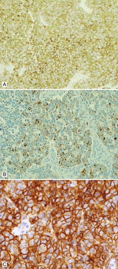
Fig 6 Small-cell carcinoma. Neuroendocrine markers: A, synaptophysin with granular staining of cytoplasm; B, chromogranin, which is less strongly positive than synaptophysin; C, CD56 with membranous staining.
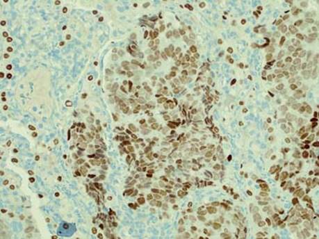
Fig 7 Small-cell carcinoma. SCLC with positive TTF-1 immunostain (nuclear); note residual positive pneumocytes.

Fig 8 Small-cell carcinoma. A case of combined small-cell carcinoma with squamous cell carcinoma component: H&E (A), and p63 immunostain (B) shows positive nuclear staining of squamous cell carcinoma cells and only rare reactivity of SCLC component.