
Definition
• A malignant soft tissue sarcoma that recapitulates the morphological and biological features of skeletal muscle
Clinical features
Epidemiology
• Primary rhabdomyosarcoma of the lung is rare and mostly confined to the pediatric population
• Some tumors reported as being associated with underlying lesions, including congenital cystic adenomatoid malformation and peripheral bronchogenic cyst, are now thought to represent pleuropulmonary blastoma, types 1 and 2
• In the adult it may rarely be seen arising from the pulmonary artery
Presentation
• Nonspecific, including coughing, dyspnea, or, rarely, spontaneous pneumothorax
• Radiographically, a single, homogeneous intraparenchymal mass on CT or MRI
Prognosis and treatment
• Surgical resection with addition of postoperative chemotherapy and radiation therapy
• Poor prognosis
Pathology
Gross
• Poorly circumscribed, fleshy, pale-tan mass with additional features for botryoid and spindle cell variants
• Botryoid rhabdomyosarcoma: polypoid mass with attached small, sessile, or pedunculated nodules
• Spindle cell rhabdomyosarcoma: firm, fibrous tumor with tan-yellow, whorled cut surfaces
Histology
• Primitive mesenchymal cells in various stages of myogenesis, including primitive round cells with amphophilic cytoplasm and central, oval nuclei, more differentiated “tadpole” cells with eosinophilic cytoplasmic inclusion, terminal differentiated rhabdomyoblasts with cytoplasmic cross-striation and multinucleation, and pleomorphic polygonal rhabdomyoblasts
• Histological architecture varies for different variants
• Embryonal: solid, sheetlike growth pattern or alternate dense cellular and loose myxoid stroma
• Botryoid: characterized by forming “cambium layer,” which is a linear condensation of tumor cells that tightly abut an epithelial surface
• Alveolar: collagenous fibrovascular septa divide the tumor cells into discrete nests; central cells in the nests are discohesive and detached from peripheral cells. Tumor cells are aligned along the septa in a picket fence pattern
• Pleomorphic: diffuse proliferation with areas of necrosis and hemorrhage
Immunopathology/special stains
• Positive for at least one or more skeletal muscle markers: myogenin, myoD1, desmin, myoglobin, myosin
• Invariably positive for vimentin and SMA
• Negative for cytokeratin and EMA
Ultrastructure
• Cytoplasmic sarcomeric differentiation with formation of Z bands
Main differential diagnoses
• Diffuse large B-cell lymphoma: diffuse proliferation of large neoplastic B lymphoid cells; positive for B-cell markers: CD45, CD20, CD19, CD79a
• Primitive neuroectodermal tumor/Ewing sarcoma: round cell sarcoma demonstrating neuroectodermal differentiation; positive for CD99, FLI1, with t(11;22)(q24;q12) translocation
• Neuroblastoma: embryonic neoplasm of sympathetic nervous system; positive for synaptophysin, chromogranin, PGP9.5
• Angiosarcoma: usually metastatic, with primitive vascular channels formed by primitive round or spindly tumor cells; tumor cells are positive for CD31, CD34, and FLI1

Fig 1 Rhabdomyosarcoma lung. Grossly, the tumor mass is well-delineated, white-tan, soft, and with myxoid degenerative areas.

Fig 2 Rhabdomyosarcoma lung. This embryonal rhabdomyosarcoma is composed of spindle cells in a myxoid stroma with a sheetlike growth pattern (A); desmin stain is positive in the tumor cells (B).
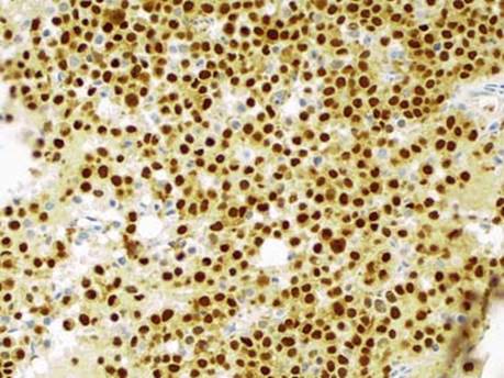
Fig 3 Rhabdomyosarcoma lung. Strong nuclear myogenin staining is seen in this alveolar rhabdomyosarcoma.