
Definition
• A rare lung tumor composed of perivascular epithelioid cells
Clinical features
Epidemiology
• Usually in adults age 40 to 50 years (range, 8-73 years)
• Slight female predominance
• Can be associated with tuberous sclerosis
Presentation
• Usually a peripheral lesion found incidentally on imaging studies
• Rarely is seen as a central lesion with symptoms such as hemoptysis, fever, chest pain, shortness of breath
Prognosis and treatment
• Usually benign with an excellent prognosis
• Treatment consists of surgical excision
Pathology
Gross
• Very well-circumscribed
• Red-tan cut surface
• No hemorrhage or necrosis
Histology
• Sheets of cells with abundant clear to slightly eosinophilic cytoplasm and well-defined cell borders
• Very vascular tumor with thin-walled sinusoidal blood vessels
• Multinucleated tumor cells
• Spider cells: large cells with lines of eosinophilic granules radiating from the nucleus
• No cytological atypia; low nuclear to cytoplasmic ratio; nucleoli may be prominent
• Low to no mitotic activity; no abnormal mitoses
• No necrosis
• Entrapped respiratory epithelium present
• May have brown lipochrome pigment (important to distinguish from melanin)
Immunopathology/special stains
• Positive for HMB45, Melan-A
• Negative for cytokeratin
• Cytoplasm is filled with glycogen that is PAS-positive and diastase-sensitive
Main differential diagnoses
• Metastatic renal cell carcinoma: positive for cytokeratin
• Clear cell carcinoid of the lung: positive for neuroendocrine markers (chromogranin, synaptophysin)
• Metastatic melanoma: positive for S100 and Mitf1
• Clear cell sarcoma: positive for S100 and HMB45
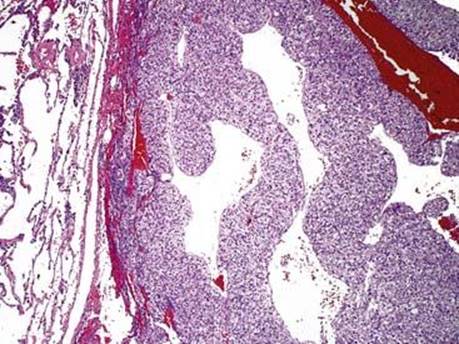
Fig 1 Perivascular epithelioid cell tumor. Well-circumscribed and composed of sheets of cells with clear cytoplasm and sinusoidal blood vessels.
(Courtesy of Dr. Tazelaar, Mayo Clinic, Scottsdale, Ariz.)
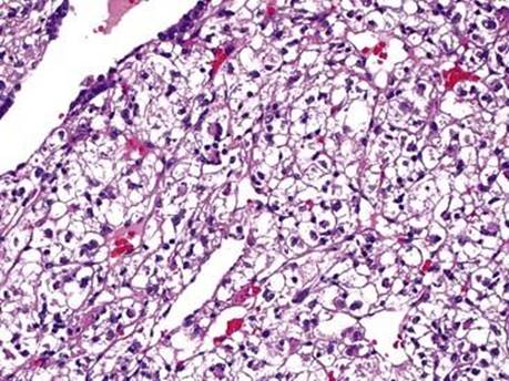
Fig 2 Perivascular epithelioid cell tumor. PEComa with delicate vascular network and epithelioid tumor cells with abundant clear cytoplasm and well-defined cell borders.
(Courtesy of Dr. Tazelaar, Mayo Clinic, Scottsdale, Ariz.)
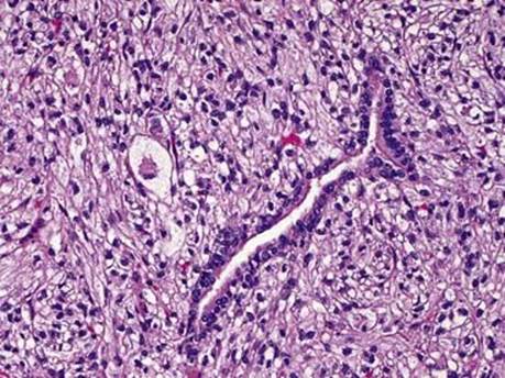
Fig 3 Perivascular epithelioid cell tumor. Entrapped respiratory mucosa and spider cells (large cells with eosinophilic granular material, to the left and upper left of the epithelium) are present.
(Courtesy of Dr. Tazelaar, Mayo Clinic, Scottsdale, Ariz.)
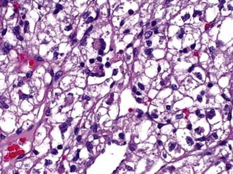
Fig 4 Perivascular epithelioid cell tumor. High-power view showing multinucleated tumor cells.
(Courtesy of Dr. Tazelaar, Mayo Clinic, Scottsdale, Ariz.)

Fig 5 Perivascular epithelioid cell tumor. Tumor cells with cytoplasmic lipochrome pigment, which is important to distinguish from melanin.
(Courtesy of Dr. Tazelaar, Mayo Clinic, Scottsdale, Ariz.)
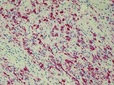
Fig 6 Perivascular epithelioid cell tumor. Tumor cells show strong expression of HMB45.
(Courtesy of Dr. Tazelaar, Mayo Clinic, Scottsdale, Ariz.)