
Definition
• Heterogeneous group of lymphoid or plasmacytic proliferations occurring in patients who have received solid organ, bone marrow (BM), or stem cell transplantation (SCT)
Pathogenesis
• Because of decreased T-cell immune surveillance as a consequence of immunosuppression
• Majority associated with Epstein-Barr virus (EBV) infection
• In SCT allograft recipients, majority are of donor origin
• In solid organ recipients, majority are of host origin
• EBV-negative PTLDs are probably caused by human herpesvirus-8 (associated with primary effusion lymphoma), other unknown viruses, undetectable EBV, or chronic antigenic stimulation
Clinical features
Epidemiology
• EBV seronegative recipients are at high risk of EBV-driven PTLD
• Children are at much higher risk of PTLD, and most are associated with posttransplantation primary EBV infection
• Incidence is low (approximately 1%) in peripheral blood, stem cell, and BM allografts and usually associated with an early onset (<6 months)
• In adult solid organ allografts, incidence partially correlates with intensity of immunosuppressive regimen:
• Lowest frequency in renal transplant recipients (<1%)
• Intermediate frequency in hepatic and cardiac recipients (1%-2%)
• Highest frequency in lung and intestinal recipients (>5%)
• In lung transplant patients, PTLDs frequently involve the allograft
Presentation
• Nonspecific symptoms with fever, malaise, lethargy, and weight loss
• Usually with lymphadenopathy and organ-specific dysfunction
• Lymph node, gastrointestinal tract, lungs, and liver are commonly involved
• Central nervous system involvement is rare
• When lung is involved, patients present with coughing, chest pain, and shortness of breath
Prognosis and treatment
• Treatment
• Early lesions and some of polymorphic PTLDs (P-PTLD) regress with reduction of immunosuppression
• The remaining P-PTLDs and most of monomorphic PTLDs (M-PTLD) require additional therapies, such as anti-CD20 monoclonal antibody immunotherapy and/or chemotherapy
• Prognosis
• Long-term survival is dependent on the particular form of the disease and other factors such as graft function
• Early PTLDs tend to have excellent prognosis with reduction of immune suppression
• Prognosis is better in children than adults
• Mortality is greater in BM/SCT allograft recipients than in solid organ recipients
Pathology
World Health Organization classification
• Early lesions
• Involve lymph nodes or tonsils and adenoids; rarely, extranodal sites
• Occur in younger patients with no prior EBV infection
• Histologically characterized by plasmacytic hyperplasia or infectious mononucleosis-like lesion with architectural preservation
• Usually with EBV+ cells by EBER in situ hybridization
• P-PTLD
• Involve lymph nodes and extranodal sites
• Younger patients with no prior EBV infection
• Destructive lesions with effacement of underlying tissue architectures
• Full range of B-cell maturation with immunoblasts, plasma cells, and small and intermediate lymphocytes
• May have geographical necrosis, atypical mitoses, and/or scattered large bizarre cells
• May have focal monomorphic areas with focal light chain restriction
• Most cases show numerous EBV+ cells by EBER in situ hybridization
• M-PTLD
• Fulfill the diagnostic criteria of B-cell or T/natural killer (NK)-cell neoplasms of immunocompetent patients
• With or without EBV positivity
• Monomorphic B-cell PTLD subtypes
– Diffuse large B-cell lymphoma
– Burkitt lymphoma
– Plasma cell myeloma
– Plasmacytoma-like lesion
• T/NK-cell PTLD: includes almost entire spectrum of T-cell and NK-cell neoplasms
• Classical Hodgkin lymphoma-type PTLD
• The least common form of PTLD
• Most common in renal allografts
• Almost always EBV positive
Lung involvement of PTLD
• PTLDs of the lung are mostly P-PTLD and M-PTLD
• Rarely involved by early lesions
Main differential diagnosis
• Acute cellular rejection
• Not a mass lesion; transbronchial biopsy is diagnostic
• Usually with perivascular, peribronchiolar, and septal mixed mononuclear infiltrates
• EBV negative

Fig 1 Posttransplant lymphoproliferative disorder. Low-power view of M-PTLD of lung in a 9-year-old patient 4.5 months after liver transplantation (for massive hepatic necrosis) showing a mass lesion extending into the pleura.
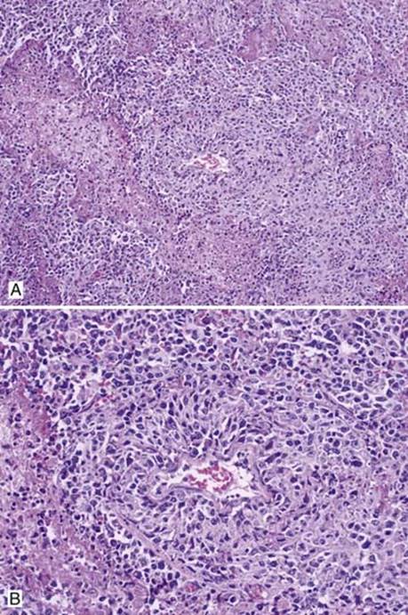
Fig 2 Posttransplant lymphoproliferative disorder. Intermediate (A) and high (B) powers showing focal necrosis and vascular involvement by PTLD.
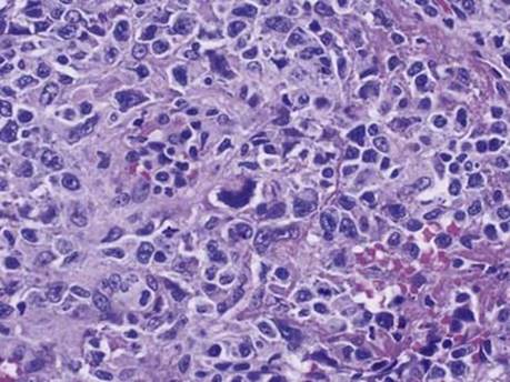
Fig 3 Posttransplant lymphoproliferative disorder. High-power view of M-PTLD showing clearly malignant cells with prominent nucleoli and atypical mitosis.
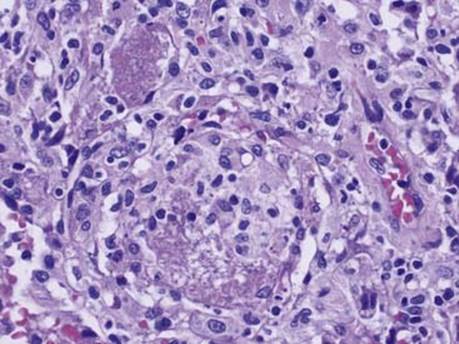
Fig 4 Posttransplant lymphoproliferative disorder. This patient also had Pneumocystis jiroveci infection in adjacent lung seen here as characteristic fibrinous exudate with punctate blue dots in alveoli, on H&E stain.
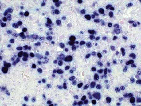
Fig 5 Posttransplant lymphoproliferative disorder. In situ hybridization for EBER shows many positive cells.