
Definition
• LIP is a diffuse interstitial process that is characterized by small cytologically bland lymphocytes and plasma cells in a predominantly interstitial pattern of distribution
Clinical features
Epidemiology
• LIP is more commonly seen in women than in men, typically between 40 and 50 years of age
• It is increasingly diagnosed in patients with altered immune states such as from autoimmune diseases (Sjögren syndrome, systemic lupus erythematosus), AIDS, chronic infections with Epstein-Barr virus, congenital immunodeficiency, and bone marrow transplantation
• Children with AIDS may present with LIP, which is considered to be an AIDS-defining illness in patients younger than 13 years
Presentation
• The most frequent presenting symptoms are a cough and/or shortness of breath
• A small subset of individuals may show systemic symptoms such as fever, weight loss, and polyclonal hypergammaglobulinemia
• Radiographically, lesions are seen as bilateral basilar patchy opacities or reticulonodular infiltrates
Prognosis and treatment
• Corticosteroids or antineoplastic agents have been used with varied success
• The clinical course for patients with LIP ranges from complete resolution to death caused by superimposed infection
• A subset of LIP cases progress to end-stage pulmonary fibrosis with honeycomb changes, imparting a worse prognosis
• The overall mortality is approximately 50% at 5 years after diagnosis
Pathology
Histology
• Characterized by a dense (“coat-sleeve”) and diffuse inflammatory infiltrate present around blood vessels in the interstitium; however, angiodestruction is not a feature
• Infiltrates consist of small, cytologically bland lymphocytes admixed with plasma cells, plasmacytoid cells, and histiocytes that distend the alveolar walls
• Multinucleated giant cells and small nonnecrotizing granulomas may be present
• Neutrophils and eosinophils are scarce
• Germinal centers may be present (20% to 30%) to a variable extent along airways and lymphatic routes, and when prominent, diffuse lymphoid hyperplasia is the preferred term
Immunopathology/special stains
• CD20+ lymphocytes represent >75% of cells; no coexpression of CD20 and CD43 in the same cell
• Immunophenotyping and gene rearrangement studies demonstrate an absence of clonality
Main differential diagnoses
• Low-grade malignant lymphoma of the bronchus-associated lymphoid tissue (BALT): often has lymphocyte invasion of epithelial structures (lymphoepithelial lesions); monoclonal, CD20+, and CD43+/-
• Nonspecific interstitial pneumonia
• Sjögren syndrome
• Hypersensitivity pneumonitis
• Atypical mycobacterial infections
• Drug reactions
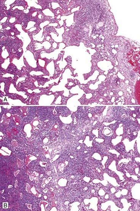
Fig 1 Lymphoid interstitial pneumonia. Variable amount of lymphoid infiltrate is present in the interstitium (A and B).
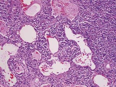
Fig 2 Lymphoid interstitial pneumonia. An inflammatory infiltrate has caused marked widening of alveolar walls.
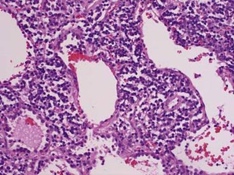
Fig 3 Lymphoid interstitial pneumonia. At high power, mature small lymphocytes and plasma cells are seen.
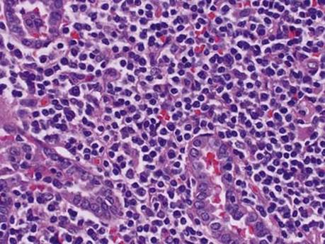
Fig 4 Lymphoid interstitial pneumonia. In this area of dense, lymphoplasmacytic infiltrate, reactive type II pneumocytes are present. Although the infiltrate surrounds the epithelial structures, no “lymphoepithelial lesions” are present.