
Definition
• Secondary carcinomas originating from extrapulmonary sites or discontinuous from a primary lung carcinoma
Clinical features
Epidemiology
• A third to a half of all malignant lung tumors are metastases because of rich pulmonary vasculature collecting entire blood volume and lymphatic fluid output
• Most common primary sites include breast, colon, kidney, stomach, thyroid, head and neck, pancreas, female genitalia, liver, prostate, adrenal
• Metastatic pulmonary disease is more commonly seen in surgical specimens because of increased palliative surgeries and overall improvement in cancer survival rates
Presentation
• History of prior malignancy
• X-ray films and CT scans are best diagnostic modalities, most commonly showing multiple well-defined rapidly enlarging nodules of mid-to-lower lobes
• Depends on extent of lung involvement and mechanism of cancer spreading:
• Hematogenous spreading to small vessels: initially asymptomatic, then develop chest pain, dyspnea, coughing, hemoptysis, wheezing
• Lymphogenous spreading: initially asymptomatic, then develop dyspnea, cor pulmonale
• Large-vessel tumor emboli: acute heart failure, rapidly developing pulmonary hypertension, pulmonary infarction, cor pulmonale, sudden death
• Pleural infringement: chest pain, Pancoast syndrome, spontaneous pneumothorax, pleural effusions
• Endobronchial metastasis: “adult-onset asthma”
Prognosis and treatment
• Prognosis varies depending on the extent of metastatic disease and tumor type, but overall long term survival is poor
• Metastasectomy in limited disease; chemotherapy and radiation for widespread disease
Pathology
Gross
• Four main macroscopic types in order of frequency:
• Multiple bilateral parenchymal well-circumscribed nodules
• Solitary nodule (peripheral or central)
• Cavitary or cystic lesion
• Polypoid endobronchial mass
• Size: average 1 to 2 cm but highly variable depending on “age” and type of metastasis from small miliary to large confluent “cannonball” masses
• Location: periphery of lower lobes is most common
• Necrosis and hemorrhage often present
Histology
• Highly variable: well-differentiated adenomatous, clear cell, papillary, signet-ring, round cell, squamous, mucinous, oncocytic, small round cell, sarcomatoid, and spindle cell
• Nodular, perivascular, lepidic, interstitial, polypoid luminal
• In cases of resection of solitary tumor, surgical margin must be carefully assessed
Immunopathology/special stains
• Highly valuable in differentiating primary lung cancer from various secondary carcinomas
• Diagnostic table for prediction of primary site of metastatic carcinoma using immunohistochemical (IHC) panel (see Table 8 in the Appendix)
Main differential diagnoses
• Primary lung carcinoma: TTF-1 positive
• Tuberculosis, sarcoidosis: presence of AFB-positive organisms, absence of cell atypia
• Interstitial lung disease: absence of cell atypia
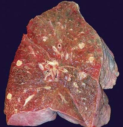
Fig 1 Metastatic carcinomas. Gross image of lung cut surface with multiple well-circumscribed parenchymal nodules of metastatic pancreatic cancer.
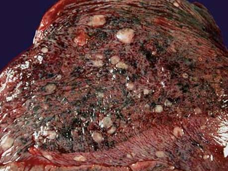
Fig 2 Metastatic carcinomas. Gross image of pleural surface showing numerous subpleural nodules of metastatic renal cell carcinoma.

Fig 3 Metastatic carcinomas. Large solitary mass of metastatic colonic adenocarcinoma with necrosis.

Fig 4 Metastatic carcinomas. Hematogenous spreading of metastatic ovarian carcinoma (A). Tumor emboli in the center show strong cytokeratin immunoreactivity (B); note positive pneumocytes.

Fig 5 Metastatic carcinomas. Low-power image of multifocal metastatic adenocarcinoma from colon.

Fig 6 Metastatic carcinomas. Well-demarcated breast cancer metastasis with minimal desmoplastic reaction.

Fig 7 Metastatic carcinomas. Subpleural peripheral tumor nests of metastatic breast carcinoma.

Fig 8 Metastatic carcinomas. Lepidic growth of pancreatic cancer imitating primary lung adenocarcinoma.

Fig 9 Metastatic carcinomas. TTF-1 immunostaining shows strong nuclear staining only in residual pneumocytes, whereas metastatic carcinoma is negative.