
Definition
• Secondary lung carcinomas from primary breast or female genitalia
Clinical features
Epidemiology
• Lungs are the most common site of metastases from breast and gynecological tract
• The frequency of lung metastasis at presentation is 10% to 15% for cervical cancer and 2% to 5% for breast, endometrial, and ovarian cancers
• The frequency of lung metastasis at autopsy: breast (60%), ovarian (45%), endometrial (40%), and cervical (30%)
Presentation
• Breast carcinoma: classically presents with lymphangitic spreading/lymphangitic carcinomatosis (LC) causing shortness of breath and dyspnea but also could manifest with:
• Malignant pleural effusions
• Direct chest extension, chest pain, and upper extremity lymphedema
• Endobronchial metastasis: “adult-onset asthma”
• Ovarian carcinoma most commonly presents with pleural effusions
• Cervical and endometrial carcinomas spread hematogenously and are initially asymptomatic, then develop chest pain, dyspnea, coughing, hemoptysis, wheezing
Prognosis and treatment
• Prognosis is dismal
• Metastasectomy in limited disease; chemotherapy and radiation for widespread disease
Pathology
Gross
• Breast carcinoma:
• LC: most common: thickening of intervascular bundles, interlobular septae and pleura, hilar, or mediastinal lymphadenopathy
• Endobronchial mass
• Solitary nodule (cannonball)
• Cervical carcinoma: cavitary nodules
• Ovarian carcinomas: pleural metastases, effusions, and thickening
• Uterine carcinomas: multiple nodules at periphery of lower lobes are most common
• Adenocarcinomas: firm, gray-tan with areas of hemorrhage and necrosis
• Any mucin-secreting tumor: wet, slimy, glistening, yellow-tan cut surface
Histology
• Variable: adenomatous or mucinous (breast, ovary, cervix), serous-papillary (ovary, uterus), endometrioid (uterus), squamous cell (cervix), solid
• Interstitial thickening and desmoplastic reaction (breast)
• Tumor emboli are common (breast, ovary)
Immunopathology/special stains
• TTF-1 is virtually negative in breast and female genital tract metastases, except for small-cell carcinomas from female genitalia
• CK7 is generally strongly positive in carcinomas from breast, ovary, uterus, and cervix
• The most useful positive immunohistochemical markers for metastatic carcinomas from breast and female genital tract are listed in Table 9 in the Appendix
Main differential diagnoses
• Primary lung carcinoma: TTF-1 positive
• Interstitial lung disease, lymphangitic lymphoma or lymphangitic sarcoidosis: absence of cell atypia

Fig 1 Metastatic carcinomas: breast and female genital tract. Lymphangitic spreading of breast adenocarcinoma with dramatic thickening of pleura, interlobular septae, perivascular and peribronchial interstitium. LC conveys very poor prognosis (half of patients die within 3 months) with rapid tumor dissemination and massive pleural effusions.

Fig 2 Metastatic carcinomas: breast and female genital tract. Retrograde LC of breast cancer, which is spreading from hilum in a radial fashion forming a fine network of connected lines along interlobular septae.

Fig 3 Metastatic carcinomas: breast and female genital tract. Nests of poorly differentiated necrotizing breast carcinoma within periarterial and peribronchiolar lymphatic spaces.

Fig 4 Metastatic carcinomas: breast and female genital tract. Same case showing intense nuclear reactivity for ER.
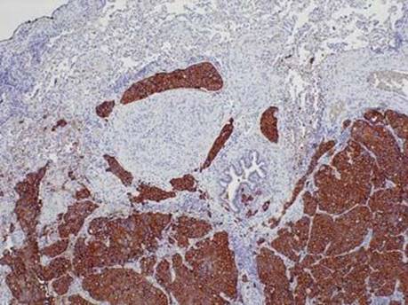
Fig 5 Metastatic carcinomas: breast and female genital tract. Same case of metastatic breast carcinoma with strong membranous expression of Her2/neu antibody.

Fig 6 Metastatic carcinomas: breast and female genital tract. The vast majority of primary breast and gynecological metastatic carcinomas are strongly positive with CK7, as shown here, which makes differential diagnosis with primary lung carcinomas difficult.
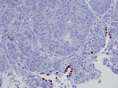
Fig 7 Metastatic carcinomas: breast and female genital tract. Negative staining with TTF-1 of breast and gynecological carcinomas is essential in differential diagnosis. Note residual positive pneumocytes.

Fig 8 Metastatic carcinomas: breast and female genital tract. Same case as in Fig 7 exhibiting scattered strong nuclear reactivity with PR antibody.