
Definition
• Secondary lung carcinomas from head and neck organs
Clinical features
Epidemiology
• Head and neck is the fifth most common primary site for lung metastases; thyroid, larynx, and oropharynx are the most common primary sites
• Incidence of lung metastases from thyroid cancers at presentation is less then 10% but at autopsy is up to 65%
• One quarter of patients with head and neck squamous cell carcinomas will eventually develop lung metastases: incidence at presentation, 4% to 10%; at autopsy, 10% to 40%
• The incidence of synchronous or metachronous independent pulmonary malignancy in patients with head and neck squamous cell carcinomas ranges from 4.5% to 14%
• Salivary gland tumors may metastasize to lungs but usually after a long period of latency
Presentation
• Metastatic thyroid carcinomas:
• Papillary: asymptomatic, slow-growing, calcified nodule; 50% of cases associated with regional lymph node involvement
• Follicular: hematogenous spreading with solitary or multiple fleshy nodules
• Medullary: miliary, firm, gray-to-yellow, well-circumscribed nodules
• Anaplastic: direct tracheal invasion
• Metastatic squamous cell carcinomas: cavitary nodules with sputum production, dyspnea, wheezing, hemoptysis, bronchopleural fistulas, and postobstruction pneumonia
• Less commonly, metastatic squamous and thyroid carcinomas present with intrathoracic nodal spreading or endobronchial masses manifesting as “adult-onset asthma”
Prognosis and treatment
• Solitary nodules either from thyroid or head and neck squamous cell carcinoma have a favorable prognosis with 20% to 50% 5-year survival after resection
• Dismal for anaplastic or disseminated metastatic disease
Pathology
Gross
• Thyroid: usually solid, white to tan, firm or fleshy, well-circumscribed nodules with calcifications, cysts, or hemorrhages
• Metastatic squamous laryngeal and oropharyngeal carcinomas: cavities ranging from 1 to 6 cm with thick, shaggy walls, necrotic material, hemorrhages, and surrounding inflammation
• Metastatic salivary gland carcinomas: circumscribed, firm, solid, white, gray, or tan; commonly with cysts, necrosis, and hemorrhages
• Parathyroid: well-circumscribed multiple firm white-to-tan nodules
Histology
• Thyroid:
• Papillary: papillary structures, psammoma bodies, and distinct nuclear features including grooving, membrane irregularities, pseudoinclusions, and nuclear overlap
• Follicular: variable, ranging from well-formed follicles with colloid to trabecular, cribriform, or solid growth pattern with eosinophilic or clear cell change, nuclear pleomorphism, prominent nucleoli, and isolated bizarre nuclei
• Medullary: variable but most commonly solid sheets of round to polygonal cells with granular amphophilic cytoplasm, vascular hyalinized stroma with amyloid production
• Anaplastic: squamoid or sarcomatoid spindle or giant cells
• Larynx, oropharynx, and salivary gland: squamous cell, adenoid cystic, or acinic cell carcinomas with typical cytological and architectural appearance
• Parathyroid: solid nests or trabecular structures composed of clear cells or eosinophilic monotonous cells
Immunopathology/special stains
• The most useful positive and negative immunohistochemical markers for head and neck metastatic carcinomas are listed in Table 12 in the Appendix
Main differential diagnoses
• Primary squamous cell lung carcinoma: no reliable feature or diagnostic marker to predict the primary site; however, multiplicity of nodules favors secondary tumor
• Primary lung carcinoma with clear cell change or large cell type: TTF-1 positive
• Tuberculosis or sarcoidosis for miliary nodules: no atypia, characteristic granulomas
• Sarcomas: pancytokeratin negative

Fig 1 Metastatic carcinomas: head and neck. Metastatic thyroid papillary carcinoma seen as a slow-growing single nodule.

Fig 2 Metastatic carcinomas: head and neck. Higher magnification of the same tumor showing psammoma bodies, nuclear crowding, and overlap (A), nuclear grooves (B), and pseudoinclusions (C), all classic features of thyroid papillary carcinoma.

Fig 3 Metastatic carcinomas: head and neck. Thyroglobulin immunostaining is highly specific for primary thyroid carcinomas of both papillary and follicular types.
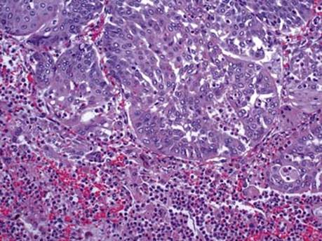
Fig 4 Metastatic carcinomas: head and neck. Metastatic moderately differentiated squamous cell carcinoma of the head and neck with associated bronchopneumonia.

Fig 5 Metastatic carcinomas: head and neck. Lymph node with metastatic squamous cell carcinoma: the majority of metastatic squamous cell carcinomas from larynx and oropharynx also present with simultaneous extensive hilar and mediastinal lymphadenopathy.

Fig 6 Metastatic carcinomas: head and neck. Metastatic Hurthle cell carcinoma displaying solid architecture with scant follicles and composed of monotonous cells with deeply eosinophilic granular cytoplasm packed with mitochondria.

Fig 7 Metastatic carcinomas: head and neck. Nearly all metastatic follicular carcinomas from thyroid are strongly positive with thyroglobulin (A) and TTF-1 (B), with the exception of poorly differentiated and anaplastic cancers.

Fig 8 Metastatic carcinomas: head and neck. Multiple variably sized nodules of metastatic parathyroid carcinoma.
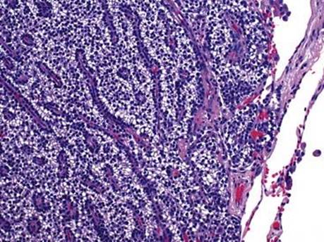
Fig 9 Metastatic carcinomas: head and neck. Characteristic trabecular and nested growth pattern of metastatic parathyroid carcinoma composed of clear cells with bland appearance resembling normal parathyroid gland.