
Definition
• Secondary lung carcinomas from primary GU tract organs and adrenal gland
Clinical features
Epidemiology
• Kidney is the most common primary GU site for lung metastases with a frequency of 5% to 33% at presentation and 55% to 75% at autopsy
• Lung metastases from the kidney may either precede the clinical detection of a primary tumor or develop several years after resection of a primary tumor
• Prostate and bladder have similar frequency of metastases at presentation (5%-15%) and autopsy (25%-38%)
• For prostate cancer, bone is almost invariably involved before the lungs
• About 7% to 10% of testicular tumors metastasize to the lungs
• Adrenal gland: true incidence of secondary adrenal carcinomas is unknown, but lung metastases to the adrenal glands are far more common than adrenal metastases to the lungs
Presentation
• Solitary nodule is the common presentation of renal cell carcinomas (RCCs), bladder carcinomas, and testicular nonseminomatous tumors, whereas prostate cancer most commonly presents with miliary nodules or lymphangitic carcinomatosis
• Second most common presentation of RCC and testicular choriocarcinomas is either small or large tumor emboli; multiple emboli cause shortness of breath, coughing, hemoptysis, and severe dyspnea; there can be pulmonary infarcts or acute heart failure, more typical for metastatic choriocarcinoma
• Thoracic lymph nodal disease with persistent coughing, wheezing, and dyspnea is also a frequent presentation for RCC, prostate cancer metastases, and testicular seminomas
• RCC, prostate cancer, and testicular cancers can also form an endobronchial mass causing coughing and dyspnea
• Adrenocortical carcinomas are often seen as a bulky mass with mediastinal lymphadenopathy
Prognosis and treatment
• Bladder and kidney solitary metastases have a relatively favorable prognosis with 50% 5-year survival on resection
• Testicular tumors have favorable prognosis with more than 60% 5-year survival for chemotherapy-sensitive tumors and 50% for chemotherapy-resistant metastases after their resection
• Prognosis is dismal for disseminated metastatic prostate cancer, especially in lymphangitic carcinomatosis
Pathology
Gross
• Kidney: solitary, well-circumscribed, white or yellow nodules; multiple variably sized emboli associated with infarcted lung areas; polypoid endobronchial mass or concentric airway narrowing
• Bladder: solitary gray firm nodules
• Prostate: enlargement of hilar or mediastinal lymph nodes, thickening of intervascular bundles and interlobular septae, or endobronchial mass
• Testis: seminomas, embryonal carcinomas, and yolk-sac tumors are well-circumscribed, fleshy tan-gray tumors with mucinous cut surface in the latter; choriocarcinomas are soft, hemorrhagic necrotic masses; teratomas show variegated appearance with cartilage, fat, cysts, hair, fluid, etc.
• Adrenal: large masses with necrosis and hemorrhage
Histology
• Metastatic RCCs most commonly display prominent cell clearing with solid architecture, blood lakes, rich vasculature network, and frequent sarcomatoid dedifferentiation
• Papillary, chromophobe, or oncocytic variants of metastatic RCC are rare
• Metastatic urothelial cell carcinomas from bladder or kidney retain characteristic transitional cell architecture but commonly show squamoid differentiation, extensive cellular dyscohesion, and necrosis
• Metastatic prostate cancers usually have acinar cribriform architecture or poorly differentiated high-grade histological features
• Testicular metastatic germ cell tumors commonly combine typical primary site morphological features with maturation effects
• Adrenocortical carcinoma: solid nests of large polygonal cells with abundant, commonly lipidized cytoplasm, large nuclei, cellular dyscohesion
Immunopathology/special stains
• TTF-1 is virtually negative in GU metastases, except in about one third of small-cell carcinomas of the bladder
• The most useful positive and negative immunohistochemical markers for GU metastatic carcinomas are listed in Table 11 in the Appendix
Main differential diagnoses
• Primary lung carcinoma with clear cell change, large-cell type or with giant cells: TTF-1 positive solitary lesion; older patients with smoking history
• Primary squamous cell lung carcinoma: no prior history of GU tumor
• Clear cell “sugar” tumor: HMB45+, MyoD1+, S100+, SMA+, absence of mitoses, necroses, and rich vasculature
• Sarcomatoid change or lymphangitic carcinomatosis: interstitial lung disease, lymphangitic lymphoma, or lymphangitic sarcoidosis; absence of cell atypia
• Metastatic melanomas: S100+, HMB45+
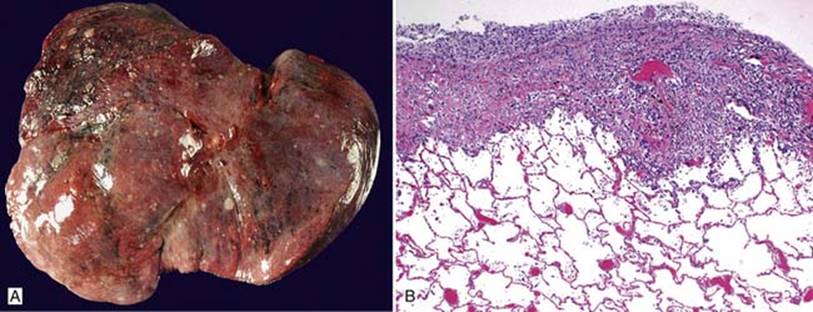
Fig 1 Metastatic carcinomas: genitourinary tract and adrenal. Numerous well-circumscribed subpleural nodules of widely metastatic RCC grossly (A) and microscopically (B).

Fig 2 Metastatic carcinomas: genitourinary tract and adrenal. Metastatic clear cell RCC with optically clear cells and thin-walled blood vessels.

Fig 3 Metastatic carcinomas: genitourinary tract and adrenal. Same tumor as Fig 2 with prominent sarcomatoid dedifferentiation, which is common in metastatic RCC to the lungs, simulating sarcoma.

Fig 4 Metastatic carcinomas: genitourinary tract and adrenal. Metastatic adrenocortical carcinosarcoma to the lungs composed of large spindly and polygonal cells with enlarged vesiculated hyperchromatic nuclei with prominent nucleoli and dense lymphocytic infiltration.

Fig 5 Metastatic carcinomas: genitourinary tract and adrenal. Same case as in Fig 4 with focal expression of calretinin.