
Definition
• Secondary melanoma originating from skin, mucosal surfaces, or orbit
Clinical features
Epidemiology
• Lungs are the second most frequent site of malignant melanoma metastases after lymph nodes, but they are the most common initial site of systemic relapse
• Lung metastases at presentation of primary tumor are rare (5%) but at autopsy are extremely common (70%-80%)
• Malignant melanoma is known for unpredictable behavior, sometimes with pulmonary involvement after a prolonged period of latency
Presentation
• History of prior malignant melanoma
• Pulmonary metastases develop 4 to 6 months after primary melanoma detection
• Most common presentations of metastatic malignant melanoma in descending order:
• Asymptomatic solitary nodule (20%-38% cases); often large (>5 cm)
• Multiple nodules, including miliary type, causing coughing, hemoptysis, and wheezing
• Intrathoracic nodal spreading with chest pain
• Endobronchial mass with dyspnea, coughing, and “adult-onset asthma”
• Rarely, as pleural effusion and lymphangitic spreading
Prognosis and treatment
• Poor prognosis: radiation therapy, chemotherapy, and immunotherapy have proved largely ineffective
Pathology
Gross
• Variable gross presentation, most commonly as solitary or multiple firm nodules that can be tan, patchy gray-brown or black due to variable pigment production
Histology
• Malignant melanoma is a great pathological mimicker and can resemble carcinoma, sarcoma, and even lymphoma
• Pigmented malignant melanoma: clusters of atypical tumor cells with prominent nesting and abundant, dark-staining cytoplasmic melanin
• Amelanotic tumors: sarcomatoid, myxoid, neuroendocrine-like, spindle-cell, balloon cell, small-cell, signet-cell, and anaplastic appearance
• Melanomas often exhibit prominent nucleoli
Immunopathology/special stains
• Highly valuable in differentiating metastatic malignant melanoma from various primary and secondary pulmonary malignancies
• The most sensitive and specific malignant melanoma markers in descending order: S100, tyrosinase, MITF, Melan-A (clone A103), MART1 (clone M2-7C10), and HMB45
Main differential diagnoses
• Carcinomas: pan-keratin+, EMA+, CEA+
• Sarcomas: negative for melanoma markers, but both sarcomas and melanomas are vimentin+
• Lymphoma: negative for melanoma markers but positive for lymphoid markers including LCA
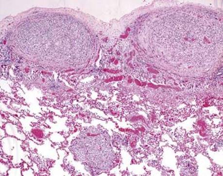
Fig 1 Metastatic melanoma. Multiple well-circumscribed subpleural and parenchymal metastases of malignant melanoma.
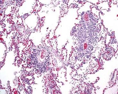
Fig 2 Metastatic melanoma. Lymphangitic spreading of malignant melanoma in perivascular spaces.
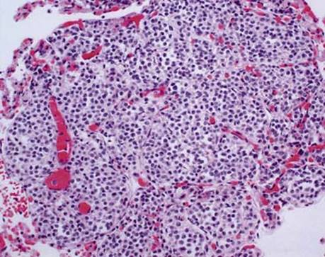
Fig 3 Metastatic melanoma. Nested pattern of growth of metastatic melanoma composed of bland appearing monotonous cells with abundant nuclear grooves.
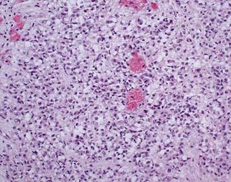
Fig 4 Metastatic melanoma. Same case as in Fig 3 with another melanoma nodule displaying prominent cellular pleomorphism with cytoplasmic clearing and signet-ring features.
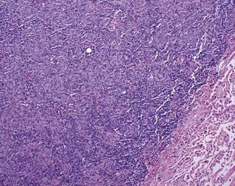
Fig 5 Metastatic melanoma. Well-circumscribed large melanoma lung metastasis with abundant lymphoplasmacytic infiltrate and sarcomatoid histological features.
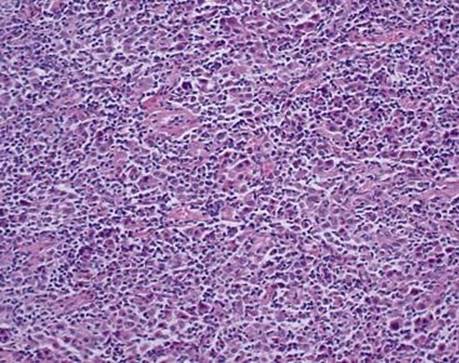
Fig 6 Metastatic melanoma. Diffuse discohesive cells of amelanotic melanoma composed of polygonal and plasmacytoid pleomorphic cells mimicking lymphoma.
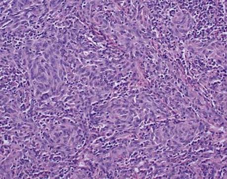
Fig 7 Metastatic melanoma. Amelanotic area of melanoma consisting of closely packed spindle cells with prominent nucleoli mimicking sarcoma.
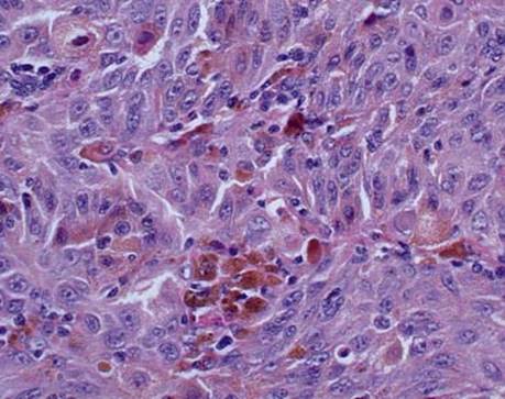
Fig 8 Metastatic melanoma. Melanin production by melanoma cells and accumulation in melanophages are very helpful but quite uncommon features at metastatic site. Note prominent nucleoli.