
Definition
• Inflammation of the pleura
Clinical features
Epidemiology
• May affect any age group
• Causes include infection, collagen vascular disease, neoplastic infiltration, uremia, trauma, iatrogenic, drug-induced, or idiopathic
Presentation
• Chest pain, shallow respiration, and evidence of pleural friction and effusion
• Sharp, sudden chest pain due to inflammation of parietal pleura (worse on deep inspiration) with localization directly over the affected area (and possible radiation of pain to the shoulder, head, or epigastrium)
Prognosis and treatment
• Treatment and prognosis based on the underlying etiology
Pathology
Histology
• Acute pleuritis:
• May be either fibrinous pleuritis or suppurative pleuritis:
– Fibrinous pleuritis is histologically characterized by fibrinous exudates on the visceral and/or parietal pleural surfaces. Acute inflammatory cells are not present or rarely seen
– Suppurative pleuritis is histologically characterized by an intense inflammatory reaction with neutrophils
• Orderly arrangement of vessels with a zonation effect, perpendicular to the pleural surface, can be appreciated
• Chronic pleuritis:
• Mild to moderate infiltration by lymphocytes and plasma cells; vascular arrangement and zonation effect similar to acute pleuritis
• Fibrosing pleuritis:
• Often associated with other pathological conditions such as tumor or cardiovascular disease (rarely cryptogenic)
• Acellular fibrous thickening of the pleura with focal plaquelike features
• Blood vessels are perpendicular to the pleural surface
• No invasive mesothelial growth or necrosis is present
• Eosinophilic pleuritis:
• Classically associated with pneumothorax, hemothorax, drugs, or infection
• Fibrous pleural thickening with mesothelial hyperplasia and infiltration by eosinophils, histiocytes, and lymphocytes
• Granulomatous pleuritis:
• Granulomas may be either necrotizing or nonnecrotizing
• Usually seen as a result of foreign body reaction or pleural involvement by sarcoidosis; rarely from Wegener granulomatosis
• Less commonly associated with infection (specifically. tuberculosis or fungal infections)
• Xanthomatous pleuritis
• Prominent accumulation of foamy macrophages and fat necrosis
• Can be associated with trauma or a complication of pancreatitis
Immunopathology/special stains
• Macrophages are CD68 positive, CD1a negative
• RCC, S100, and Melan-A negative
• Polyclonal lymphocytes and plasma cells
Main differential diagnoses
• Malignant lymphoma
• Desmoplastic malignant mesothelioma
• Langerhans cell histiocytosis
• Renal cell carcinoma or malignant melanoma

Fig 1 Pleuritis. In this patient with fibrinous pleuritis, note markedly reactive mesothelial cells entrapped within the fibrin, which may be mistaken for malignancy.
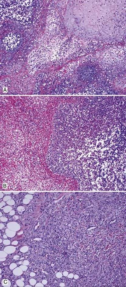
Fig 2 Pleuritis. Acute suppurative pleuritis with an extensive inflammatory cell exudate and necrosis (A). Layers of fibrin are seen admixed with neutrophils (B). Granulation tissue extending into underlying fat (C).

Fig 3 Pleuritis. Fibrosing pleuritis with fibrin layer at the top and granulation tissue underneath (A). Note paucity of inflammatory cells. Blood vessels within fibrosis are evenly spaced, parallel to each other, and oriented perpendicular to the surface (these features differentiate it from desmoplastic mesothelioma) (B).

Fig 4 Pleuritis. Eosinophilic pleuritis is seen in this patient with spontaneous pneumothorax. Note the presence of scattered eosinophils and few plasma cells. There is intravascular margination of neutrophils.

Fig 5 Pleuritis. Talc pleurodesis had been performed in this patient with recurrent pleural effusion. Numerous talc crystals, which are strongly birefringent under polarized light (A), are seen within multinucleated foreign body giant cells within the pleura (B). Underlying lung is seen on the right.

Fig 6 Pleuritis. Sarcoidosis involving the pleura. Note characteristic perigranulomatous fibrosis.

Fig 7 Pleuritis. This patient with tuberculosis of the pleura had both necrotizing and nonnecrotizing granulomas on biopsy. Only a single acid-fast bacillus could be identified after examining multiple AFB stained slides (not illustrated here).

Fig 8 Pleuritis. Granulomatous pleuritis with nonnecrotizing granulomas and marked chronic inflammation.