
Definition
• A reactive mesothelial proliferation that may mimic mesothelioma or metastatic carcinoma
Clinical features
Etiology
• Common causes include infection, trauma, pneumothorax, surgery, connective tissue disease, pulmonary infarcts, drug reactions, subpleural lung carcinomas, nonspecific inflammation
Presentation
• Commonly seen as pleural effusion; other presentations vary by underlying cause
Prognosis and treatment
• Treat underlying cause
• Pleurocentesis for pleural effusion
• Prognosis varies by underlying cause; usually much better than malignant mesothelioma
Pathology
Histology
• Flat, single layer or multilayer mesothelial cell growth, sometimes with papillary formation
• Features that may mimic a neoplasm include high cellularity, cytological atypia, high mitotic activity, papillary formation, necrosis, and entrapment of mesothelial cells within fibrous tissue mimicking invasion
• Reactive mesothelial hyperplasias tend to have a uniformity of growth that have regular sheets and sweeping fascicles of cells that respect mesothelial boundaries
Immunopathology/special stains
• Positive for mesothelial markers (calretinin, WT-1, CK5/6, and D2-40)
• Pankeratins are used to highlight the uniform growth of reactive mesothelial cells
• Desmin is often positive
• EMA, p53, and GLUT-1 are usually negative
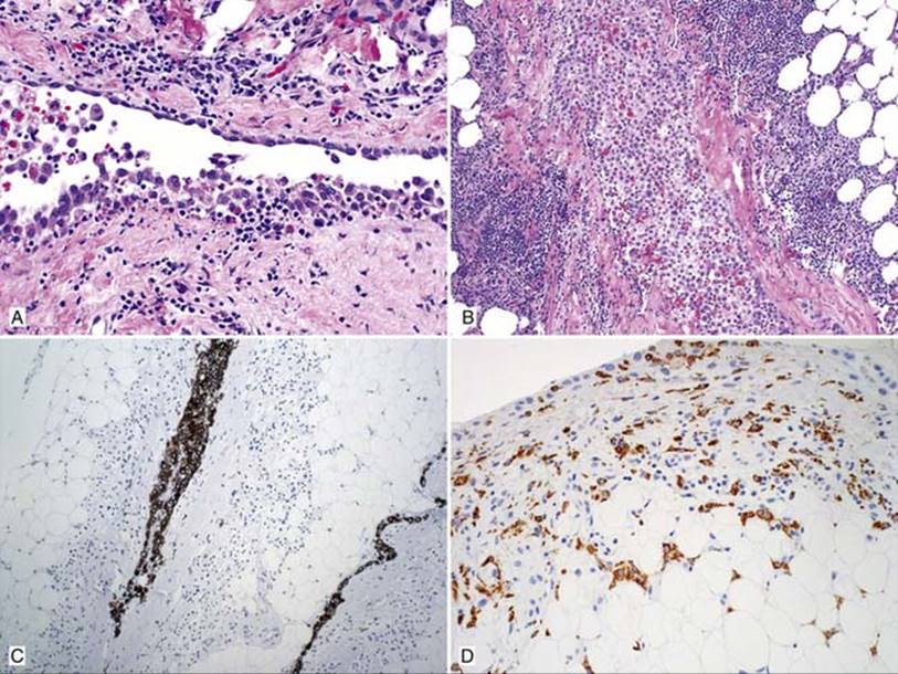
Fig 1 Mesothelial hyperplasia. Reactive mesothelial hyperplasia showing single (top) to multiple (bottom) layers of reactive mesothelial cells with inflammation and hemosiderin (A); showing sheet of benign mesothelial cells (center), which appears to be within fibrous tissue (B); however, this appearance is due to poor orientation. Note associated chronic inflammation with lymphocytes, plasma cells, and histiocytes; CAM5.2 is very useful for demonstrating linear growth of mesothelial cells without invasion (C); and CD68 confirms the mixed macrophages and those within the adipose tissue (D).
Main differential diagnoses
• Epithelioid mesothelioma (see Web Table 2 on the Expert Consult website):
• Key feature is to demonstrate stromal/fat invasion
• Mitotic activity or cytological atypia is not useful in distinguishing them
• Metastatic carcinoma: positive carcinoma markers (MOC31, BG8, BerEp4) and negative mesothelial markers
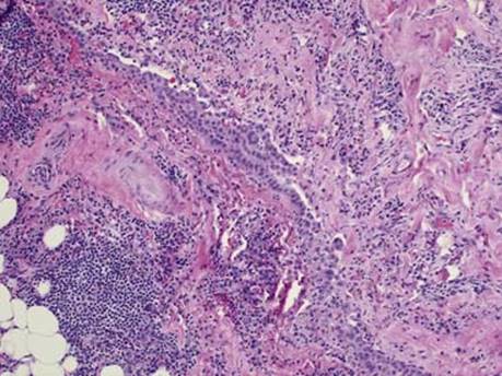
Fig 2 Mesothelial hyperplasia. Reactive mesothelial hyperplasia with fibrous tissue mimicking invasion.
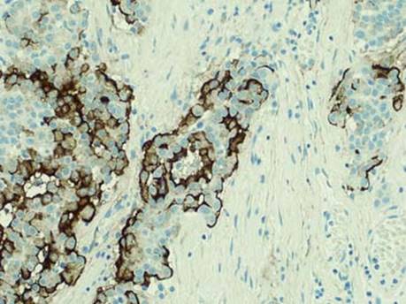
Fig 3 Mesothelial hyperplasia. Epithelioid malignant mesothelioma is positive for EMA immunostain with a membranous staining pattern, whereas reactive mesothelium would be negative.
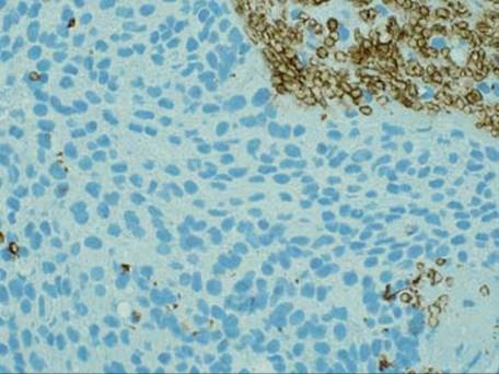
Fig 4 Mesothelial hyperplasia. Reactive mesothelial hyperplasia is negative for GLUT-1 immunostain. As a positive internal control, red blood cells (top right) are strongly positive.
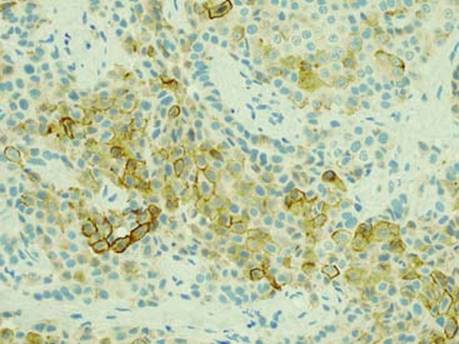
Fig 5 Mesothelial hyperplasia. Epithelioid malignant mesothelioma is positive for GLUT-1 immunostain with a membranous staining pattern.
WEB TABLE 2 Reactive Mesothelial Hyperplasia Versus Epithelioid Mesothelioma
|
MESOTHELIAL HYPERPLASIA |
EPITHELIOID MESOTHELIOMA |
|
|
Uniformity of growth (highlighted with cytokeratin staining) |
Uniform: regular sheets and sweeping fascicles; respect each other’s boundaries |
Disorganized growth: haphazardly intersecting proliferation |
|
Invasion |
Absence of stromal/fat invasion (pitfall: entrapment can mimic invasion) |
Stromal/fat invasion usually apparent (highlighted with cytokeratin staining) |
|
Architecture |
Simple papillae; single cell layers; loose sheets of cells without stroma or prominent cellularity within mesothelial space (not in stroma) |
Complex papillae, tubules, cellular stratification; expansile nodules surrounded by stroma; sometimes “bulky tumor” may involve the mesothelial space without obvious invasion |
|
Inflammation |
Common |
Usually minimal |
|
Necrosis |
Rare |
Present occasionally |
|
EMA |
Usually negative |
Often positive |
|
p53 |
Usually negative |
Often positive |
|
GLUT-1 |
Negative |
Often positive |
|
Desmin |
Often positive |
Often negative |