
Definition
• A variant of malignant mesothelioma (MM) composed of spindle cells
Clinical features
Epidemiology
• Less common than epithelioid and biphasic variants
Presentation
• Same as other pleural mesotheliomas
Prognosis and treatment
• Worse prognosis than epithelioid and biphasic variants
• Usually not treated with surgical resection because of poor outcome
Pathology
Histology
• Malignant spindle cells growing in short fascicles, typically with a storiform pattern or a haphazard distribution, within a fibrous stroma
• Mostly bland cytological features; tumor cells may be plump, with relatively uniform nuclei
• Mitotic rate varies; may demonstrate anaplastic and bizarre giant cells, similar to pleomorphic sarcoma
• In a small percentage of cases, areas of osteosarcomatous or chondrosarcomatous differentiation may be present
Immunopathology/special stains
• Initial panel should include cytokeratins, calretinin, and D2-40
• All mesothelial markers may be negative
• Multiple cytokeratin antibodies including AE1/AE3, CAM5.2 (or CK18), and CK7 should be used, as expression can be focal, weak, and variable
• Osteosarcomatous and chondrosarcomatous areas often stain negatively for cytokeratins
• May stain positive for vimentin, desmin, actin, or S100
• 90% to 100% of sarcomatoid malignant mesotheliomas have homozygous deletion of p16
Main differential diagnoses
• Sarcomatoid carcinoma of lung involving pleura
• Pleomorphic sarcoma
• Osteosarcoma
• Chondrosarcoma
• Solitary fibrous tumor: usually localized; CD34 positive
• Metastatic sarcomatoid renal cell carcinoma
• Metastasizes to pleura and grows in a pseudomesotheliomatous sarcomatoid pattern
• Both are keratin positive
• Metastatic sarcomatoid renal cell carcinomas are negative for calretinin and D2-40
• Clinical and radiological correlation may be helpful
• Synovial sarcoma of the pleura:
• Usually seen as localized solid tumors; sometimes can be seen with diffuse pleural thickening similar to MM
• It should be considered when there is a highly cellular neoplasm with very little cytoplasm (resulting in nuclear overlapping), focal hemangiopericytoma-like blood vessels, and limited keratin expression
• The diagnosis can be confirmed by molecular testing for X;18 translocation
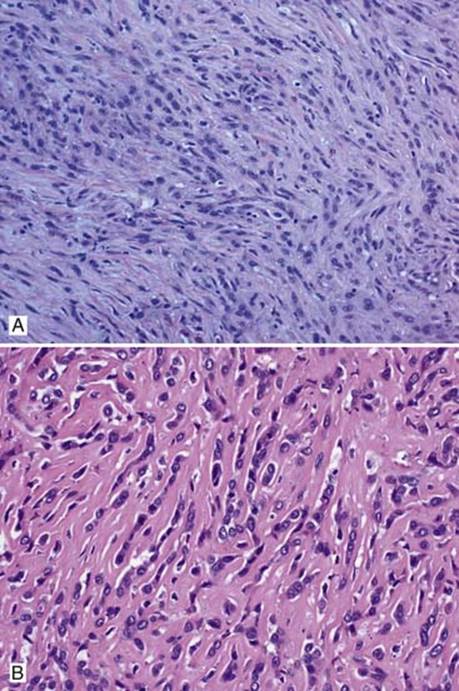
Fig 1 Sarcomatoid malignant mesothelioma. A, Sarcomatoid MM with neoplastic bland spindle cells growing in short fascicles with a haphazard or storiform pattern. B, Epithelioid MM infiltrating in cords with dense fibrosis, mimicking sarcomatoid mesothelioma.

Fig 2 Sarcomatoid malignant mesothelioma. The degree of pleomorphism is variable in sarcomatoid MM; mild to moderate in A and severe with bizarre anaplastic giant cells, mimicking pleomorphic sarcoma (B).

Fig 3 Sarcomatoid malignant mesothelioma. Sarcomatoid MM with bone formation.

Fig 4 Sarcomatoid malignant mesothelioma. Sarcomatoid MM with necrosis (lower right corner).
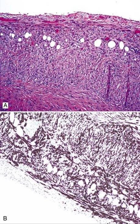
Fig 5 Sarcomatoid malignant mesothelioma. Sarcomatoid MM infiltrating adipose tissue (A), confirmed by pancytokeratin CAM5.2 immunostain (B).
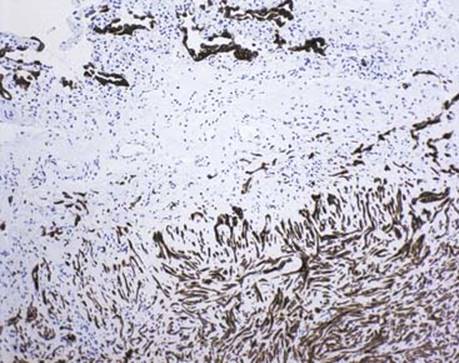
Fig 6 Sarcomatoid malignant mesothelioma. CAM5.2 immunostain shows sarcomatoid MM (lower right portion) involving visceral pleura without invasion of the lung parenchyma (upper left portion:pneumocytes with CAM5.2 positivity).

Fig 7 Sarcomatoid malignant mesothelioma. Sarcomatoid MM with positive GLUT-1 staining, which is helpful in distinguishing from pleural fibrosis.
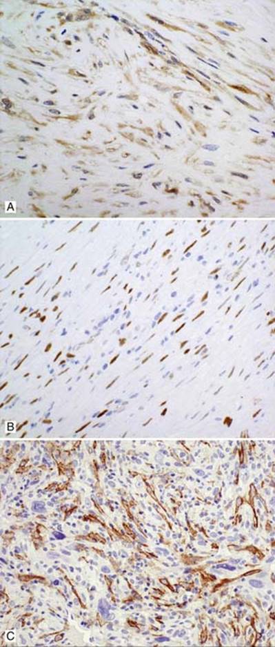
Fig 8 Sarcomatoid malignant mesothelioma. Sarcomatoid MM with positive staining for calretinin (A), WT-1 (B), and D2-40 (C).