
Definition
• Fibroblastic neoplasm with abundant collagen fibers and hemangiopericytoma (HPC)-like blood vessels
Clinical features
Epidemiology
• Rare
• Occurs in adults aged 20 to 70 years (average, 50 years)
Presentation
• Slowly growing, well-defined mass
• Most common symptoms are coughing, chest pain, and dyspnea
• May cause paraneoplastic syndromes:
• Doege-Potter syndrome: hypoglycemia secondary to production of insulin-like growth factor
• Pierre Marie-Bamberger syndrome: hypertrophic osteoarthropathy commonly seen as finger clubbing occurs in up to 35% of patients with SFT
Prognosis and treatment
• Usually behaves in a benign fashion; 10% to 15% are aggressive
• Metastases can occur to the lungs, liver, bone, or brain
• Treatment consists of surgical excision
• Patients must have follow-up because of an inability to predict which tumors will behave more aggressively
Pathology
Gross
• Most commonly attached to the visceral pleura; however, they can be attached to the parietal pleura, within the lung parenchyma, in the pericardium, or, rarely, within the heart
• Well-circumscribed, partially encapsulated
• Average size of 5 to 8 cm (range, 1-25 cm)
• Cut surface is multinodular, firm, and white
• Usually lacks necrosis
• Malignant tumors may have hemorrhage and necrosis and may locally invade surrounding tissue
Histology
• Benign SFT:
• Alternating hypocellular and hypercellular areas
• Branching HPC-like vessels
• Thick bands of collagen; may be keloid-like collagen
• Fibroblast-like cells not arranged in any pattern
• Round to spindle nuclei with vesicular chromatin
• Low mitotic activity
• May contain multinucleated cells or adipocytes
• Malignant SFT:
• Hypercellular tumor with cytological atypia
• Necrosis often present
• Increased mitotic activity (≥4 mitoses per high-power fields)
• Infiltrative growth pattern
• May have myxoid areas
Immunopathology/special stains
• Tumor cells are CD34+ and CD99+
• One third of cases are positive for at least one of the following: epithelial membrane antigen (EMA), Bcl-2, and smooth muscle actin
Main differential diagnosis
• Sarcomatous carcinoma: keratin positive, CD34 negative
• Sarcomatous mesothelioma: keratin usually positive, CD34 negative
• Includes other tumors with HPC-like vessels:
• HPC
• Myofibroma
• Synovial sarcoma: often biphasic tumor, keratin, EMA, and TLE-1 positive
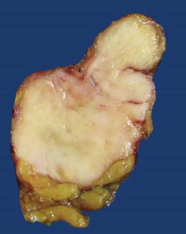
Fig 1 Solitary fibrous tumor. Gross image of benign SFT, which is well-circumscribed with a firm yellow-white cut surface.
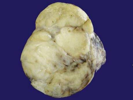
Fig 2 Solitary fibrous tumor. Gross image of a formalin fixed malignant SFT that has a gray-white, less homogenous cut surface.
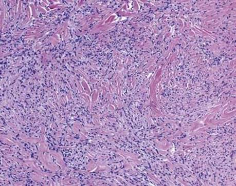
Fig 3 Solitary fibrous tumor. Benign SFT; high-power view of area with ropey collagen.
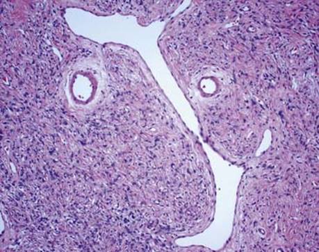
Fig 4 Solitary fibrous tumor. Benign SFT; high-power view of a characteristic branching HPC-like vessel.
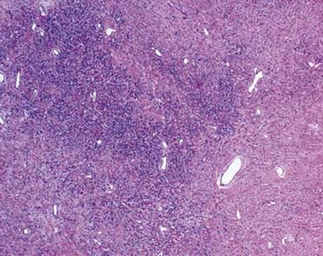
Fig 5 Solitary fibrous tumor. Benign SFT; intermediate-power view showing hypocellular and hypercellular areas of spindled cells.
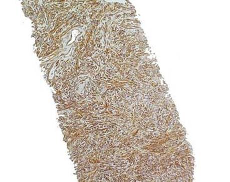
Fig 6 Solitary fibrous tumor. Core needle biopsy of benign SFT; CD34 stain is strongly positive.
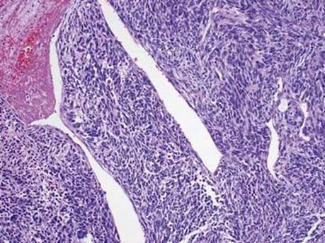
Fig 7 Solitary fibrous tumor. Malignant SFT; hypercellular tumor with cellular atypia and HPC-like vessels.
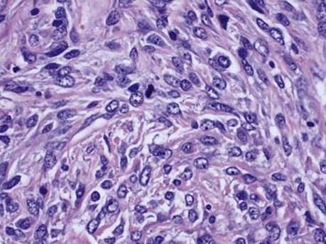
Fig 8 Solitary fibrous tumor. Malignant SFT; high-power view showing tumor cells with high nuclear cytoplasmic ratio, mitotic figure, and interspersed collagen.