
Definition
• Rare, slow-growing plaquelike fibrous lesion of the visceral pleura
Pathogenesis
• The pathogenesis is unknown
• It is postulated to represent a late sclerosing stage of inflammatory myofibroblastic tumor (IMT)
Clinical features
Epidemiology
• Lesions most commonly affect children and young adults and arise in subcutaneous and deep soft tissue of the extremities
• Rare cases have been reported in the mediastinum, pleura, and visceral peritoneum
Presentation
• Most often asymptomatic but may present with chest pain
• Radiologically appear as well-demarcated (<12 cm in diameter) single or multiple pleural-based masses
Prognosis and treatment
• Surgical excision is adequate; low frequency of local recurrence
Pathology
Histology
• Well-circumscribed, unencapsulated masses of hyalinized, paucicellular, collagenized fibrous tissue without laminar architecture
• Possibly scant lymphoid aggregates, giant cells, or necrosis and typical psammomatous and dystrophic calcification
• Lesions do not involve underlying lung parenchyma
Immunopathology/special stains
• Vimentin positive
• ALK-1, P80, CD34, and keratin negative
Main differential diagnoses
• Solitary fibrous tumor
• Pleural plaques
• Chronic fibrous pleuritis
• Calcified fibrous plaques
• IMT

Fig 1 Calcifying pseudotumor of pleura. Low power shows well-circumscribed fibrous tumor within which are embedded rounded foci of calcifications.
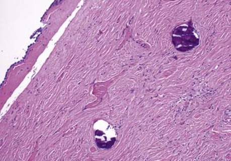
Fig 2 Calcifying pseudotumor of pleura. Higher power shows surface mesothelial lining overlying the tumor.

Fig 3 Calcifying pseudotumor of pleura. Highest power shows bland fibrous tissue containing characteristic psammomatous calcifications.