
Refer to Pulmonary Synovial Sarcoma for additional details.
Definition
• A mesenchymal tumor displaying variable epithelial differentiation
• Has characteristic chromosomal translocation t(X;18)(p11;q11)
Clinical features
Epidemiology
• Most often affects young to middle-aged adults
• Biphasic tumors tend to occur in younger patients
Presentation
• Chest pain, pleural effusion, dyspnea, dysphagia, pneumothorax
Prognosis and treatment
• Generally poor prognosis; surgical and adjuvant therapies are main treatments
Pathology
Gross
• Usually seen as a solitary, solid tumor
• May be seen as diffuse pleural thickening that mimics mesothelioma
Histology
• The histological features of pleural SS are identical to its pulmonary counterpart (refer to Pulmonary Synovial Sarcoma section)
• The most common histological type is biphasic
Immunopathology/special stains
• Immunohistochemical profile is identical to its pulmonary counterpart
Main differential diagnoses
• Malignant mesothelioma
• Sarcomatoid carcinoma from the lung
• Solitary fibrous tumor
• Metastatic SS

Fig 1 Pleural synovial sarcoma. Biphasic SS comprises epithelial and spindle cell components; the epithelial cells have ovoid to round nuclei and abundant eosinophilic cytoplasm. The tumor cells form a cribriform glandular structure; the spindle cell component is composed of monotonous plump fusiform cells in a myxoid stroma; two components are closely intermingled.

Fig 2 Pleural synovial sarcoma. In this biphasic tumor the epithelioid cells form solid islands.
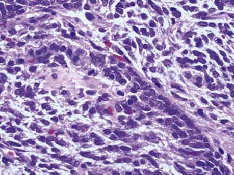
Fig 3 Pleural synovial sarcoma. High power view of SS: epithelioid cells are arranged in a cord, with round nuclei, adjacent to oval and spindled cells. The spindle tumor cells are uniform and have ovoid nuclei with fine chromatin, inconspicuous nucleoli, and scant cytoplasm.
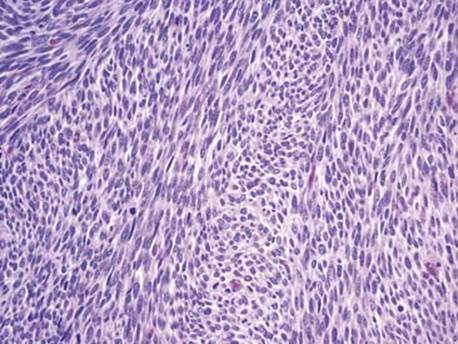
Fig 4 Pleural synovial sarcoma. Monophasic spindle cell SS is composed of intersecting fascicles of uniform spindle cells with ovoid nuclei and inconspicuous nucleoli; frequent mitotic figures are present.

Fig 5 Pleural synovial sarcoma. EMA staining of spindle cells in monophasic SS.