
Definition
• A primitive, polyphenotypic, and highly aggressive neoplasm of serosal surfaces with extremely rare reports of pleural involvement
Clinical features
Epidemiology
• Very few cases of pleural DSRCT reported in the literature; lesions mainly occur in abdominal, pelvic, and paratesticular locations
• Peak incidence in the second and third decades; male predominance (4:1)
Presentation
• Patients present with chest pain and radiological evidence of pleural effusion
• Most present with nodular masses encasing the lungs, although case reports of presentation with pleural studding or as pedunculated lesions have been reported
• Paravertebral lesions may lead to scoliosis
Prognosis and treatment
• Treatment is catered to stage of disease, varying from chemoradiation therapy to surgical management.
• Lower stage cases are managed surgically followed by radiation and chemotherapy
• There are no data to support a benefit of tumor debulking in higher stage cases
• Because of its rare incidence, a prognosis for pleural DSRCT is not available in the literature. However, in its usual locations, it is an aggressive tumor, often unresectable at presentation; most patients die within the first 5 years
Pathology
Histology
• Nests of small-to-intermediate round cells with scant cytoplasm and nuclear molding within a dense fibrous or hypercellular spindle stroma
• Larger cells with rhabdoid features, multinucleated sarcomatoid cells, larger epithelioid cells with foci of anaplasia, and signet-ring cells may be seen
Immunopathology/special stains
• Keratin, EMA, WT1 (nuclear), desmin (dotlike) and vimentin positive
• t (11;22) (p13:q12) that causes an EWS-WT1 fusion can be demonstrated by cytogenetics
Main differential diagnoses
• Extraskeletal Ewing sarcoma and primitive neuroectodermal tumor
• Alveolar rhabdomyosarcoma
• Desmoplastic round cell mesothelioma; rare in children
• Small-cell carcinoma; rare in children

Fig 1 Desmoplastic small round cell tumor of the pleura. Low power (A) and high power (B) show nests of small round blue cells with characteristic desmoplastic stroma separating the tumor nests. Note focal necrosis.
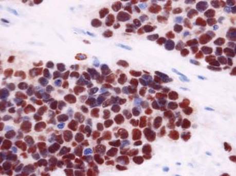
Fig 2 Desmoplastic small round cell tumor of the pleura. WT-1 staining reveals strong nuclear positivity in the tumor cells.