
Definition
• PEL is a rare, aggressive, non-Hodgkin lymphoma (NHL) that affects body cavities and occurs mainly, but not exclusively, in HIV-positive patients
• PEL is defined by the presence of Kaposi sarcoma herpes virus (KSHV)/human herpes virus-8 (HHV-8)
Pathogenesis
• PEL represents the malignant counterpart of a B cell that has reached a mature stage of development and is shifting toward terminal plasma cell differentiation
• The exact mechanism by which KSHV/HHV-8 promotes oncogenesis in PEL is unclear
• Three latent gene products that are thought to play significant roles are latent nuclear antigen-1 (LANA1; ORF73), viral cyclin (v-Cyc, ORF72), and viral FLICE inhibitory protein (v-FLIP, ORF71)
• Infection by Epstein-Barr virus (EBV) is reported in a large proportion of PEL cases
• Moreover, PELs are devoid of the chromosomal translocations associated with other aggressive B-cell NHLs
Clinical features
Epidemiology
• PEL accounts for approximately 4% of AIDS-related lymphomas but only for 0.3% of aggressive lymphomas in HIV-negative patients
• PEL may also occur in other immunodeficiency states such as after solid organ transplantation
Presentation
• The classic patient is an HIV-positive middle-aged homosexual male
• Typically, patients with PEL present with effusions in the pleural, pericardial, and/or abdominal cavities, usually in the absence of an obvious tumor mass, lymphadenopathy, or hepatosplenomegaly
• Although the majority of PELs occur exclusively as lymphomatous effusions, some patients may either subsequently develop or initially present with solid tissue masses, so-called extracavitary PEL
Prognosis and treatment
• The prognosis of PEL is poor: median survival is less than 6 months
• Standard Ann Arbor staging for PEL is not relevant because, by definition, all cases of PEL are stage IV. Similarly, the International Prognostic Index has not been validated in a cohort of patients with PEL
• The strongest predictor of poor clinical outcome in the population of HIV-infected individuals is the absence of highly active antiretroviral therapy (HAART) before PEL diagnosis
• The significant improvement of AIDS-related lymphoma prognosis observed in the HAART era also applies to the PEL setting. However, the improvement in patients with PEL is below that reported in patients with HIV infection and diffuse large B-cell lymphoma
• Despite the improvement in therapeutic strategies during the last few years, there is no evidence of a cure for PEL patients; chemotherapy regimens have been similar to other aggressive NHLs
Pathology
Histology
• The neoplastic cells are pleomorphic and show a range of cytomorphological appearances from features of large immunoblastic cells to those of anaplastic cells
• These cells frequently display a certain degree of plasma cell differentiation
Immunopathology/special stains
• The lymphoma cells typically express CD45 but are usually negative for B-cell markers (such as CD19, CD20, CD79a, and PAX5), in spite of being a B-cell lymphoma
• They usually lack expression of both immunoglobulin heavy and light chains and OCT2
• Tumor cells show a high proliferative index (almost 100% Ki-67 positive)
• Activation markers (such as CD30) and markers associated with plasma cell differentiation (such as CD138/Syndecan-1, MUM1/IRF4, and BLIMP1) are expressed in most cases
• Detecting evidence of viral infection by KSHV/HHV-8 in the neoplastic cells is essential for the diagnosis of PEL. Immunostaining for ORF73/LANA1 is the standard diagnostic assay
• A majority of cases show coinfection of neoplastic cells with EBV, as can be detected by in situ hybridization (using EBER probe); immunostains for latent membrane proteins are usually negative
• Despite its frequent indeterminate phenotype, PEL is consistently represented by a monoclonal B-cell population on immunogenotypic studies
Main differential diagnoses
• Serous effusions, particularly pleural effusions, are a frequent complication of a range of lymphoma subtypes such as Hodgkin lymphoma, Burkitt lymphoma, T-lymphoblastic lymphoma, and anaplastic large-cell lymphoma. PEL can be readily differentiated from these by its immunoprofile and positivity for KSHV/HHV-8–associated LANA-1
• Primary involvement of serous cavities without solid masses can be seen in both extranodal large-cell lymphoma and extranodal Burkitt lymphoma, both of which are negative for KSHV/HHV-8 and are positive for B-cell markers. In addition, Burkitt lymphoma is typically positive for c-MYC
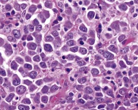
Fig 1 Primary effusion lymphoma. The neoplastic cells are large and pleomorphic with ample cytoplasm and prominent nucleoli. Some cells show plasmacytoid differentiation.

Fig 2 Primary effusion lymphoma. There is diffuse nuclear positivity for ORF73/LANA1.

Fig 3 Primary effusion lymphoma. In situ hybridization for EBV (using EBER probe) is positive.