
Three compartments
• Thymic epithelial compartment
• The site of T-cell maturation
• Composed of thymic epithelial cells and thymic lymphocytes (thymocytes), which are arranged into two morphologically distinctive areas (cortex and medulla)
• Cortex contains numerous lymphocytes with scattered epithelial cells
• Medulla contains more epithelial cells and fewer lymphocytes
• Perivascular space compartment
• Contains blood vessels
• More prominent with aging and in thymic diseases
• Replaced with fat and lymphocytes in normal involuted thymus
• Stromal compartment
• Contains other cell types, such as neuroendocrine cells, Langerhans cells, and myoid cells
Organotypic features of mature thymus of childhood and adolescence
• Fibrous capsule
• Lobular architecture
• Sharp separation of cortex and medulla
• Dual cell population of epithelial cells and thymocytes
• Thymocytes (thymic T lymphocytes)
– The predominant cell population in the cortex
– Varying in size and maturation and forming a gradient from outer cortex to corticomedullary junction and into medulla; the outer cortical thymocytes are larger and mitotically active lymphoblasts and the inner cells are small mature T lymphocytes
• Epithelial cells
– Epithelial cells in the cortex
– Scattered but form a loose network to provide a specialized microenvironment for T-cell maturation and differentiation
– Large plump cells with round to oval nuclei, vesicular chromatin, and abundant cytoplasm
– Epithelial cells in medulla
– Abundant
– Some form “Hassall corpuscles”
– Concentric keratinized structures
– Reactive changes to inflammation
– May undergo cystic degeneration, dystrophic calcification, or infiltration of inflammatory cells and accumulation of foamy macrophages
• Perivascular spaces
• May contain lymphoid follicles with germinal centers
– Prominent in patients with thymic hyperplasia, myasthenia gravis, and other immune-mediated diseases
– Also present in normal thymus without clinical significance
Normal physiological age-induced thymic involution
Definition
• The process of thymus aging after it reaches its maximum weight at puberty (approximately 30 g)
Gradual atrophic changes of different compartments of the thymus
• Epithelial compartment becomes progressively reduced
• Decrease in the number of thymocytes
• Epithelial cells become small, oval- to spindle-shaped with hyperchromatic nuclei and scant cytoplasm
• Residual epithelial elements form small, microscopic, rosettelike epithelial structures
• Residual Hassall corpuscles undergo dilatation and form abundant cystic structures
• The perivascular spaces become more prominent and are replaced with adipose tissue
Organotypic features of involuted thymus of adults
• Mature adipose tissue or collagenized stroma
• Microscopic epithelial remnants with islands or anastomosing strands of epithelial cells mixed with small lymphocytes and abortive Hassall corpuscles
• Hassall corpuscles may have calcifications or cystic degeneration
Immunopathology/special stains
• Thymic epithelial cells:
• Positive for cytokeratins (e.g., AE1/AE3, CAM5.2, and CK7, except CK20)
• Positive for p63 and CK5/6
• Negative for CD5 and CD70
• Outer cortical T lymphoblasts are CD1a+, CD4+, CD8+, CD99+, TdT+, and CD5+; have high Ki-67 index (approximately 90%)
• Mature T lymphocytes in inner cortex and medulla are CD3+, CD5+, CD1a–, CD99–, and TdT– and have low proliferation

Fig 1 Normal thymus. Normal lobular architecture of thymus low (A) and medium (B) powers; note clear separation of cortex and medulla
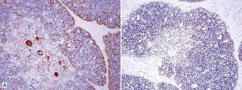
Fig 2 Normal thymus. Low-power immunostains of normal thymus highlight the loose epithelial networks in cortex and Hassall corpuscles in medulla: AE1/AE3 (A) and CAM5.2 (B).
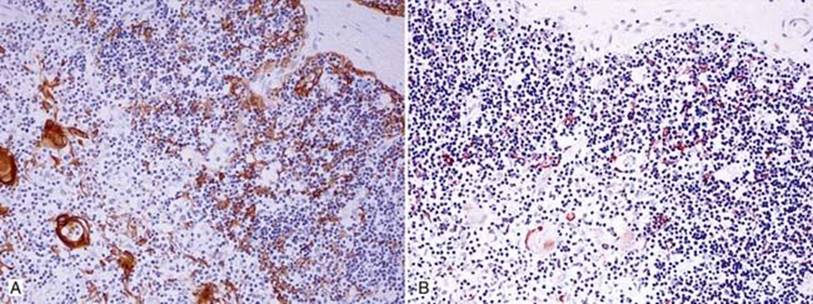
Fig 3 Normal thymus. Medium-power immunostains of normal thymus AE1/AE3 (A) and CAM5.2 (B); note the loose epithelial network.

Fig 4 Normal thymus. Normal involuted thymus: low power (A); note the residual microscopic islands of thymic remnants in a background of adipose tissue; medium power (B); note the residual epithelial elements with rosette formation.
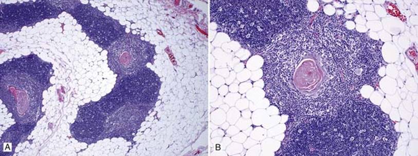
Fig 5 Normal thymus. Normal involuted thymus: low (A) and medium (B) powers; note clear corticomedullary separation and prominent Hassall corpuscles in medulla.

Fig 6 Normal thymus. Normal involuted thymus: low (A) and medium (B) powers; note calcified Hassall corpuscles.