
Definition
• Unilocular thymic cyst (UTC) is a single epithelial-lined cyst within thymic parenchyma
• Multilocular thymic cyst (MTC) consists of multiple epithelial-lined cystic spaces within the thymus
Clinical features
Epidemiology
• Thymic cysts are rare lesions, representing only 5% of all mediastinal cysts
• UTCs are a developmental remnant of the third branchial pouch and are usually seen in pediatric patients
• MTCs occur more often in adults and are acquired or reactive in nature
• MTCs can be associated with HIV, Sjögren’s syndrome, previous thoracic surgery, or neoplasms
Presentation
• Usually asymptomatic anterior mediastinal lesions found incidentally on imaging
• Because of pressure on surrounding structures, they can cause superior vena cava syndrome, Horner’s syndrome, cardiac compression, dyspnea, dysphagia
Prognosis and treatment
• If there is no associated neoplasm, these are benign lesions with an excellent prognosis
• Exceptionally, malignant transformation to squamous cell carcinoma can occur
• Treatment consists of surgical excision
Pathology
Gross
• UTC
• Unilocular, thin-walled, translucent cyst filled with clear fluid
• MTC
• Large size, sometimes with extension into surrounding tissue
• Multiple cysts filled with cloudy or bloody fluid
• Thick, fibrous septae separate the cystic spaces
Histology
• UTC
• Cyst may be lined by squamous, cuboidal, or columnar epithelium
• Thymic tissue is present within the cyst wall
• Lacks inflammation
• MTC
• Cysts are usually lined by squamous epithelium but can have ciliated columnar epithelium or exhibit pseudoepitheliomatous hyperplasia
• Fibrosis and inflammatory infiltrate in cyst walls
• Residual thymic tissue within wall of cyst
• Other features that may be present include:
– Cholesterol cleft granulomas
– Dilated Hassall corpuscles
– Lymphoid hyperplasia
– Hemorrhage
• Thymomas and carcinomas can occur within MTCs. Basaloid carcinoma of the thymus has a predilection for arising within MTCs
Immunopathology/special stains
• Not contributory
Main differential diagnoses
• Nodular sclerosis Hodgkin lymphoma with cystic change
• Seminoma with cystic change
• Bronchogenic cyst: lined by ciliated respiratory epithelium with bronchial glands, cartilage, and/or smooth muscle
• Pericardial cyst: lined by a single layer of mesothelium
• Cystic lymphangioma
• Cystic thymoma
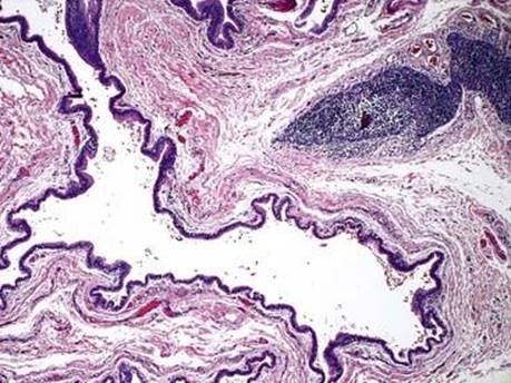
Fig 1 Unilocular and multilocular thymic cysts. MTC: low power showing small amount of thymic tissue within cyst wall.
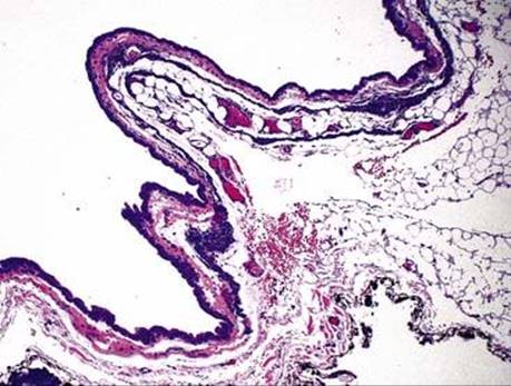
Fig 2 Unilocular and multilocular thymic cysts. Different case of UTC with compressed atrophic thymic tissue in the cyst wall.
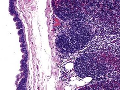
Fig 3 Unilocular and multilocular thymic cysts. MTC lined by pseudostratified columnar epithelium with thymic tissue (not atrophic) in the cyst wall.
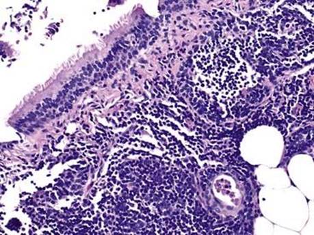
Fig 4 Unilocular and multilocular thymic cysts. High-power view of MTC showing the ciliated pseudostratified columnar epithelium and thymic tissue.

Fig 5 Unilocular and multilocular thymic cysts. MTC with thick fibrous septae between cystic spaces.
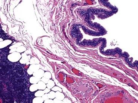
Fig 6 Unilocular and multilocular thymic cysts. MTC with pseudostratified columnar lining and thymic tissue and fat within the wall.

Fig 7 Unilocular and multilocular thymic cysts. UTC with single cystic space compressed by surrounding thymic tissue.